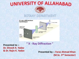
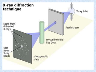
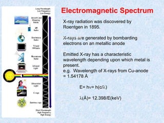
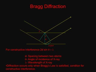
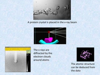
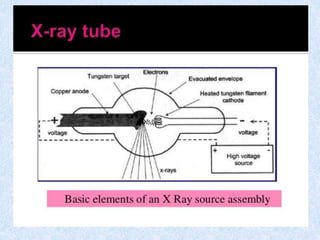
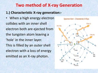
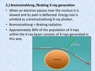
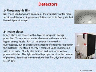
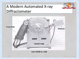
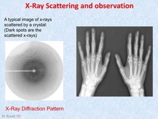
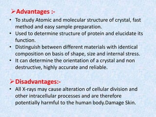
Presented by :- Faraz Ahmad Khan
(M.Sc. 3rd Semester)
“ X - Ray Diffraction ”Presented to :-
Dr. Dinesh K. Yadav
& Dr. Rajiv K. Yadav
Introduction
• X-Ray diffraction is a method of X-Ray
Crystallography, in which a beam of X-rays strikes
a sample (crystalline solid), land on a piece of a
film or other detector to produce scattered
beams.
• It is novel & non destructive method of chemical
analysis and a variety of X-Ray techniques in
practice.
• These are: 1.) X-Ray Absorption; 2.) X-Ray
Diffraction; 3.) X-Ray Fluorescence
• “Every crystalline substance gives a pattern;
the same substance always gives the same
pattern; and in a mixture of substance each
produces its pattern independently of the
others.”
• The X-Ray diffraction pattern of a pure
substance is, therefore, like a fingerprint of
the substance. It is based on the scattering of
x-rays by crystals.
• It shows the arrangement of atoms with in a
crystal.
Electromagnetic Spectrum
10-1 to 10 nm
400 to 700 nm
10-4 to 10 -1 nm
10 to 400 nm
700 to 104 nm
X-ray radiation was discovered by
Roentgen in 1895.
X-rays are generated by bombarding
electrons on an metallic anode
Emitted X-ray has a characteristic
wavelength depending upon which metal is
present.
e.g. Wavelength of X-rays from Cu-anode
= 1.54178 Å
E= hn= h(c/l)
l(Å)= 12.398/E(keV)
NMR
10 um - 10 mm
Bragg Diffraction
d
q
q
d sinq
For constructive interference 2d sin q = l
d- Spacing between two atoms
q- Angle of incidence of X-ray
l- Wavelength of X-ray
•Diffraction occurs only when Bragg’s Law is satisfied, conditon for
constructive interference.
A protein crystal is placed in the x-ray beam
The x-rays are
diffracted by the
electron clouds
around atoms
The atomic structure
can be deduced from
the data
Specimen Preparation
• Powders :-
0.1 micro m < particle size < 40 micro m
(Peak broadining) (Less diffraction)
• Bulks:- Smooth surface after polishing, specimen
should be thermal annealed to eliminate any surface
deformation induced during polishing.
Production of X-Rays
• X-rays are produced whenever high speed
electrons collide with a metal target.
• A source of electrons-hot Tungsten filament, a
high accelerating voltage between the
cathode(W) and the anode and the metal
target Cu, Al, Mo, Mg.
• The anode is a water cooled block of Cu
containing desired target metal.
Two method of X-ray Generation
1.) Characteristic X-ray generation:-
• When a high energy electron
collides with an inner shell
electron both are ejected from
the tungsten atom leaving a
'hole' in the inner layer.
This is filled by an outer shell
electron with a loss of energy
emitted as an X-ray photon.
2.) Bremsstrahlung /Braking X-ray generation
• When an electron passes near the nucleus it is
slowed and its path is deflected. Energy lost is
emitted as a bremsstrahlung X-ray photon.
• Bremsstrahlung = Braking radiation
• Approximately 80% of the population of X-rays
within the X-ray beam consists of X-rays generated in
this way.
1- Photographic film
Not much used anymore because of the availability of far more
sensitive detectors. Superior resolution due to its fine grain, but
limited dynamic range.
2- Image plates
Image plates are coated with a layer of inorganic storage
phosphor. X-ray photons excite electrons in the material to
higher energy levels. Part of the energy is emitted as
fluorescence, but an appreciable amount of energy is retained in
the material. The stored energy is released upon illumination
with a red laser. Blue light is emitted and measured with a
photomultiplier. The light emitted is proportional to the number
of photons. Ten times more sensitive than film, dynamic range
(1:104-105)
Detectors
S. Doublié © 2000
X-Ray Scattering and observation
A typical image of x-rays
scattered by a crystal:
(Dark spots are the
scattered x-rays)
X-Ray Diffraction Pattern
M. Rould ‘02
Advantages :-
• To study Atomic and molecular structure of crystal, fast
method and easy sample preparation.
• Used to determine structure of protein and elucidate its
function.
• Distinguish between different materials with identical
composition on basis of shape, size and internal stress.
• It can determine the orientation of a crystal and non
destructive, highly accurate and reliable.
Disadvantages:-
• All X-rays may cause alteration of cellular division and
other intracellular processes and are therefore
potentially harmful to the human body.Damage Skin.
Referances
• Principles and Techniques of Biochemistry and
Molecular Biology by Wilson and Walker.
• Fundamentals and Techniques of Biophysics
and Molecular Biology by Pranav Kumar.
X ray diffraction

X ray diffraction

  • 1.
    Presented by :-Faraz Ahmad Khan (M.Sc. 3rd Semester) “ X - Ray Diffraction ”Presented to :- Dr. Dinesh K. Yadav & Dr. Rajiv K. Yadav
  • 2.
    Introduction • X-Ray diffractionis a method of X-Ray Crystallography, in which a beam of X-rays strikes a sample (crystalline solid), land on a piece of a film or other detector to produce scattered beams. • It is novel & non destructive method of chemical analysis and a variety of X-Ray techniques in practice. • These are: 1.) X-Ray Absorption; 2.) X-Ray Diffraction; 3.) X-Ray Fluorescence
  • 3.
    • “Every crystallinesubstance gives a pattern; the same substance always gives the same pattern; and in a mixture of substance each produces its pattern independently of the others.” • The X-Ray diffraction pattern of a pure substance is, therefore, like a fingerprint of the substance. It is based on the scattering of x-rays by crystals. • It shows the arrangement of atoms with in a crystal.
  • 5.
    Electromagnetic Spectrum 10-1 to10 nm 400 to 700 nm 10-4 to 10 -1 nm 10 to 400 nm 700 to 104 nm X-ray radiation was discovered by Roentgen in 1895. X-rays are generated by bombarding electrons on an metallic anode Emitted X-ray has a characteristic wavelength depending upon which metal is present. e.g. Wavelength of X-rays from Cu-anode = 1.54178 Å E= hn= h(c/l) l(Å)= 12.398/E(keV) NMR 10 um - 10 mm
  • 6.
    Bragg Diffraction d q q d sinq Forconstructive interference 2d sin q = l d- Spacing between two atoms q- Angle of incidence of X-ray l- Wavelength of X-ray •Diffraction occurs only when Bragg’s Law is satisfied, conditon for constructive interference.
  • 7.
    A protein crystalis placed in the x-ray beam The x-rays are diffracted by the electron clouds around atoms The atomic structure can be deduced from the data
  • 8.
    Specimen Preparation • Powders:- 0.1 micro m < particle size < 40 micro m (Peak broadining) (Less diffraction) • Bulks:- Smooth surface after polishing, specimen should be thermal annealed to eliminate any surface deformation induced during polishing.
  • 9.
    Production of X-Rays •X-rays are produced whenever high speed electrons collide with a metal target. • A source of electrons-hot Tungsten filament, a high accelerating voltage between the cathode(W) and the anode and the metal target Cu, Al, Mo, Mg. • The anode is a water cooled block of Cu containing desired target metal.
  • 11.
    Two method ofX-ray Generation 1.) Characteristic X-ray generation:- • When a high energy electron collides with an inner shell electron both are ejected from the tungsten atom leaving a 'hole' in the inner layer. This is filled by an outer shell electron with a loss of energy emitted as an X-ray photon.
  • 12.
    2.) Bremsstrahlung /BrakingX-ray generation • When an electron passes near the nucleus it is slowed and its path is deflected. Energy lost is emitted as a bremsstrahlung X-ray photon. • Bremsstrahlung = Braking radiation • Approximately 80% of the population of X-rays within the X-ray beam consists of X-rays generated in this way.
  • 13.
    1- Photographic film Notmuch used anymore because of the availability of far more sensitive detectors. Superior resolution due to its fine grain, but limited dynamic range. 2- Image plates Image plates are coated with a layer of inorganic storage phosphor. X-ray photons excite electrons in the material to higher energy levels. Part of the energy is emitted as fluorescence, but an appreciable amount of energy is retained in the material. The stored energy is released upon illumination with a red laser. Blue light is emitted and measured with a photomultiplier. The light emitted is proportional to the number of photons. Ten times more sensitive than film, dynamic range (1:104-105) Detectors S. Doublié © 2000
  • 15.
    X-Ray Scattering andobservation A typical image of x-rays scattered by a crystal: (Dark spots are the scattered x-rays) X-Ray Diffraction Pattern M. Rould ‘02
  • 16.
    Advantages :- • Tostudy Atomic and molecular structure of crystal, fast method and easy sample preparation. • Used to determine structure of protein and elucidate its function. • Distinguish between different materials with identical composition on basis of shape, size and internal stress. • It can determine the orientation of a crystal and non destructive, highly accurate and reliable. Disadvantages:- • All X-rays may cause alteration of cellular division and other intracellular processes and are therefore potentially harmful to the human body.Damage Skin.
  • 17.
    Referances • Principles andTechniques of Biochemistry and Molecular Biology by Wilson and Walker. • Fundamentals and Techniques of Biophysics and Molecular Biology by Pranav Kumar.