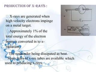
SlideShare a Scribd company logo
X-Ray Diffraction
Prashant Pandey
M.Pharm (Pharmaceutics)
INTRODUCTION:
X-RAY
o X-rays
:
were discovered in
1895 by the German physicist
Wilhelm Conrad
named
Röntgen
because
and
their
the
were so
nature
time.
was unknown at
Wilhelm Conrad Röntgen
(1845-1923)
.
o He was awarded the Nobel
prize for physics in 1901. Wilhelm Conrad Röntgen
(1845-1923)
X-ray :
 Type of high-energy radiation.In low doses,
x-rays are used to diagnose diseases by
making pictures of the inside of the body.In
high doses, x-rays are used to treat cancer.
 A relatively high-energy photon having a
wavelength in the approximate range from 0.01
to 10 nanometers.
 A stream of such photons,used for their penetrating power in
radiography,radiology,radiotherapy,and scientific research.Often used in
the plural.Also called as Roentgen ray.
The penetrating power
types of x-rays :
of x-rays depends on energy .There are two
1) Hard x-rays
2) soft x-rays
low energy
from about 12 to 120 keV (0.10 to 0.01 nm
1) Hard x-rays :
X-rays
wavelength) as "hard" rays, due to their penetrating abilities.Hard X-rays can
penetrate solid objects,and their most common use is to take images of the
inside of objects in diagnostic radiography and crystallography.As a result, the
term X-ray is used to refer to a radiographic image produced using this
method.
2) soft x-rays :
X-rays from about 0.12 to 12 keV (10 to 0.10 nm wavelength)
are classified as "soft" X-rays. Soft X-rays hardly penetrate matter at
all,the attenuation length of 600 eV (~ 2 nm) X-rays in water is less than 1
micrometer.
DIFFRACTION :
• Diffraction is a wave
apparent bending and
phenomenon in which the
spreading of waves when they
meet an obstruction.
• Italian scientist Francesco Maria
Grimaldi coined the word "diffraction"
and was the first to record accurate
observations of the phenomenon in
1665.
• Diffraction occurs with electromagnetic waves, such as
light and radio waves, and also in sound waves and water
waves.
• The most conceptually simple example of diffraction is
double-slit diffraction.
t
.
History of X-Ray Diffraction
1895 - X-rays discovered by Roentgen.
1914 - First diffraction pattern of a crystal made by
Knipping and von Laue
1915 - Theory to determine crystal structure from
diffraction pattern developed by Bragg.
1953 - DNA structure solved by Watson and Crick.
Now - Diffraction improved by computer technology
methods used to determine atomic structures
in medical applications.
and
Diffraction of x-rays :
 Like other types of electromagnetic radiation,when x-
radiation
vector of
atoms of
passes through a sample of matter,the electric
the radiation interacts with the electrons in the
the matter to produce scattering.
 When x-rays are scattered by the ordered
environment in a crystal,constructive and destructive
interference occurs among the scattered rays because
the distances between the scatterin centers are of the
same order of magnitude as the wavelength of the
radiation.Diffraction is the result.
Bragg’s law :
• English physicists Sir W.H. Bragg and his
son Sir W.L. Bragg developed a relationship
in 1913 to explain why the cleavage faces
crystals appear to reflect X-ray beams at
certain angles of incidence (theta, θ).This
observation is an example of X-ray wave
interference.
of
Sir William
Henry
Bragg(1862-
1942)
William
Lawrence
Bragg(1890-
1971)
the
• 1915,the father and son were awarded the noble prize for
physics” For there services in the analysis of crystal structure by
means of x-rays”
 When an x-ray beam strikes a crystal surface at some angle θ,part of
the beam is scattered by the layer of atoms at the surface.
 The unscattered part of the beam penetrates to the second layer of
atoms where again a fraction is scattered,and the remainder passes
on to the third layer and so on.
 The cumulative effect of this scattering from the regularly spaced
centers of the crystal is diffraction of the beam in much the same way
visible radiation is diffracted by a reflection gating
as
The requirements of x-ray diffraction are :
1)The spacing between layers of atoms must be roughly the same as
the wavelength of the radiation.
2)The scattering centres must be spatially distributed in a highly
regular way.
 In 1912,W.L.Bragg treated the diffraction of x-rays by
crystals as shown in figure above.
 Here,a narrow beam of radiation strikes the crystal
surface at angle θ,scattering occurs as a result of
interaction of the radiation with atoms located at O,Pand
R.If the distance
AP+PC=nλ
 Where n is an integer,the scattered radiation will be in
phase at OCD,and the crystal will appear to reflect the X-
radiation
Equ (1)
 Where d is the interplanar distance of the crystal.Thus,the
conditions for constructive interference of the beam at angle
θ are
Equ (2)
 Equation(2) is the fundamentally important Bragg
equation.
Note : X-rays appear to be reflected from the crystal only if the angle of
incidence satisfies the condition
Sin θ = nλ/2d
nλ = 2d sin θ
AP=PC=d sin θ
INSTRUMENTATION
The instrumentation consists of the following parts,they
are :
 Production of x-rays
 Collimator
 Monochromators
 Detector
PRODUCTION OF X-RAYS :
X-rays are generated when
high velocity electrons impinge
on a metal target.
Approximately 1% of the
total energy of the electron
beam is converted in to x-
radiation.
The remainder being dissipated as heat.
Many types of x-ray tubes are available which
used to producing x-rays.
Selection Of Target Material :
1)Select the target material whose atomic number should be
greater than that of elements being examined in the sample.
2)The energy of x-rays emitted by the target material should be
greater than that required to excite the elements being irradiated.
Generally the target gets very hot in use. The problem has
been solved to some extent by cooling the tube with
water.Another method to solve the problem is to rotate the
target at high speed so that the production of localized
heating is reduced
The main advantage of this method is It permits the
production of x-rays of intensity much greater than that
obtainable from stationary target.
One more disadvantage of the most x-ray tubes is that there
is lack of focusing of the electrons so that whole surface
becomes a source of x-rays.
COLLIMATOR :
 In order to get a narrow beam of x-rays, the x-rays
generated by the target material are allowed to pass
through a collimator which consists of two sets of
closely packed metal plates separated by a small gap.
 The collimator absorbs all the x-rays except the
narrow beam that passes between the gap.
MONOCHROMATORS :
In order to do monochromatisation
available.These are :
of x-rays,two methods are
1) Filter
2)Crystal Monochromators
a) Filter :
A filter is a window of material that absorbs
undesirable radiation but allows the radiation of required
wavelength to pass.This method makes use of large
difference in the mass absorption co-efficient on either
side of an absorption edge.
Example : Zirconium filter which is used for molybdenum
radiation
b) Crystal Monochromator :
A crystal monochromator is made up of a suitable crystalline
material positioned in the X-ray beam so that the angle of the
reflecting planes satisfied the Bragg’s equation(nλ=2d sin θ) for
the required wavelength.
The beam is split up by the crystalline material in to the
component wavelengths in the same way as a prism splits up
the white light into a rainbow.Such as crystalline
called an analyzing crystal.
substance is
Crystal monochromators are of two types :
1) Flat crystal monochromator and
2) Curved crystal monochromator
Curved crystal monochromator
Flat crystal monochromator
DETECTORS :
• The x-ray intensities can be measured and
recorded either by photographic or counter methods.
• Both these types of methods depends upon ability of x-
rays to ionize matter and differ only in the subsequent
fate of electrons produced by the ionizing process.
A) Photographic method :
In order to record position and
a plane cylindrical film is used.
The film after exposing to x-rays is
intensity of x-ray beam
developed,the blackening
of the developed field is expressed in terms of density units
given by
D
D=logIo/I
D is related to the total x-ray energy that causes the
blackening of the photographic film and measured by
densitometer.
B) Counter methods :
These are of many types, like
a.Geiger-muller tube counter
b.Proportional counter
c.Scintillation counter
d.Solid-state semiconductor detector
e.Semiconductor detector
a)Geiger-muller tube counter :
The Geiger tube is filled with an inert gas like
argon and central wire anode is maintained at a
positive potential of 800 to 2500 V.When an x-ray
is entering the Geiger tube,this ray undergoes
collision with the filling gas,resulting in the
production of an ion pair;the electron produced
moves towards the central anode while the
positive ion moves towards the outer
electrode.Theelectron is accelerated by the
potential gradient and causes the ionization of a
large number of argon atoms,resulting the
production of an avalanche of electrons that are
travelling towards the central anode.This results
an output pulse of 1 to 10V which can be
measured very easily by employing a simple
circuit.
in
Advantages :
1) The Geiger tube is inexpensive
2) Relatively trouble-free detector
3)This tube gives the highest signal for a given X-ray intensity
Disadvantages :
1) The Geiger tube is used for counting low rates
2)The efficiency of a Geiger tube falls off rapidly at wavelengths
below 1Ǻ
3)As the magnitude of the output pulse doesnot depend upon the
energy of the X-ray which causes ionization,a Geiger tube cannot
be used to measure the energy of the ionizing radiation
b) Proportional counter:
Construction is similar to Geiger-tube counter
only but proportional counter is filled with a
heavier gas like xenon or krypton.
Heavier gas is preferred because it is easily
ionised.
 The main disadvantage of proportional counter is that
the associated electronic circuit is complex and
expensive.
Thus,the advantages of the proportional counters
outweight the extra cost.
c) Scintillation detector :
 In this detector, there is a large sodium iodide crystal
activated with a small amount of thallium.
 When x-rays incident upon crystal, the pulses of visible
light are emitted which can be detected by photomultiplier
tube.
 The scintillation detector is particularly usefull for
measuring X-rays of short wavelengths but its usefulness
drops off X-rays of longer wavelengths.
 Crystals used in scintillation detectors include sodium
iodide,anthracene,naphthalene and p-terphenol in xylene.
d) Solid -state semi-conductor detector :
• In this type of detector,the electrons produced by x-ray beam
are promoted in to conduction bands and the current which
flows is directly proportional to the incident x-ray energy..
• The main disadvantage of a solid-state-semi-conductor is
that the semi-conductor is to be maintained at very low
temperatures to minimize the noise and prevent deterioration
in the detector characteristics
d) Semi-conductor detectors :
When x-ray falls on a semiconductor or a silicon
lithium-drifted detector,it generates an electron(-e) and
a hole(+e) in a fashion analogous to the formation of a
primary ion pair in a proportional counter.
 The principle is similar to that of gas ionization
detector as used in a proportional counter, except that
the materials used are in a solid state.
X-RAY DIFFRACTION METHODS
These are generally used for investigating
structures and crystal structures of various
the internal
solid compounds.
These are :
a) Laue-photographic method
b) Bragg’s x-ray spectrometer method
c) Rotating crystal method
d) Powder method
LAUE METHOD :
Diffraction of crystals by two methods :
a) Transmission method :
• In the transmission Laue method, the film is placed behind
the crystal to record beams which
crystal.
are transmitted through the
• One side of the cone of Laue
reflections is defined by the
transmitted beam. The film
intersects the cone, with the
diffraction spots generally lying on
an ellipse
b)Back-reflection Laue Method
• In the back-reflection method, the film is placed between the x-
ray source and the crystal. The beams
backward direction are recorded.
which are diffracted in a
• One side of the cone of Laue
reflections
transmitted
is defined by the
film
the
beam. The
with
intersects the cone,
diffraction spots generally lying
on an hyperbola.
Bragg’s x-ray spectrometer method:
This method is based on Bragg’s law, bragg analysed the
structures of Nacl,KaI and ZnS.
APPLICATIONS :
 Structure of crystals
 polymer characterisation
 particle size determination
 soil classification based on crystallinity
 Analysis of industrial dusts can be effected
and their relationship to industrial disease
ascertained
 corrosion products can be studied
Advantages and disadvantages of X-rays
Advantages :
X-ray is the cheapest, the most convenient and widely used
method.
X-rays are not absorbed very much by air, so the specimen
need not be in an evacuated chamber.
Disadvantage :
They do not interact very strongly with lighter elements.
:
xray .pdf

More Related Content

What's hot

Scintillation Counter and Semiconductor Detector
Scintillation Counter and Semiconductor DetectorScintillation Counter and Semiconductor Detector
X ray diffraction ppt
X ray diffraction pptX ray diffraction ppt
X ray diffraction ppt
VanithaVaniN1
 
Production of x rays
Production of x raysProduction of x rays
Production of x raysDeepaGautam
 
PPT of X Ray
PPT of X RayPPT of X Ray
PPT of X Ray
Rupesh Kumar
 
A presentation on X-Ray applications in various fields .
A presentation on X-Ray applications in various fields .A presentation on X-Ray applications in various fields .
A presentation on X-Ray applications in various fields .
Himanshu Dixit
 
Betatron ppt suya
Betatron ppt suyaBetatron ppt suya
Betatron ppt suya
SuyaPadhraHaridha
 
Interaction of Radiation with Matter
Interaction of  Radiation with  MatterInteraction of  Radiation with  Matter
Interaction of Radiation with MatterArnab Bose
 
Particles (alpha beta gamma)
Particles (alpha beta gamma)Particles (alpha beta gamma)
Particles (alpha beta gamma)
Institute of Physics
 
Betatron ppt
Betatron pptBetatron ppt
Betatron ppt
Oriental
 
X-ray diffraction
X-ray  diffractionX-ray  diffraction
X-ray diffraction
Gulfam Hussain
 
Introduction Quantitative X-Ray Diffraction Methods
Introduction Quantitative X-Ray Diffraction MethodsIntroduction Quantitative X-Ray Diffraction Methods
Introduction Quantitative X-Ray Diffraction MethodsJosé da Silva Rabelo Neto
 
Radiation detection and measurement
Radiation detection and measurement Radiation detection and measurement
Radiation detection and measurement Shahid Younas
 
X ray diffraction
X ray diffractionX ray diffraction
X ray diffraction
Priyanka Jaiswal
 
Wilson cloud chamber by hassan ali
Wilson cloud chamber by hassan aliWilson cloud chamber by hassan ali
Wilson cloud chamber by hassan ali
hassan4360
 
Scanning Electron microscopy
Scanning Electron microscopy Scanning Electron microscopy
Scanning Electron microscopy
sagar sen sen
 

What's hot (20)

Scintillation Counter and Semiconductor Detector
Scintillation Counter and Semiconductor DetectorScintillation Counter and Semiconductor Detector
Scintillation Counter and Semiconductor Detector
 
X ray diffraction ppt
X ray diffraction pptX ray diffraction ppt
X ray diffraction ppt
 
Production of x rays
Production of x raysProduction of x rays
Production of x rays
 
PPT of X Ray
PPT of X RayPPT of X Ray
PPT of X Ray
 
A presentation on X-Ray applications in various fields .
A presentation on X-Ray applications in various fields .A presentation on X-Ray applications in various fields .
A presentation on X-Ray applications in various fields .
 
Betatron ppt suya
Betatron ppt suyaBetatron ppt suya
Betatron ppt suya
 
Interaction of Radiation with Matter
Interaction of  Radiation with  MatterInteraction of  Radiation with  Matter
Interaction of Radiation with Matter
 
Particles (alpha beta gamma)
Particles (alpha beta gamma)Particles (alpha beta gamma)
Particles (alpha beta gamma)
 
Betatron ppt
Betatron pptBetatron ppt
Betatron ppt
 
X-ray diffraction
X-ray  diffractionX-ray  diffraction
X-ray diffraction
 
Introduction Quantitative X-Ray Diffraction Methods
Introduction Quantitative X-Ray Diffraction MethodsIntroduction Quantitative X-Ray Diffraction Methods
Introduction Quantitative X-Ray Diffraction Methods
 
Chapter 6 x ray
Chapter 6 x rayChapter 6 x ray
Chapter 6 x ray
 
Bragg’s law
Bragg’s lawBragg’s law
Bragg’s law
 
Radiation detection and measurement
Radiation detection and measurement Radiation detection and measurement
Radiation detection and measurement
 
Laser physics lect1 (1)
Laser physics lect1 (1)Laser physics lect1 (1)
Laser physics lect1 (1)
 
X rays
X raysX rays
X rays
 
X ray diffraction
X ray diffractionX ray diffraction
X ray diffraction
 
Wilson cloud chamber by hassan ali
Wilson cloud chamber by hassan aliWilson cloud chamber by hassan ali
Wilson cloud chamber by hassan ali
 
Scanning Electron microscopy
Scanning Electron microscopy Scanning Electron microscopy
Scanning Electron microscopy
 
X ray diffraction
X ray diffractionX ray diffraction
X ray diffraction
 

Similar to xray .pdf

X-Ray Spectroscopy.pptx
X-Ray Spectroscopy.pptxX-Ray Spectroscopy.pptx
X-Ray Spectroscopy.pptx
KarismaDash1
 
X ray crystallography
X ray crystallographyX ray crystallography
X ray crystallography
Rajput1998
 
B.Tech sem I Engineering Physics U-IV Chapter 2-X-Rays
B.Tech sem I Engineering Physics U-IV Chapter 2-X-RaysB.Tech sem I Engineering Physics U-IV Chapter 2-X-Rays
B.Tech sem I Engineering Physics U-IV Chapter 2-X-Rays
Abhi Hirpara
 
B.tech sem i engineering physics u iv chapter 2-x-rays
B.tech sem i engineering physics u iv chapter 2-x-raysB.tech sem i engineering physics u iv chapter 2-x-rays
B.tech sem i engineering physics u iv chapter 2-x-rays
Rai University
 
X – ray diffraction by iswar hazarika
X – ray diffraction by iswar hazarikaX – ray diffraction by iswar hazarika
x ray crystallography & diffraction
x ray crystallography & diffractionx ray crystallography & diffraction
x ray crystallography & diffraction
Arman Dalal
 
XRD(X RAY DIFFRACTION).pptx
XRD(X RAY DIFFRACTION).pptxXRD(X RAY DIFFRACTION).pptx
XRD(X RAY DIFFRACTION).pptx
KhushiManiktala
 
Analysis ppt xrd
Analysis ppt xrdAnalysis ppt xrd
Analysis ppt xrd
pooja joshi
 
X- ray crystallography
X- ray crystallographyX- ray crystallography
X- ray crystallography
Ishu Sharma
 
X ray crystallography
X ray crystallographyX ray crystallography
X ray crystallography
Ishu Sharma
 
X ray crystallography
X ray crystallographyX ray crystallography
X ray crystallography
ROHIT PAL
 
Radiation physics in Dental Radiology...
Radiation physics in Dental Radiology...Radiation physics in Dental Radiology...
Radiation physics in Dental Radiology...
navyadasi1992
 
X ray crystallography for mpharm
X ray crystallography for mpharm X ray crystallography for mpharm
X ray crystallography for mpharm
Martin Jacob
 
Basics of radiation and production of x rays
Basics of radiation and production of x raysBasics of radiation and production of x rays
Basics of radiation and production of x rays
dbc9427
 
x-ray-lecture-1-1.pptx
x-ray-lecture-1-1.pptxx-ray-lecture-1-1.pptx
x-ray-lecture-1-1.pptx
LokendrasinghChoudha
 
Analysis of pharmaceuticals by mani
Analysis of pharmaceuticals by mani Analysis of pharmaceuticals by mani
Analysis of pharmaceuticals by mani
ManiKandan1405
 
Xrd presentation [autosaved] [autosaved] copy (2)
Xrd presentation [autosaved] [autosaved]   copy (2)Xrd presentation [autosaved] [autosaved]   copy (2)
X ray machine vaibhav
X ray machine  vaibhavX ray machine  vaibhav
X ray machine vaibhav
vaibhav salunke
 
Radiation physics
Radiation physicsRadiation physics
Radiation physics
Dr Kumar
 

Similar to xray .pdf (20)

X-Ray Spectroscopy.pptx
X-Ray Spectroscopy.pptxX-Ray Spectroscopy.pptx
X-Ray Spectroscopy.pptx
 
X ray crystallography
X ray crystallographyX ray crystallography
X ray crystallography
 
B.Tech sem I Engineering Physics U-IV Chapter 2-X-Rays
B.Tech sem I Engineering Physics U-IV Chapter 2-X-RaysB.Tech sem I Engineering Physics U-IV Chapter 2-X-Rays
B.Tech sem I Engineering Physics U-IV Chapter 2-X-Rays
 
B.tech sem i engineering physics u iv chapter 2-x-rays
B.tech sem i engineering physics u iv chapter 2-x-raysB.tech sem i engineering physics u iv chapter 2-x-rays
B.tech sem i engineering physics u iv chapter 2-x-rays
 
X – ray diffraction by iswar hazarika
X – ray diffraction by iswar hazarikaX – ray diffraction by iswar hazarika
X – ray diffraction by iswar hazarika
 
x ray crystallography & diffraction
x ray crystallography & diffractionx ray crystallography & diffraction
x ray crystallography & diffraction
 
XRD(X RAY DIFFRACTION).pptx
XRD(X RAY DIFFRACTION).pptxXRD(X RAY DIFFRACTION).pptx
XRD(X RAY DIFFRACTION).pptx
 
Analysis ppt xrd
Analysis ppt xrdAnalysis ppt xrd
Analysis ppt xrd
 
"X ray crystallography" - Tathagata Pradhan , Department of Pharmaceutical Ch...
"X ray crystallography" - Tathagata Pradhan , Department of Pharmaceutical Ch..."X ray crystallography" - Tathagata Pradhan , Department of Pharmaceutical Ch...
"X ray crystallography" - Tathagata Pradhan , Department of Pharmaceutical Ch...
 
X- ray crystallography
X- ray crystallographyX- ray crystallography
X- ray crystallography
 
X ray crystallography
X ray crystallographyX ray crystallography
X ray crystallography
 
X ray crystallography
X ray crystallographyX ray crystallography
X ray crystallography
 
Radiation physics in Dental Radiology...
Radiation physics in Dental Radiology...Radiation physics in Dental Radiology...
Radiation physics in Dental Radiology...
 
X ray crystallography for mpharm
X ray crystallography for mpharm X ray crystallography for mpharm
X ray crystallography for mpharm
 
Basics of radiation and production of x rays
Basics of radiation and production of x raysBasics of radiation and production of x rays
Basics of radiation and production of x rays
 
x-ray-lecture-1-1.pptx
x-ray-lecture-1-1.pptxx-ray-lecture-1-1.pptx
x-ray-lecture-1-1.pptx
 
Analysis of pharmaceuticals by mani
Analysis of pharmaceuticals by mani Analysis of pharmaceuticals by mani
Analysis of pharmaceuticals by mani
 
Xrd presentation [autosaved] [autosaved] copy (2)
Xrd presentation [autosaved] [autosaved]   copy (2)Xrd presentation [autosaved] [autosaved]   copy (2)
Xrd presentation [autosaved] [autosaved] copy (2)
 
X ray machine vaibhav
X ray machine  vaibhavX ray machine  vaibhav
X ray machine vaibhav
 
Radiation physics
Radiation physicsRadiation physics
Radiation physics
 

Recently uploaded

Lateral Ventricles.pdf very easy good diagrams comprehensive
Lateral Ventricles.pdf very easy good diagrams comprehensiveLateral Ventricles.pdf very easy good diagrams comprehensive
Lateral Ventricles.pdf very easy good diagrams comprehensive
silvermistyshot
 
Observation of Io’s Resurfacing via Plume Deposition Using Ground-based Adapt...
Observation of Io’s Resurfacing via Plume Deposition Using Ground-based Adapt...Observation of Io’s Resurfacing via Plume Deposition Using Ground-based Adapt...
Observation of Io’s Resurfacing via Plume Deposition Using Ground-based Adapt...
Sérgio Sacani
 
Comparing Evolved Extractive Text Summary Scores of Bidirectional Encoder Rep...
Comparing Evolved Extractive Text Summary Scores of Bidirectional Encoder Rep...Comparing Evolved Extractive Text Summary Scores of Bidirectional Encoder Rep...
Comparing Evolved Extractive Text Summary Scores of Bidirectional Encoder Rep...
University of Maribor
 
Deep Behavioral Phenotyping in Systems Neuroscience for Functional Atlasing a...
Deep Behavioral Phenotyping in Systems Neuroscience for Functional Atlasing a...Deep Behavioral Phenotyping in Systems Neuroscience for Functional Atlasing a...
Deep Behavioral Phenotyping in Systems Neuroscience for Functional Atlasing a...
Ana Luísa Pinho
 
PRESENTATION ABOUT PRINCIPLE OF COSMATIC EVALUATION
PRESENTATION ABOUT PRINCIPLE OF COSMATIC EVALUATIONPRESENTATION ABOUT PRINCIPLE OF COSMATIC EVALUATION
PRESENTATION ABOUT PRINCIPLE OF COSMATIC EVALUATION
ChetanK57
 
In silico drugs analogue design: novobiocin analogues.pptx
In silico drugs analogue design: novobiocin analogues.pptxIn silico drugs analogue design: novobiocin analogues.pptx
In silico drugs analogue design: novobiocin analogues.pptx
AlaminAfendy1
 
如何办理(uvic毕业证书)维多利亚大学毕业证本科学位证书原版一模一样
如何办理(uvic毕业证书)维多利亚大学毕业证本科学位证书原版一模一样如何办理(uvic毕业证书)维多利亚大学毕业证本科学位证书原版一模一样
如何办理(uvic毕业证书)维多利亚大学毕业证本科学位证书原版一模一样
yqqaatn0
 
What is greenhouse gasses and how many gasses are there to affect the Earth.
What is greenhouse gasses and how many gasses are there to affect the Earth.What is greenhouse gasses and how many gasses are there to affect the Earth.
What is greenhouse gasses and how many gasses are there to affect the Earth.
moosaasad1975
 
Unveiling the Energy Potential of Marshmallow Deposits.pdf
Unveiling the Energy Potential of Marshmallow Deposits.pdfUnveiling the Energy Potential of Marshmallow Deposits.pdf
Unveiling the Energy Potential of Marshmallow Deposits.pdf
Erdal Coalmaker
 
platelets_clotting_biogenesis.clot retractionpptx
platelets_clotting_biogenesis.clot retractionpptxplatelets_clotting_biogenesis.clot retractionpptx
platelets_clotting_biogenesis.clot retractionpptx
muralinath2
 
Chapter 12 - climate change and the energy crisis
Chapter 12 - climate change and the energy crisisChapter 12 - climate change and the energy crisis
Chapter 12 - climate change and the energy crisis
tonzsalvador2222
 
DMARDs Pharmacolgy Pharm D 5th Semester.pdf
DMARDs Pharmacolgy Pharm D 5th Semester.pdfDMARDs Pharmacolgy Pharm D 5th Semester.pdf
DMARDs Pharmacolgy Pharm D 5th Semester.pdf
fafyfskhan251kmf
 
Orion Air Quality Monitoring Systems - CWS
Orion Air Quality Monitoring Systems - CWSOrion Air Quality Monitoring Systems - CWS
Orion Air Quality Monitoring Systems - CWS
Columbia Weather Systems
 
in vitro propagation of plants lecture note.pptx
in vitro propagation of plants lecture note.pptxin vitro propagation of plants lecture note.pptx
in vitro propagation of plants lecture note.pptx
yusufzako14
 
role of pramana in research.pptx in science
role of pramana in research.pptx in sciencerole of pramana in research.pptx in science
role of pramana in research.pptx in science
sonaliswain16
 
3D Hybrid PIC simulation of the plasma expansion (ISSS-14)
3D Hybrid PIC simulation of the plasma expansion (ISSS-14)3D Hybrid PIC simulation of the plasma expansion (ISSS-14)
3D Hybrid PIC simulation of the plasma expansion (ISSS-14)
David Osipyan
 
Introduction to Mean Field Theory(MFT).pptx
Introduction to Mean Field Theory(MFT).pptxIntroduction to Mean Field Theory(MFT).pptx
Introduction to Mean Field Theory(MFT).pptx
zeex60
 
nodule formation by alisha dewangan.pptx
nodule formation by alisha dewangan.pptxnodule formation by alisha dewangan.pptx
nodule formation by alisha dewangan.pptx
alishadewangan1
 
Leaf Initiation, Growth and Differentiation.pdf
Leaf Initiation, Growth and Differentiation.pdfLeaf Initiation, Growth and Differentiation.pdf
Leaf Initiation, Growth and Differentiation.pdf
RenuJangid3
 
Toxic effects of heavy metals : Lead and Arsenic
Toxic effects of heavy metals : Lead and ArsenicToxic effects of heavy metals : Lead and Arsenic
Toxic effects of heavy metals : Lead and Arsenic
sanjana502982
 

Recently uploaded (20)

Lateral Ventricles.pdf very easy good diagrams comprehensive
Lateral Ventricles.pdf very easy good diagrams comprehensiveLateral Ventricles.pdf very easy good diagrams comprehensive
Lateral Ventricles.pdf very easy good diagrams comprehensive
 
Observation of Io’s Resurfacing via Plume Deposition Using Ground-based Adapt...
Observation of Io’s Resurfacing via Plume Deposition Using Ground-based Adapt...Observation of Io’s Resurfacing via Plume Deposition Using Ground-based Adapt...
Observation of Io’s Resurfacing via Plume Deposition Using Ground-based Adapt...
 
Comparing Evolved Extractive Text Summary Scores of Bidirectional Encoder Rep...
Comparing Evolved Extractive Text Summary Scores of Bidirectional Encoder Rep...Comparing Evolved Extractive Text Summary Scores of Bidirectional Encoder Rep...
Comparing Evolved Extractive Text Summary Scores of Bidirectional Encoder Rep...
 
Deep Behavioral Phenotyping in Systems Neuroscience for Functional Atlasing a...
Deep Behavioral Phenotyping in Systems Neuroscience for Functional Atlasing a...Deep Behavioral Phenotyping in Systems Neuroscience for Functional Atlasing a...
Deep Behavioral Phenotyping in Systems Neuroscience for Functional Atlasing a...
 
PRESENTATION ABOUT PRINCIPLE OF COSMATIC EVALUATION
PRESENTATION ABOUT PRINCIPLE OF COSMATIC EVALUATIONPRESENTATION ABOUT PRINCIPLE OF COSMATIC EVALUATION
PRESENTATION ABOUT PRINCIPLE OF COSMATIC EVALUATION
 
In silico drugs analogue design: novobiocin analogues.pptx
In silico drugs analogue design: novobiocin analogues.pptxIn silico drugs analogue design: novobiocin analogues.pptx
In silico drugs analogue design: novobiocin analogues.pptx
 
如何办理(uvic毕业证书)维多利亚大学毕业证本科学位证书原版一模一样
如何办理(uvic毕业证书)维多利亚大学毕业证本科学位证书原版一模一样如何办理(uvic毕业证书)维多利亚大学毕业证本科学位证书原版一模一样
如何办理(uvic毕业证书)维多利亚大学毕业证本科学位证书原版一模一样
 
What is greenhouse gasses and how many gasses are there to affect the Earth.
What is greenhouse gasses and how many gasses are there to affect the Earth.What is greenhouse gasses and how many gasses are there to affect the Earth.
What is greenhouse gasses and how many gasses are there to affect the Earth.
 
Unveiling the Energy Potential of Marshmallow Deposits.pdf
Unveiling the Energy Potential of Marshmallow Deposits.pdfUnveiling the Energy Potential of Marshmallow Deposits.pdf
Unveiling the Energy Potential of Marshmallow Deposits.pdf
 
platelets_clotting_biogenesis.clot retractionpptx
platelets_clotting_biogenesis.clot retractionpptxplatelets_clotting_biogenesis.clot retractionpptx
platelets_clotting_biogenesis.clot retractionpptx
 
Chapter 12 - climate change and the energy crisis
Chapter 12 - climate change and the energy crisisChapter 12 - climate change and the energy crisis
Chapter 12 - climate change and the energy crisis
 
DMARDs Pharmacolgy Pharm D 5th Semester.pdf
DMARDs Pharmacolgy Pharm D 5th Semester.pdfDMARDs Pharmacolgy Pharm D 5th Semester.pdf
DMARDs Pharmacolgy Pharm D 5th Semester.pdf
 
Orion Air Quality Monitoring Systems - CWS
Orion Air Quality Monitoring Systems - CWSOrion Air Quality Monitoring Systems - CWS
Orion Air Quality Monitoring Systems - CWS
 
in vitro propagation of plants lecture note.pptx
in vitro propagation of plants lecture note.pptxin vitro propagation of plants lecture note.pptx
in vitro propagation of plants lecture note.pptx
 
role of pramana in research.pptx in science
role of pramana in research.pptx in sciencerole of pramana in research.pptx in science
role of pramana in research.pptx in science
 
3D Hybrid PIC simulation of the plasma expansion (ISSS-14)
3D Hybrid PIC simulation of the plasma expansion (ISSS-14)3D Hybrid PIC simulation of the plasma expansion (ISSS-14)
3D Hybrid PIC simulation of the plasma expansion (ISSS-14)
 
Introduction to Mean Field Theory(MFT).pptx
Introduction to Mean Field Theory(MFT).pptxIntroduction to Mean Field Theory(MFT).pptx
Introduction to Mean Field Theory(MFT).pptx
 
nodule formation by alisha dewangan.pptx
nodule formation by alisha dewangan.pptxnodule formation by alisha dewangan.pptx
nodule formation by alisha dewangan.pptx
 
Leaf Initiation, Growth and Differentiation.pdf
Leaf Initiation, Growth and Differentiation.pdfLeaf Initiation, Growth and Differentiation.pdf
Leaf Initiation, Growth and Differentiation.pdf
 
Toxic effects of heavy metals : Lead and Arsenic
Toxic effects of heavy metals : Lead and ArsenicToxic effects of heavy metals : Lead and Arsenic
Toxic effects of heavy metals : Lead and Arsenic
 

xray .pdf

  • 2. INTRODUCTION: X-RAY o X-rays : were discovered in 1895 by the German physicist Wilhelm Conrad named Röntgen because and their the were so nature time. was unknown at Wilhelm Conrad Röntgen (1845-1923) . o He was awarded the Nobel prize for physics in 1901. Wilhelm Conrad Röntgen (1845-1923)
  • 3. X-ray :  Type of high-energy radiation.In low doses, x-rays are used to diagnose diseases by making pictures of the inside of the body.In high doses, x-rays are used to treat cancer.  A relatively high-energy photon having a wavelength in the approximate range from 0.01 to 10 nanometers.  A stream of such photons,used for their penetrating power in radiography,radiology,radiotherapy,and scientific research.Often used in the plural.Also called as Roentgen ray.
  • 4. The penetrating power types of x-rays : of x-rays depends on energy .There are two 1) Hard x-rays 2) soft x-rays low energy from about 12 to 120 keV (0.10 to 0.01 nm 1) Hard x-rays : X-rays wavelength) as "hard" rays, due to their penetrating abilities.Hard X-rays can penetrate solid objects,and their most common use is to take images of the inside of objects in diagnostic radiography and crystallography.As a result, the term X-ray is used to refer to a radiographic image produced using this method. 2) soft x-rays : X-rays from about 0.12 to 12 keV (10 to 0.10 nm wavelength) are classified as "soft" X-rays. Soft X-rays hardly penetrate matter at all,the attenuation length of 600 eV (~ 2 nm) X-rays in water is less than 1 micrometer.
  • 5. DIFFRACTION : • Diffraction is a wave apparent bending and phenomenon in which the spreading of waves when they meet an obstruction. • Italian scientist Francesco Maria Grimaldi coined the word "diffraction" and was the first to record accurate observations of the phenomenon in 1665.
  • 6. • Diffraction occurs with electromagnetic waves, such as light and radio waves, and also in sound waves and water waves. • The most conceptually simple example of diffraction is double-slit diffraction. t
  • 7. . History of X-Ray Diffraction 1895 - X-rays discovered by Roentgen. 1914 - First diffraction pattern of a crystal made by Knipping and von Laue 1915 - Theory to determine crystal structure from diffraction pattern developed by Bragg. 1953 - DNA structure solved by Watson and Crick. Now - Diffraction improved by computer technology methods used to determine atomic structures in medical applications. and
  • 8. Diffraction of x-rays :  Like other types of electromagnetic radiation,when x- radiation vector of atoms of passes through a sample of matter,the electric the radiation interacts with the electrons in the the matter to produce scattering.  When x-rays are scattered by the ordered environment in a crystal,constructive and destructive interference occurs among the scattered rays because the distances between the scatterin centers are of the same order of magnitude as the wavelength of the radiation.Diffraction is the result.
  • 9. Bragg’s law : • English physicists Sir W.H. Bragg and his son Sir W.L. Bragg developed a relationship in 1913 to explain why the cleavage faces crystals appear to reflect X-ray beams at certain angles of incidence (theta, θ).This observation is an example of X-ray wave interference. of Sir William Henry Bragg(1862- 1942) William Lawrence Bragg(1890- 1971) the • 1915,the father and son were awarded the noble prize for physics” For there services in the analysis of crystal structure by means of x-rays”
  • 10.  When an x-ray beam strikes a crystal surface at some angle θ,part of the beam is scattered by the layer of atoms at the surface.  The unscattered part of the beam penetrates to the second layer of atoms where again a fraction is scattered,and the remainder passes on to the third layer and so on.  The cumulative effect of this scattering from the regularly spaced centers of the crystal is diffraction of the beam in much the same way visible radiation is diffracted by a reflection gating as The requirements of x-ray diffraction are : 1)The spacing between layers of atoms must be roughly the same as the wavelength of the radiation. 2)The scattering centres must be spatially distributed in a highly regular way.
  • 11.  In 1912,W.L.Bragg treated the diffraction of x-rays by crystals as shown in figure above.  Here,a narrow beam of radiation strikes the crystal surface at angle θ,scattering occurs as a result of interaction of the radiation with atoms located at O,Pand R.If the distance AP+PC=nλ
  • 12.  Where n is an integer,the scattered radiation will be in phase at OCD,and the crystal will appear to reflect the X- radiation Equ (1)  Where d is the interplanar distance of the crystal.Thus,the conditions for constructive interference of the beam at angle θ are Equ (2)  Equation(2) is the fundamentally important Bragg equation. Note : X-rays appear to be reflected from the crystal only if the angle of incidence satisfies the condition Sin θ = nλ/2d nλ = 2d sin θ AP=PC=d sin θ
  • 13. INSTRUMENTATION The instrumentation consists of the following parts,they are :  Production of x-rays  Collimator  Monochromators  Detector
  • 14. PRODUCTION OF X-RAYS : X-rays are generated when high velocity electrons impinge on a metal target. Approximately 1% of the total energy of the electron beam is converted in to x- radiation. The remainder being dissipated as heat. Many types of x-ray tubes are available which used to producing x-rays.
  • 15. Selection Of Target Material : 1)Select the target material whose atomic number should be greater than that of elements being examined in the sample. 2)The energy of x-rays emitted by the target material should be greater than that required to excite the elements being irradiated. Generally the target gets very hot in use. The problem has been solved to some extent by cooling the tube with water.Another method to solve the problem is to rotate the target at high speed so that the production of localized heating is reduced
  • 16. The main advantage of this method is It permits the production of x-rays of intensity much greater than that obtainable from stationary target. One more disadvantage of the most x-ray tubes is that there is lack of focusing of the electrons so that whole surface becomes a source of x-rays.
  • 17. COLLIMATOR :  In order to get a narrow beam of x-rays, the x-rays generated by the target material are allowed to pass through a collimator which consists of two sets of closely packed metal plates separated by a small gap.  The collimator absorbs all the x-rays except the narrow beam that passes between the gap.
  • 18. MONOCHROMATORS : In order to do monochromatisation available.These are : of x-rays,two methods are 1) Filter 2)Crystal Monochromators
  • 19. a) Filter : A filter is a window of material that absorbs undesirable radiation but allows the radiation of required wavelength to pass.This method makes use of large difference in the mass absorption co-efficient on either side of an absorption edge. Example : Zirconium filter which is used for molybdenum radiation
  • 20. b) Crystal Monochromator : A crystal monochromator is made up of a suitable crystalline material positioned in the X-ray beam so that the angle of the reflecting planes satisfied the Bragg’s equation(nλ=2d sin θ) for the required wavelength. The beam is split up by the crystalline material in to the component wavelengths in the same way as a prism splits up the white light into a rainbow.Such as crystalline called an analyzing crystal. substance is Crystal monochromators are of two types : 1) Flat crystal monochromator and 2) Curved crystal monochromator
  • 21. Curved crystal monochromator Flat crystal monochromator
  • 22. DETECTORS : • The x-ray intensities can be measured and recorded either by photographic or counter methods. • Both these types of methods depends upon ability of x- rays to ionize matter and differ only in the subsequent fate of electrons produced by the ionizing process.
  • 23. A) Photographic method : In order to record position and a plane cylindrical film is used. The film after exposing to x-rays is intensity of x-ray beam developed,the blackening of the developed field is expressed in terms of density units given by D D=logIo/I D is related to the total x-ray energy that causes the blackening of the photographic film and measured by densitometer.
  • 24. B) Counter methods : These are of many types, like a.Geiger-muller tube counter b.Proportional counter c.Scintillation counter d.Solid-state semiconductor detector e.Semiconductor detector
  • 25. a)Geiger-muller tube counter : The Geiger tube is filled with an inert gas like argon and central wire anode is maintained at a positive potential of 800 to 2500 V.When an x-ray is entering the Geiger tube,this ray undergoes collision with the filling gas,resulting in the production of an ion pair;the electron produced moves towards the central anode while the positive ion moves towards the outer electrode.Theelectron is accelerated by the potential gradient and causes the ionization of a large number of argon atoms,resulting the production of an avalanche of electrons that are travelling towards the central anode.This results an output pulse of 1 to 10V which can be measured very easily by employing a simple circuit. in
  • 26. Advantages : 1) The Geiger tube is inexpensive 2) Relatively trouble-free detector 3)This tube gives the highest signal for a given X-ray intensity Disadvantages : 1) The Geiger tube is used for counting low rates 2)The efficiency of a Geiger tube falls off rapidly at wavelengths below 1Ǻ 3)As the magnitude of the output pulse doesnot depend upon the energy of the X-ray which causes ionization,a Geiger tube cannot be used to measure the energy of the ionizing radiation
  • 27. b) Proportional counter: Construction is similar to Geiger-tube counter only but proportional counter is filled with a heavier gas like xenon or krypton. Heavier gas is preferred because it is easily ionised.  The main disadvantage of proportional counter is that the associated electronic circuit is complex and expensive. Thus,the advantages of the proportional counters outweight the extra cost.
  • 28. c) Scintillation detector :  In this detector, there is a large sodium iodide crystal activated with a small amount of thallium.  When x-rays incident upon crystal, the pulses of visible light are emitted which can be detected by photomultiplier tube.  The scintillation detector is particularly usefull for measuring X-rays of short wavelengths but its usefulness drops off X-rays of longer wavelengths.  Crystals used in scintillation detectors include sodium iodide,anthracene,naphthalene and p-terphenol in xylene.
  • 29. d) Solid -state semi-conductor detector : • In this type of detector,the electrons produced by x-ray beam are promoted in to conduction bands and the current which flows is directly proportional to the incident x-ray energy.. • The main disadvantage of a solid-state-semi-conductor is that the semi-conductor is to be maintained at very low temperatures to minimize the noise and prevent deterioration in the detector characteristics
  • 30. d) Semi-conductor detectors : When x-ray falls on a semiconductor or a silicon lithium-drifted detector,it generates an electron(-e) and a hole(+e) in a fashion analogous to the formation of a primary ion pair in a proportional counter.  The principle is similar to that of gas ionization detector as used in a proportional counter, except that the materials used are in a solid state.
  • 31. X-RAY DIFFRACTION METHODS These are generally used for investigating structures and crystal structures of various the internal solid compounds. These are : a) Laue-photographic method b) Bragg’s x-ray spectrometer method c) Rotating crystal method d) Powder method
  • 32. LAUE METHOD : Diffraction of crystals by two methods : a) Transmission method : • In the transmission Laue method, the film is placed behind the crystal to record beams which crystal. are transmitted through the • One side of the cone of Laue reflections is defined by the transmitted beam. The film intersects the cone, with the diffraction spots generally lying on an ellipse
  • 33. b)Back-reflection Laue Method • In the back-reflection method, the film is placed between the x- ray source and the crystal. The beams backward direction are recorded. which are diffracted in a • One side of the cone of Laue reflections transmitted is defined by the film the beam. The with intersects the cone, diffraction spots generally lying on an hyperbola.
  • 34. Bragg’s x-ray spectrometer method: This method is based on Bragg’s law, bragg analysed the structures of Nacl,KaI and ZnS.
  • 35.
  • 36.
  • 37. APPLICATIONS :  Structure of crystals  polymer characterisation  particle size determination  soil classification based on crystallinity  Analysis of industrial dusts can be effected and their relationship to industrial disease ascertained  corrosion products can be studied
  • 38. Advantages and disadvantages of X-rays Advantages : X-ray is the cheapest, the most convenient and widely used method. X-rays are not absorbed very much by air, so the specimen need not be in an evacuated chamber. Disadvantage : They do not interact very strongly with lighter elements. :