X-ray powder diffraction (XRD) is a technique used to identify crystalline materials by analyzing the scattering pattern of monochromatic X-rays through a powdered sample. XRD works by generating X-rays that interact with the sample, producing constructive interference when conditions satisfy Bragg's law. This diffraction pattern is unique to each mineral and can be used for identification. XRD has various applications including phase identification, determination of unit cell dimensions, and characterization of materials. It provides a rapid and accurate method for mineral identification but requires access to reference patterns for comparison.
Wilhelm Roentgen discovered x-rays in 1895. X-rays are generated when electrons collide with a metal target in a vacuum tube. When x-rays hit the planes of atoms in a crystal, they cause diffraction patterns that can be used to determine the crystal structure. X-ray diffraction analysis involves directing a beam of x-rays at a sample and measuring the angles and intensities of the diffracted beams to determine the sample's crystal structure, phase composition, and structural properties like lattice parameters. It is a non-destructive technique used to identify crystalline materials and phases.
X-Ray Diffraction (XRD) is a technique used to analyze the crystal structure of materials. X-rays are produced when high-energy electrons strike a metal target, and are collimated and passed through a monochromator to produce a narrow beam. When the beam interacts with a crystalline sample, diffraction occurs according to Bragg's law. The diffraction pattern is measured by detectors and analyzed to determine properties such as lattice parameters and crystal structure. Common applications of XRD include identifying crystalline phases, measuring strain, and analyzing thin film materials.
X-ray crystallography uses x-ray diffraction patterns to determine the atomic structure of crystals. Bragg's law describes how x-rays interact with a crystal lattice and produce diffraction patterns. Common x-ray crystallography methods include Laue photography, rotating crystal, and powder methods. Applications include determining the structure of molecules, polymers, metals, and pharmaceutical compounds as well as particle size analysis and medical imaging.
X-ray fluorescence (XRF) spectrometry is an elemental analysis technique that uses X-rays to identify and quantify elements in a sample. When an inner shell electron is ejected from an atom by an X-ray source, an outer shell electron fills its place and emits a fluorescent X-ray that is characteristic of that element. By measuring the intensities of emitted X-ray energies, the elements present in a sample can be determined. XRF spectrometers consist of an X-ray source, sample holder, detector, and analyzer to measure fluorescent X-rays. XRF analysis is non-destructive, rapid, and widely used in industries like petroleum, mining, metallurgy, and cer
This document provides an overview of X-ray fluorescence (XRF) spectroscopy. It describes how XRF works by exciting a sample with X-rays which causes the emission of secondary X-rays unique to the elemental composition of the sample. The document discusses the basic components of an XRF spectrometer and the principles behind XRF analysis. It also outlines the history of XRF development and provides examples of its applications in fields like geology, metallurgy, and more.
In mineral science, there are several analytical instruments used for various purpose, viz…
Scanning electron microscopy
X-ray diffraction
Transmission electron microscopy
X-ray fluorescence
Flame atomic absorption spectroscopy
Electron microprobe analysis
Secondary ion mass spectrometry
Atomic force microscopy
X-ray powder diffraction is a technique used to identify unknown crystalline materials by analyzing the scattering pattern produced when a beam of X-rays interacts with a powdered sample. It can provide information on a material's unit cell dimensions and phase identification. A powder diffractometer directs a monochromatic X-ray beam at a sample and measures the intensities of the diffracted beams. Common types include Bragg-Brentano and powder camera diffractometers. X-ray powder diffraction is a powerful and rapid technique for mineral identification and characterization of crystalline materials.
This document discusses X-ray diffraction, including its basic principles and applications. It describes how X-rays are produced via bombardment of a metal target, and how they interact with crystal structures to produce diffraction patterns. Bragg's law is explained as relating the diffraction angle to the wavelength and interplanar spacing. The key methods of X-ray diffraction analysis are powder diffraction and single crystal diffraction using Bragg spectrometers or rotating crystal cameras. Its applications include mineral identification, crystalline structure determination, and measurement of crystallite size.
The document discusses the key components and functioning of a diffractometer used in X-ray crystallography. It describes the X-ray tube, optics, goniometer, sample holder, detector and how they are used to produce and analyze diffracted X-rays. It also explains Bragg's law which governs X-ray diffraction from crystal planes and is important for analyzing diffraction patterns. Different X-ray diffraction methods including Laue, rotating crystal and powder methods are also summarized.
X-ray diffraction is a technique used to analyze the crystal structure of materials. When X-rays strike a crystalline material, they cause the atoms to diffract in predictable patterns. By analyzing these diffraction patterns, properties of the crystal such as its d-spacing and unit cell parameters can be determined. Powder XRD is commonly used, where a sample is finely powdered and exposed to monochromatic X-rays, producing a characteristic diffraction pattern that can identify unknown crystalline materials.
X-ray fluorescence is a technique used to analyze the elemental composition of materials. It works by using X-rays to excite electrons in the inner shells of atoms within a sample. This causes the emission of characteristic X-rays from the outer electron shells filling the inner shell vacancies. The energies of these emitted X-rays are analyzed to identify the elemental composition of the sample. Common applications of this technique include analysis of materials in forensic investigations and archaeological specimens to determine their elemental makeup.
The document provides an overview of x-ray powder diffraction, including the fundamental principles of how it works, how data is obtained using an x-ray powder diffractometer, and its applications. X-ray powder diffraction utilizes x-rays and Bragg's law of diffraction to analyze the crystalline structure of materials by producing a diffraction pattern that can be used to identify unknown compounds and determine unit cell parameters. It is a powerful technique commonly used for chemical analysis and phase identification in fields such as pharmaceuticals, materials science, and mineralogy.
The document discusses X-ray fluorescence (XRF) theory and applications. XRF involves bombarding a sample with X-rays, which causes fluorescent X-rays to be emitted from the sample that are characteristic of its elemental composition. This allows for both qualitative and quantitative elemental analysis. Key advantages of XRF include rapid, nondestructive analysis of major and trace elements in various materials. Common applications include analysis of soils, minerals, metals, and more in fields like geology, archaeology, and environmental analysis.
EDAX or energy dispersive x-ray analysis is a technique used to identify the elemental composition of a specimen. It works as part of a scanning electron microscope by using an electron beam that excites electrons in an atom, ejecting them and creating an x-ray. The x-rays are measured by an energy-dispersive spectrometer to determine the number and energy of the x-rays emitted from the specimen. The EDAX instrument consists of an electron beam source, x-ray detector, analyzer, and pulse processor which work together to collect and analyze the x-rays for elemental analysis of materials. EDAX has applications in fields like medicine, industry, agriculture, and more.
X-ray fluorescence (XRF) spectrometry is a technique used for elemental analysis. There are two main types of XRF spectrometers: energy-dispersive (ED) and wavelength-dispersive (WD). ED spectrometers use a detector to measure the energy of emitted X-rays, producing a spectrum. WD spectrometers use crystals to diffract and measure wavelengths of emitted X-rays. XRF can be used to identify elements in materials like metals, glass, ceramics, and paintings.
X-ray diffraction is a technique used to analyze the crystal structure of materials. When x-rays interact with the electrons in a crystal, they produce a unique diffraction pattern. Bragg's law describes the angles and wavelengths that produce constructive interference and diffraction peaks. X-ray diffraction instruments contain an x-ray source, sample holder, and detector. The document discusses several methods for collecting and analyzing x-ray diffraction data, including Laue, Bragg, rotating crystal, and powder methods. Applications include determining crystal structures, analyzing polymers and metals, and characterizing particles.
A diffractometer is an instrument that analyzes the structure of materials by measuring the scattering pattern produced when beams of radiation like X-rays interact with the material. X-ray diffraction is based on constructive interference of monochromatic X-rays with a crystalline sample. Key components of a diffractometer include an X-ray tube, sample holder that can be rotated, and detector. Diffractometers are used to identify crystalline phases, determine structural properties, and analyze both organic and inorganic materials.
This document discusses various methods for growing single crystals, including from liquid solutions and vapor phases. It describes flux growth which uses a solvent with a different composition than the melt to produce crystals. Temperature gradient methods involve heating reactants in water or steam under high pressure and temperature to control solubility. Vapor phase methods like epitaxial growth deposit thin crystal layers on substrates for electronic devices. The substrate's surface lattice parameters must match the growing crystal to within a few percent for oriented crystal growth.
Its a theoretical content for Pharmacy graduates, post graduates in pharmacy and Doctor of Pharmacy And also M Sc Instrumentation, UG and PG of Ayurveda medical students, MS etc.
X-ray powder diffraction is a technique used to analyze the crystal structure of materials. Finely powdered samples are bombarded with X-rays, and the resulting diffraction pattern is analyzed. Each material produces a unique pattern that can be used for identification. The instrument works by generating monochromatic X-rays that diffract off the sample, producing concentric cones. The pattern is recorded and analyzed to determine properties like unit cell dimensions. Common applications include phase identification, purity analysis, and structure determination of minerals, compounds, and alloys. The technique is rapid, non-destructive, and requires only small sample amounts.
The document summarizes a seminar on X-ray diffraction (XRD) techniques. It introduces Bragg's law which relates the wavelength of X-rays to the diffraction pattern produced when X-rays interact with a crystal lattice. Three common XRD methods are described: the Laue method for single crystals, the rotating crystal method, and the powder method. Applications of XRD include determining crystal structures of minerals, metals, and biological molecules. Limitations are that it has a detection limit of 2% for mixed materials and peak overlap issues.
The document discusses X-ray diffraction and crystallography. It begins by providing background on the discovery of X-rays by Wilhelm Röntgen in 1895. It then describes how X-rays are produced and their properties, including their short wavelength and ability to penetrate materials. A key section explains how X-ray diffraction occurs when crystals act as diffraction gratings for X-rays due to their periodic structure. Bragg's law is also summarized, relating the diffraction condition to the wavelength and angle of the X-rays scattering from the crystal lattice planes. The document overall provides an introduction to X-ray diffraction techniques used to study crystal structures.
X-ray powder diffraction is a nondestructive technique used to characterize both organic and inorganic materials. It can be used to identify crystal phases, perform quantitative analysis, and determine structural imperfections in samples from fields like geology, polymers, pharmaceuticals, and forensics. In geology specifically, XRD is widely used for quantitative analysis and can identify clay-rich minerals and other fine-grained minerals that are difficult to analyze optically, providing information about mineral composition and properties.
X-Ray Diffraction (XRD) is a technique used in the pharmaceutical industry to analyze crystal structures. It involves using x-rays to diffract off the planes of atoms in a crystal. By analyzing the angles and intensities of the diffracted x-rays, properties of the crystal such as its unit cell, polymorphism, purity, and compatibility with excipients can be determined. These properties have important implications for drug development, manufacturing, and quality control in the pharmaceutical industry.
This document provides an overview of X-ray spectroscopy techniques, including X-ray absorption and fluorescence. It discusses the production of X-rays, the principles of X-ray absorption spectroscopy and X-ray fluorescence spectroscopy, and their applications. Key topics covered include X-ray sources like X-ray tubes and synchrotrons, Beer's law and how it relates to X-ray absorption, X-ray absorption near edge structure (XANES) and extended X-ray absorption fine structure (EXAFS), and the use of these techniques in fields like chemistry, physics and materials science.
Scanning Electron Microscope- Energy - Dispersive X -Ray Microanalysis (Sem E...Nani Karnam Vinayakam
The document discusses scanning electron microscopy (SEM) and energy-dispersive X-ray spectroscopy (EDX or EDS) analysis. It provides details on:
- How an SEM works by scanning a sample with a focused electron beam and detecting signals from electron interactions with the sample.
- The components of an SEM including the electron gun, detectors for secondary electrons and backscattered electrons.
- How EDX analysis identifies elements by measuring the energy of X-rays emitted when electrons change energy levels.
- Parameters that affect EDX analysis such as count rate, accelerating voltage, and take-off angle.
This document provides an overview of X-ray diffraction (XRD). It begins with a brief introduction and description of the basic components and operating procedure for an XRD machine. It then discusses the shut down procedure and how to analyze an XRD pattern, including identifying the significance of peaks and applications of XRD. Key applications mentioned are identifying crystalline phases, determining structural properties, and measuring thin film thickness. References for further reading are also provided.
X-Ray Crystallography and Derivation of Braggs's lawShihabPatel
This document provides an overview of x-ray diffraction, including how x-rays are produced, Bragg's law which describes x-ray diffraction, different x-ray diffraction methods like rotating crystal technique and powder diffraction, types of crystals, and applications of x-ray diffraction like material identification and particle size determination. It contains sections on the production of x-rays, Bragg's law, rotating crystal technique, x-ray powder diffraction, types of crystals, and applications of x-ray diffraction.
X-ray diffraction is a technique that uses X-rays to determine the atomic and molecular structure of crystals. When X-rays hit a crystal, they cause the atoms to diffract into specific patterns determined by Bragg's law. By analyzing these diffraction patterns, information about the crystal structure such as lattice parameters and spacing between atomic planes can be determined. Common applications of XRD include identifying materials and determining their purity, structure, and properties.
X-ray crystallography uses x-ray diffraction patterns to determine the atomic structure of crystals. X-rays are produced using an x-ray tube and passed through a monochromator to produce a single wavelength. The x-rays are then directed at a crystal sample, which causes the beams to diffract into specific directions based on the crystal structure. Detectors measure the intensities and angles of the diffracted beams, which are used to reconstruct the three-dimensional electron density and atomic positions in the crystal. X-ray crystallography has applications in determining crystal structures, polymer characterization, and analyzing materials.
X-ray diffraction is a technique used to analyze the crystal structure of materials. It works by firing X-rays at a crystalline sample and measuring the angles and intensities of the diffracted X-rays. This diffraction pattern acts as a "fingerprint" identifying the sample. Bragg's law describes how the diffraction pattern relates to the spacing of planes in the crystal lattice. XRD is used to determine properties like lattice parameters, grain size, strain, phase composition and crystal orientation. It has applications in fields like materials science, pharmaceuticals, and forensics.
X-ray diffraction is a technique used to analyze the crystal structure of materials. When X-rays strike a crystalline material, they cause the planes of atoms to interfere with one another and produce a distinct diffraction pattern. This pattern can be used like a fingerprint to identify crystalline phases and determine structural properties such as lattice parameters and grain size. X-ray diffraction is a non-destructive technique widely used for applications including phase identification, structural analysis, and thin film measurement. Modern automated X-ray diffractometers have made the technique faster and more accurate.
Qualification of Phillips X’pert MPD Diffractometer for XRD Jacob Johnson
This document summarizes the qualification of a Phillips X'pert Diffractometer for applications in X-ray diffraction and reflection. It describes how the instrument can be configured for powder diffraction and thin film reflectance measurements. It also outlines the X-ray generation process using a hot cathode tube, detection using a xenon gas chamber, calibration techniques, and how Bragg's law and thin film reflectance principles apply to analyzing diffraction and reflectance data to characterize crystal structures and thin film properties. Future work is proposed on analyzing multi-layer thin films and improving crystallography of impure samples.
This document discusses X-ray diffraction (XRD) techniques and their application to materials characterization. XRD works on Bragg's law to detect crystalline structures by measuring diffraction patterns from samples bombarded with X-rays. Key applications of XRD include phase identification, crystal structure determination, and measuring properties like crystal size and strain. The document outlines the components of an XRD system and how diffraction data is collected, indexed, and compared to standards to analyze materials. Limitations include issues with non-homogeneous samples and challenges in analyzing complex crystal structures.
CHARACTERIZATION OF CRYSTALLINE AND PARTIALLY CRYSTALLINE SOLIDS BY X-RAY POWDER DIFFRACTION (XRPD)
USP <941>
Every crystalline phase of a given substance produces a characteristic X-ray diffraction pattern.
Diffraction patterns can be obtained from a randomly oriented crystalline powder composed of crystallites (crystalline regions within a particle) or crystal fragments of finite size.
Essentially three types of information can be derived from a powder diffraction pattern:
The angular position of diffraction lines (depending on geometry and size of the unit cell).
The intensities of diffraction lines (depending mainly on atom type and arrangement and preferred orientation within the sample.
Diffraction line profiles (depending on instrumental resolution, crystallite size, strain, and specimen thickness).
X-ray diffraction is used to identify clay minerals by analyzing their atomic structure. When X-rays interact with the planes of atoms in a crystalline structure, they are either scattered or diffracted based on Bragg's law. This produces a unique diffraction pattern that can be used to identify different minerals. Modern X-ray diffractometers work by generating X-rays, directing them at a sample, detecting the diffracted X-rays, and analyzing the diffraction pattern to identify the minerals present in the sample.
This document provides an overview of x-ray diffraction (XRD) and how it can be used to analyze crystalline materials. It discusses how XRD works based on Bragg's law and diffraction of x-rays by crystal lattice planes. The document also describes how an XRD pattern acts as a "fingerprint" that can be used to identify unknown crystalline phases. Finally, it lists several applications of XRD such as qualitative and quantitative analysis of materials, determining crystal orientations, and measuring properties like crystallite size and thin film thickness.
This document discusses X-ray diffraction (XRD), including its principle, concept, instrumentation, methods, and applications. XRD is a technique used for phase identification of crystalline materials based on constructive interference of X-rays diffracted from a crystalline sample. It can provide information on unit cell dimensions. The key components of an XRD instrument are an X-ray tube, sample holder, detector, and analyzer. Common methods include Bragg's method, Laue's method, and powder diffraction. XRD has applications in determining crystal structure, polymer characterization, and particle size analysis.
X-ray diffraction is a technique used to analyze the crystal structure of materials. Wilhelm Röntgen discovered X-rays in 1895, and Max von Laue discovered X-ray diffraction by crystals in 1912. Bragg's law, discovered in 1913, forms the basis for analyzing diffraction patterns to determine crystal structures. While traditionally useful, X-ray diffraction faces challenges in analyzing nanostructures due to their lack of long-range order and increased defects. Advances in detection technology and techniques have helped make X-ray diffraction applicable to characterizing nanomaterials.
X-ray crystallography is a technique used to determine the atomic and molecular structure of crystals. When X-rays interact with a crystal, the atomic planes cause the X-rays to diffract into specific directions. This diffraction pattern can be used to reveal the crystal structure. Bragg's law describes the angles for diffraction based on the wavelength and spacing of atomic planes. X-ray crystallography techniques include Laue and rotating crystal methods and are used in applications such as determining molecular structures, analyzing protein interactions, and evaluating drug purity and stability.
X-ray crystallography is a scientific technique used to determine the atomic and molecular structure of crystals. When x-rays strike a crystal, the beam diffracts into specific directions. This diffraction pattern can be analyzed to reveal the nature and structure of the crystal lattice. Bragg's law defines the relationship between x-ray wavelength, diffraction angle, and interplanar spacing and is used to calculate crystal structures from diffraction data. X-ray crystallography is widely used to determine protein structures and has applications in pharmaceuticals, materials science, and other fields.
X-ray Crystallography is a scientific method used to determine the arrangement of atoms of a crystalline solid in three dimension. It is based on x ray diffraction. Reveals structure of a crystal at atomic level.
X-ray diffraction (XRD) is a technique used to analyze the atomic and molecular structure of materials. It works by directing a beam of X-rays at a crystalline sample; the X-rays cause the atoms in the sample to diffract according to Bragg's law. This allows researchers to determine the sample's crystal structure, including properties like interplanar spacing. XRD is a common non-destructive characterization method in materials science, as it can identify unknown materials and analyze properties like grain size and stress levels. The document provides details on how XRD works, how X-rays are produced, Bragg's law, and experimental techniques like the Laue, rotating crystal, and powder methods.
1. The document discusses X-ray diffraction (XRD) and energy dispersive X-ray spectroscopy (EDX) techniques. It provides details on how XRD works using Bragg's law and can be used to determine crystal structure.
2. The morphology and composition of polypyrrole films decorated with copper nanoparticles were analyzed using scanning electron microscopy (SEM) and EDX. SEM images show the copper nanoparticles have different morphologies as deposition time increases.
3. EDX results show that the atomic percentage of copper in the films increases with more cycles of copper oxide deposition. The films were tested for their electrocatalytic activity towards glucose, and the polypyrrole/copper nanoparticle films
X-ray diffraction can be used to analyze crystalline materials. When X-rays interact with the periodic atomic structures in a crystal, they produce a specific diffraction pattern. This pattern provides information about the crystal such as its unit cell, phases present, structural defects, and more. Both linear and area detectors are used to collect diffraction data, which is then analyzed to determine characteristics of the material being studied.
The document discusses various active beamlines at synchrotron facilities around the world. It describes what a beamline is and the typical segments it contains, including optics cabins which house components like beam position monitors, filters, slits, and primary optics like monochromators and mirrors. Specific beamlines are highlighted, such as the PROXIMA-1 beamline at SOLEIL, which is used for macromolecular crystallography to determine protein structures. Detectors play an important role in experiments like X-ray diffraction.
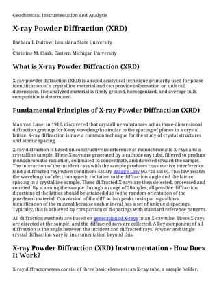
1. Geochemical Instrumentation and Analysis
X-ray Powder Diffraction (XRD)
Barbara L Dutrow, Louisiana State University
,
Christine M. Clark, Eastern Michigan University
What is X-ray Powder Diffraction (XRD)
X-ray powder diffraction (XRD) is a rapid analytical technique primarily used for phase
identification of a crystalline material and can provide information on unit cell
dimensions. The analyzed material is finely ground, homogenized, and average bulk
composition is determined.
Fundamental Principles of X-ray Powder Diffraction (XRD)
Max von Laue, in 1912, discovered that crystalline substances act as three-dimensional
diffraction gratings for X-ray wavelengths similar to the spacing of planes in a crystal
lattice. X-ray diffraction is now a common technique for the study of crystal structures
and atomic spacing.
X-ray diffraction is based on constructive interference of monochromatic X-rays and a
crystalline sample. These X-rays are generated by a cathode ray tube, filtered to produce
monochromatic radiation, collimated to concentrate, and directed toward the sample.
The interaction of the incident rays with the sample produces constructive interference
(and a diffracted ray) when conditions satisfy Bragg's Law (nλ=2d sin θ). This law relates
the wavelength of electromagnetic radiation to the diffraction angle and the lattice
spacing in a crystalline sample. These diffracted X-rays are then detected, processed and
counted. By scanning the sample through a range of 2θangles, all possible diffraction
directions of the lattice should be attained due to the random orientation of the
powdered material. Conversion of the diffraction peaks to d-spacings allows
identification of the mineral because each mineral has a set of unique d-spacings.
Typically, this is achieved by comparison of d-spacings with standard reference patterns.
All diffraction methods are based on generation of X-rays in an X-ray tube. These X-rays
are directed at the sample, and the diffracted rays are collected. A key component of all
diffraction is the angle between the incident and diffracted rays. Powder and single
crystal diffraction vary in instrumentation beyond this.
X-ray Powder Diffraction (XRD) Instrumentation - How Does
It Work?
X-ray diffractometers consist of three basic elements: an X-ray tube, a sample holder,
2. Bruker's X-ray
Diffraction D8-
Discover instrument.
Details
X-ray powder diffractogram. Peak positions occur where the X-ray beam has
been diffracted by the crystal lattice. The unique set of d-spacings derived
from this patter can be used to 'fingerprint' the mineral. Details
and an X-ray detector. X-rays are generated in a cathode ray tube by
heating a filament to produce electrons, accelerating the electrons
toward a target by applying a voltage, and bombarding the target
material with electrons. When electrons have sufficient energy to
dislodge inner shell electrons of the target material, characteristic X-
ray spectra are produced. These spectra consist of several components,
the most common being Kα and Kβ. Kα consists, in part, of Kα1 and Kα2.
Kα1 has a slightly shorter wavelength and twice the intensity as Kα2.
The specific wavelengths are characteristic of the target material (Cu,
Fe, Mo, Cr). Filtering, by foils or crystal monochrometers, is required to
produce monochromatic X-rays needed for diffraction. Kα1and Kα2 are sufficiently close
in wavelength such that a weighted average of the two is used. Copper is the most
common target material for single-crystal diffraction, with CuKα radiation = 1.5418Å.
These X-rays are collimated and directed onto the sample. As the sample and detector
are rotated, the intensity of the reflected X-rays is recorded. When the geometry of the
incident X-rays impinging the sample satisfies the Bragg Equation, constructive
interference occurs and a peak in intensity occurs. A detector records and processes this
X-ray signal and converts the signal to a count rate which is then output to a device such
as a printer or computer monitor.
The geometry of an X-ray
diffractometer is such that the
sample rotates in the path of the
collimated X-ray beam at an
angle θ while the X-ray detector
is mounted on an arm to collect
the diffracted X-rays and rotates
at an angle of 2θ. The
instrument used to maintain the
angle and rotate the sample is
termed a goniometer. For
typical powder patterns, data is
collected at 2θ from ~5° to 70°,
angles that are preset in the X-
ray scan.
Applications
X-ray powder diffraction is most widely used for the identification of unknown
crystalline materials (e.g. minerals, inorganic compounds). Determination of unknown
solids is critical to studies in geology, environmental science, material science,
engineering and biology.
Other applications include:
characterization of crystalline materials
identification of fine-grained minerals such as clays and mixed layer clays that are
3. difficult to determine optically
determination of unit cell dimensions
measurement of sample purity
With specialized techniques, XRD can be used to:
determine crystal structures using Rietveld refinement
determine of modal amounts of minerals (quantitative analysis)
characterize thin films samples by:
determining lattice mismatch between film and substrate and to inferring
stress and strain
determining dislocation density and quality of the film by rocking curve
measurements
measuring superlattices in multilayered epitaxial structures
determining the thickness, roughness and density of the film using glancing
incidence X-ray reflectivity measurements
make textural measurements, such as the orientation of grains, in a polycrystalline
sample
Strengths and Limitations of X-ray Powder Diffraction
(XRD)?
Strengths
Powerful and rapid (< 20 min) technique for identification of an unknown mineral
In most cases, it provides an unambiguous mineral determination
Minimal sample preparation is required
XRD units are widely available
Data interpretation is relatively straight forward
Limitations
Homogeneous and single phase material is best for identification of an unknown
Must have access to a standard reference file of inorganic compounds (d-spacings,
hkls)
Requires tenths of a gram of material which must be ground into a powder
For mixed materials, detection limit is ~ 2% of sample
For unit cell determinations, indexing of patterns for non-isometric crystal systems
is complicated
Peak overlay may occur and worsens for high angle 'reflections'
User's Guide - Sample Collection and Preparation
Determination of an unknown requires: the material, an instrument for grinding, and a
sample holder.
4. Packing of fine
powder into a
sample holder.
Details
Obtain a few tenths of a gram (or more) of the material, as pure as possible
Grind the sample to a fine powder, typically in a fluid to minimize inducing extra
strain (surface energy) that can offset peak positions, and to randomize orientation.
Powder less than ~10 μm(or 200-mesh) in size is preferred
Place into a sample holder or onto the sample surface:
smear uniformly onto a glass slide, assuring a flat upper
surface
pack into a sample container
sprinkle on double sticky tape
Typically the substrate is amorphous to avoid interference
Care must be taken to create a flat upper surface and to achieve a
random distribution of lattice orientations unless creating an oriented smear.
For analysis of clays which require a single orientation, specialized techniques for
preparation of clay samples are given by USGS.
For unit cell determinations, a small amount of a standard with known peak positions
(that do not interfere with the sample) can be added and used to correct peak positions.
Data Collection, Results and Presentation
Data Collection The intensity of diffracted X-rays is continuously recorded as the
sample and detector rotate through their respective angles. A peak in intensity occurs
when the mineral contains lattice planes with d-spacings appropriate to diffract X-rays
at that value of θ. Although each peak consists of two separate reflections (Kα1 and Kα2),
at small values of 2θ the peak locations overlap with Kα2 appearing as a hump on the
side of Kα1. Greater separation occurs at higher values of θ. Typically these combined
peaks are treated as one. The 2λ position of the diffraction peak is typically measured as
the center of the peak at 80% peak height.
Data Reduction
Results are commonly presented as peak positions at 2θ and X-ray counts (intensity) in
the form of a table or an x-y plot (shown above). Intensity (I) is either reported as peak
height intensity, that intensity above background, or as integrated intensity, the area
under the peak. The relative intensity is recorded as the ratio of the peak intensity to
that of the most intense peak (relative intensity = I/I1 x 100 ).
Determination of an Unknown
The d-spacing of each peak is then obtained by solution of the Bragg equation for the
appropriate value of λ. Once all d-spacings have been determined, automated
search/match routines compare the ds of the unknown to those of known materials.
Because each mineral has a unique set of d-spacings, matching these d-spacings provides
an identification of the unknown sample. A systematic procedure is used by ordering the
d-spacings in terms of their intensity beginning with the most intense peak. Files of d-
spacings for hundreds of thousands of inorganic compounds are available from the
International Centre for Diffraction Data as the Powder Diffraction File (PDF). Many
other sites contain d-spacings of minerals such as the American Mineralogist Crystal
5. Structure Database. Commonly this information is an integral portion of the software
that comes with the instrumentation.
Determination of Unit Cell Dimensions
For determination of unit cell parameters, each reflection must be indexed to a specific
hkl.
Literature
The following literature can be used to further explore X-ray Powder Diffraction (XRD)
Bish, DL and Post, JE, editors. 1989. Modern Powder Diffraction. Reviews in
Mienralogy, v. 20. Mineralogical Society of America.
Cullity, B. D. 1978. Elements of X-ray diffraction. 2nd ed. Addison-Wesley, Reading,
Mass.
Klug, H. P., and L. E. Alexander. 1974. X-ray diffraction procedures for
polycrystalline and amorphous materials. 2nd ed. Wiley, New York.
Moore, D. M. and R. C. Reynolds, Jr. 1997. X-Ray diffraction and the identification
and analysis of clay minerals. 2nd Ed. Oxford University Press, New York.
Related Links
For more information about X-ray Powder Diffraction (XRD) follow the links below.
For more information on XRD Methods, visit the USGS website
For additional information on X-ray basics; Materials Research Lab, University of
California- Santa Barbara
Rigaku Journal; an on-line journal that describes and demonstrates a wide range of
applications using Xray diffraction.
Cambridge University X-ray Tutorial
International Union of Crystallography (IUCr) Teaching Pamphlets
Introduction to X-ray Diffraction–University of California, Santa Barbara
Introduction to Crystallography–from LLNL
X-ray Crystallography Lecture Notes–from Steve Nelson, Tulane University
Reciprocal Net–part of the National Science Digital Library. Use the "Learn About"
link to find animations of the structures of common molecules (including
minerals), crystallography learning resources (tutorials, databases and software),
resources on crystallization, and tutorials on symmetry and point groups.
Online Xray Technician Schools
Teaching Activities and Resources
Teaching activities, labs, and resources pertaining to X-ray Powder Diffraction (XRD).
X-ray techniques lab exercises from the SERC Teaching Mineralogy Collections
6. Weathering of Igneous, Metamorphic, and Sedimentary Rocks in a Semi-Arid
Climate - An Engineering Application of Petrology - This problem develops skills in
X-ray diffraction analysis as applied to clay mineralogy, reinforces lecture material
on the geochemistry of weathering, and demonstrates the role of petrologic
characterization in site engineering.
Teaching Guide to X-ray Diffraction at Cambridge
A Powerpoint presentation on use of XRD in Soil Science (PowerPoint 1.6MB Sep7
07) by Melody Bergeron, Image and Chemical Analysis Laboratory at Montana
State University.
Brady, John B., and Boardman, Shelby J., 1995, Introducing Mineralogy Students to
X-ray Diffraction Through Optical Diffraction Experiments Using Lasers. Jour. Geol.
Education, v. 43 #5, 471-476.
Brady, John B., Newton, Robert M., and Boardman, Shelby J., 1995, New Uses for
Powder X-ray Diffraction Experiments in the Undergraduate Curriculum. Jour.
Geol. Education, v. 43 #5, 466-470.
Dutrow, Barb, 1997, Better Living Through Minerals X-ray Diffraction of Household
Products, in: Brady, J., Mogk, D., and Perkins D. (eds.) Teaching Mineralogy,
Mineralogical Society of America, p. 349-359.
Hovis, Guy, L., 1997, Determination of Chemical Composition, State of Order, Molar
Volume, and Density of a Monoclinic Alkali Feldspar Using X-ray Diffraction, in:
Brady, J., Mogk, D., and Perkins D. (eds.) Teaching Mineralogy, Mineralogical
Society of America, p. 107-118.
Brady, John B., 1997, Making Solid Solutions with Alkali Halides (and Breaking
Them) , in: Brady, J., Mogk, D., and Perkins D. (eds.) Teaching Mineralogy,
Mineralogical Society of America, p. 91-95.
Perkins, Dexter, III, and Sorensen, Paul, Mineral Synthesis and X-ray Diffraction
Experiments, in: Brady, J., Mogk, D., and Perkins D. (eds.) Teaching Mineralogy,
Mineralogical Society of America, p. 81-90.
Hollecher, Kurt, A Long-Term Mineralogy Practical Exam, in: Brady, J., Mogk, D.,
and Perkins D. (eds.) Teaching Mineralogy, Mineralogical Society of America, p. 43-
46.
Moecher, David, 2004, Characterization and Identification of Mineral Unknowns: A
Mineralogy Term Project, Jour. Geoscience Education, v 52 #1, p. 5-9.
Hluchy, M.M., 1999, The Value of Teaching X-ray Techniques and Clay Mineralogy
to Undergraduates, Jour. Geoscience Education, v. 47, p. 236-240.