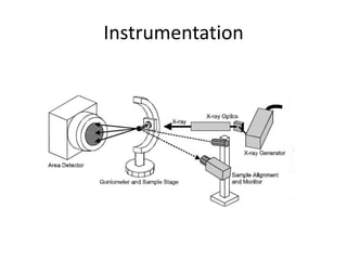
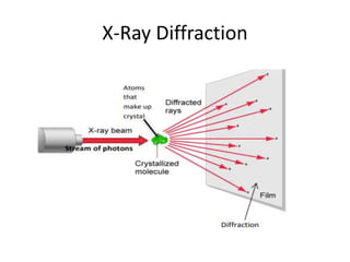
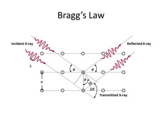
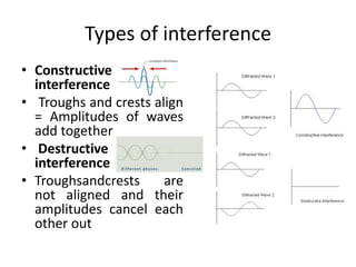
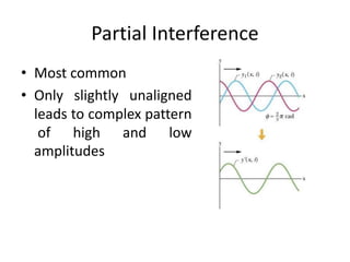
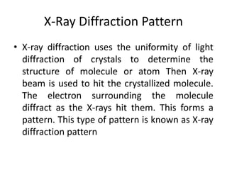
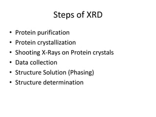
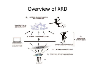
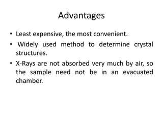
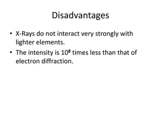
X-RAY Crystallography
By Mehwish Nawaz
Introduction
• X-ray Crystallography is a scientific method of
determining the precise positions/arrangements
of atoms in a crystal where beams of X-ray
strikes a crystal and causes the beam of light to
diffract into many specific directions.
Electromagnetic Spectrum
X-Ray
Cont…
• A technique used to determine the atomic and
molecular structure of a crystal, in which the crystalline
atoms cause a beam of incident X-rays to diffract into
many specific directions.
• The atomic planes of a crystal cause an incident beam
of X- rays to interfere with one another as they leave
the crystal. The phenomenon is called X-ray diffraction.
A stream of X-rays directed at a crystal diffract and
scatter as they encounter atoms.
• The scattered rays interfere with each other and
produce spots of different intensities that can be
recorded on film.
X-Ray Diffraction Pattern
• X-ray diffraction or X-ray crystallography uses
the uniformity of light diffraction of crystals to
determine the structure of molecule or atom
Then X-ray beam is used to hit the crystallized
molecule.
• The electron surrounding the molecule
diffract as the X-rays hit them. This forms a
pattern. This type of pattern is known as X-ray
diffraction pattern
Principle
• The principle is based on principle of diffraction
• The crystal is made to strike against x-ray beam.
• Due to striking the atoms present in crystal
diffracts the x-ray beam into different direction.
• The angle and intensity of this diffraction rays is
analog to spatial arrangement of atom in crystal.
• By studying these angle, the 3D structure of any
crystal can be determine.
Instrumentation
• X-ray Tube
The source of X rays
• Incident-beam optics
Condition the X- ray beam before it hits the sample
• The goniometer
The platform that holds and moves the sample, and
detector as well as the sample & sample holder
• Receiving-side optics
• Condition the X- ray beam after it has encountered the
sample
• Detector
Count the number of X rays scattered by the sample
Instrumentation
X-Ray Diffraction
Bragg’s Law
• Bragg’s law states that when the x-ray is
incident onto a crystal surface, its angle of
incidence, θ, will reflect back with a same
angle of scattering, θ.
• And, when the path difference, d is equal to a
whole number, n, of wavelength, a
constructive interference will occur.
Bragg’s Law
Bragg’s Law
• Bragg’s Law
nλ = 2d sinƟ
where:
• λ is the wavelength of the x-ray,
• d is the spacing of the crystal layers (path
difference),
• θ is the incident angle (the angle between
incident ray and the scatter plane), and
• n is an integer
Bragg’s Law
Types of interference
• Constructive
interference
• Troughs and crests align
= Amplitudes of waves
add together
• Destructive
interference
• Troughsandcrests are
not aligned and their
amplitudes cancel each
other out
Partial Interference
• Most common
• Only slightly unaligned
leads to complex pattern
of high and low
amplitudes
X-Ray Diffraction Pattern
• X-ray diffraction uses the uniformity of light
diffraction of crystals to determine the
structure of molecule or atom Then X-ray
beam is used to hit the crystallized molecule.
The electron surrounding the molecule
diffract as the X-rays hit them. This forms a
pattern. This type of pattern is known as X-ray
diffraction pattern
Electron density map
• In crystallography the crystalline atoms cause
a beam of incident X-rays to diffract into many
specific directions. Then crystallographer can
produce a three-dimensional picture of the
density of electrons within the crystal.
• From this electron density, the mean positions
of the atoms in the crystal can be determined.
X-ray crystallography can locate every atom in
a zeolite, an aluminosilicate
Steps of XRD
• Protein purification
• Protein crystallization
• Shooting X-Rays on Protein crystals
• Data collection
• Structure Solution (Phasing)
• Structure determination
First step
• The process begins by crystallizing a protein of interest.
• 4 critical steps are taken to achieve protein
crystallization:
• Purify the protein.
• Determine the purity of the protein and if not pure
(usually >99%), then must undergo further purification.
• Protein must be precipitated by dissolving it in an
appropriate solvent(water- buffer soln. w/ organic salt
such as 2-methyl-2,4-pentanediol).
• The solution has to be brought to supersaturation by
adding a salt to the concentrated solution of the
protein. Let the actual crystals grow.
Protein Sample for Crystallization
• Pure and homogenous (identified by SDS-PAGE, Mass Spec. etc.)
• Properly folded Stable for at least few days in its crystallization condition (dynamic
light scattering)
Conditions that Effect Crystallization
• pH (buffer) Detergent
• Protein Concentration Temperature
• Pressure Precipitant
• Size and shape of the drops
• Salt (Sodium Chloride, Ammonium Chloride etc.)
Reason to crystallize a protein?
• Researchers crystallize an atom or molecule,
because the precise position of each atom in a
molecule can only be determined if the
molecule is crystallized.
• If the molecule or atom is not in a crystallized
form, the X-rays will diffract unpredictably and
the data retrieved will be too difficult if not
impossible to understand.
Second Step
• X-rays are generated by bombarding electrons
on an metallic anode.
• Then, the x-rays are shot at the protein crystal
resulting in some of the x-rays going through
the crystal and the rest being scattered in
various directions.
• The crystal is rotated so that the x-rays are
able to hit the protein from all sides and
angles (Goniometer).
Production of X-Rays
Third Step
• An electron density map is created based on
the measured intensities of the diffraction
pattern on the film.
• A Fourier Transform can be applied to the
intensities on the film to reconstruct the
electron density distribution of the crystal.
• The mapping gives a three-dimensional
representation of the electron densities
observed through the x-ray crystallography.
Overview of XRD
Structure Solution (Phasing)
• A typical diffraction
pattern from a protein
crystal
• The 3D structure
obtained above is the
electron density map
of the crystal.
GOAL= From Diffraction Data to Electron
Density
Fourier Transform
• A Fourier transform is done to extract the
frequency-domain spectrum from the raw time-
domain spectrum.
• Because we have different waves of X-rays
superimposed on one another during diffraction,
it is difficult to isolate the contribution of each
diffraction event to determine the lattice
structure. Therefore a mathematical tool known
as the Fourier transform is used.
Resolution needed for the
interpretation of electron density map
• Resolution needs to be taken into the
following account:
• A resolution of 5Å - 10Å can reveal the
structure of polypeptide chains,
• 3Å - 4Å of groups of atoms,
• 1Å - 1.5Å of individual atoms.
Advantages
• Least expensive, the most convenient.
• Widely used method to determine crystal
structures.
• X-Rays are not absorbed very much by air, so
the sample need not be in an evacuated
chamber.
Disadvantages
• X-Rays do not interact very strongly with
lighter elements.
• The intensity is 108 times less than that of
electron diffraction.
Applications
• To identify crystalline phases and orientation
• To determine structural properties
• To measure thickness of thin films and multi-
layers
• To determine atomic arrangement
• Detection limits: ~3% in a two phase mixture;
can be~0.1% with synchrotron radiation

X Ray Crystallography

  • 1.
  • 2.
    Introduction • X-ray Crystallographyis a scientific method of determining the precise positions/arrangements of atoms in a crystal where beams of X-ray strikes a crystal and causes the beam of light to diffract into many specific directions.
  • 3.
  • 4.
    Cont… • A techniqueused to determine the atomic and molecular structure of a crystal, in which the crystalline atoms cause a beam of incident X-rays to diffract into many specific directions. • The atomic planes of a crystal cause an incident beam of X- rays to interfere with one another as they leave the crystal. The phenomenon is called X-ray diffraction. A stream of X-rays directed at a crystal diffract and scatter as they encounter atoms. • The scattered rays interfere with each other and produce spots of different intensities that can be recorded on film.
  • 5.
    X-Ray Diffraction Pattern •X-ray diffraction or X-ray crystallography uses the uniformity of light diffraction of crystals to determine the structure of molecule or atom Then X-ray beam is used to hit the crystallized molecule. • The electron surrounding the molecule diffract as the X-rays hit them. This forms a pattern. This type of pattern is known as X-ray diffraction pattern
  • 6.
    Principle • The principleis based on principle of diffraction • The crystal is made to strike against x-ray beam. • Due to striking the atoms present in crystal diffracts the x-ray beam into different direction. • The angle and intensity of this diffraction rays is analog to spatial arrangement of atom in crystal. • By studying these angle, the 3D structure of any crystal can be determine.
  • 7.
    Instrumentation • X-ray Tube Thesource of X rays • Incident-beam optics Condition the X- ray beam before it hits the sample • The goniometer The platform that holds and moves the sample, and detector as well as the sample & sample holder • Receiving-side optics • Condition the X- ray beam after it has encountered the sample • Detector Count the number of X rays scattered by the sample
  • 8.
  • 9.
  • 10.
    Bragg’s Law • Bragg’slaw states that when the x-ray is incident onto a crystal surface, its angle of incidence, θ, will reflect back with a same angle of scattering, θ. • And, when the path difference, d is equal to a whole number, n, of wavelength, a constructive interference will occur.
  • 11.
  • 12.
    Bragg’s Law • Bragg’sLaw nλ = 2d sinƟ where: • λ is the wavelength of the x-ray, • d is the spacing of the crystal layers (path difference), • θ is the incident angle (the angle between incident ray and the scatter plane), and • n is an integer
  • 13.
  • 14.
    Types of interference •Constructive interference • Troughs and crests align = Amplitudes of waves add together • Destructive interference • Troughsandcrests are not aligned and their amplitudes cancel each other out
  • 15.
    Partial Interference • Mostcommon • Only slightly unaligned leads to complex pattern of high and low amplitudes
  • 16.
    X-Ray Diffraction Pattern •X-ray diffraction uses the uniformity of light diffraction of crystals to determine the structure of molecule or atom Then X-ray beam is used to hit the crystallized molecule. The electron surrounding the molecule diffract as the X-rays hit them. This forms a pattern. This type of pattern is known as X-ray diffraction pattern
  • 17.
    Electron density map •In crystallography the crystalline atoms cause a beam of incident X-rays to diffract into many specific directions. Then crystallographer can produce a three-dimensional picture of the density of electrons within the crystal. • From this electron density, the mean positions of the atoms in the crystal can be determined. X-ray crystallography can locate every atom in a zeolite, an aluminosilicate
  • 18.
    Steps of XRD •Protein purification • Protein crystallization • Shooting X-Rays on Protein crystals • Data collection • Structure Solution (Phasing) • Structure determination
  • 19.
    First step • Theprocess begins by crystallizing a protein of interest. • 4 critical steps are taken to achieve protein crystallization: • Purify the protein. • Determine the purity of the protein and if not pure (usually >99%), then must undergo further purification. • Protein must be precipitated by dissolving it in an appropriate solvent(water- buffer soln. w/ organic salt such as 2-methyl-2,4-pentanediol). • The solution has to be brought to supersaturation by adding a salt to the concentrated solution of the protein. Let the actual crystals grow.
  • 20.
    Protein Sample forCrystallization • Pure and homogenous (identified by SDS-PAGE, Mass Spec. etc.) • Properly folded Stable for at least few days in its crystallization condition (dynamic light scattering) Conditions that Effect Crystallization • pH (buffer) Detergent • Protein Concentration Temperature • Pressure Precipitant • Size and shape of the drops • Salt (Sodium Chloride, Ammonium Chloride etc.)
  • 21.
    Reason to crystallizea protein? • Researchers crystallize an atom or molecule, because the precise position of each atom in a molecule can only be determined if the molecule is crystallized. • If the molecule or atom is not in a crystallized form, the X-rays will diffract unpredictably and the data retrieved will be too difficult if not impossible to understand.
  • 22.
    Second Step • X-raysare generated by bombarding electrons on an metallic anode. • Then, the x-rays are shot at the protein crystal resulting in some of the x-rays going through the crystal and the rest being scattered in various directions. • The crystal is rotated so that the x-rays are able to hit the protein from all sides and angles (Goniometer).
  • 23.
  • 24.
    Third Step • Anelectron density map is created based on the measured intensities of the diffraction pattern on the film. • A Fourier Transform can be applied to the intensities on the film to reconstruct the electron density distribution of the crystal. • The mapping gives a three-dimensional representation of the electron densities observed through the x-ray crystallography.
  • 25.
  • 26.
    Structure Solution (Phasing) •A typical diffraction pattern from a protein crystal • The 3D structure obtained above is the electron density map of the crystal. GOAL= From Diffraction Data to Electron Density
  • 27.
    Fourier Transform • AFourier transform is done to extract the frequency-domain spectrum from the raw time- domain spectrum. • Because we have different waves of X-rays superimposed on one another during diffraction, it is difficult to isolate the contribution of each diffraction event to determine the lattice structure. Therefore a mathematical tool known as the Fourier transform is used.
  • 28.
    Resolution needed forthe interpretation of electron density map • Resolution needs to be taken into the following account: • A resolution of 5Å - 10Å can reveal the structure of polypeptide chains, • 3Å - 4Å of groups of atoms, • 1Å - 1.5Å of individual atoms.
  • 29.
    Advantages • Least expensive,the most convenient. • Widely used method to determine crystal structures. • X-Rays are not absorbed very much by air, so the sample need not be in an evacuated chamber.
  • 30.
    Disadvantages • X-Rays donot interact very strongly with lighter elements. • The intensity is 108 times less than that of electron diffraction.
  • 31.
    Applications • To identifycrystalline phases and orientation • To determine structural properties • To measure thickness of thin films and multi- layers • To determine atomic arrangement • Detection limits: ~3% in a two phase mixture; can be~0.1% with synchrotron radiation