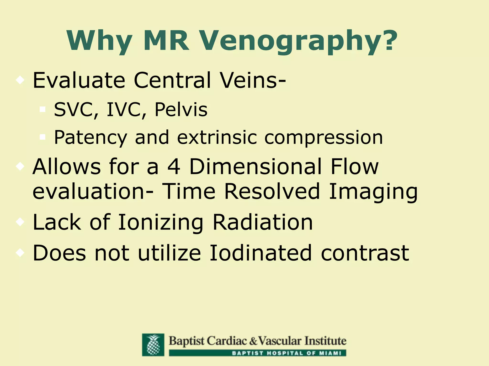
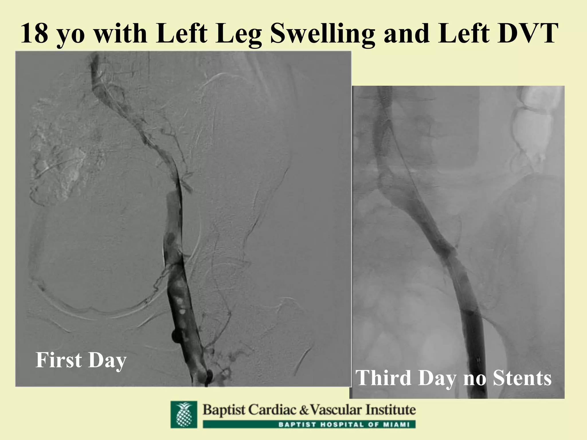
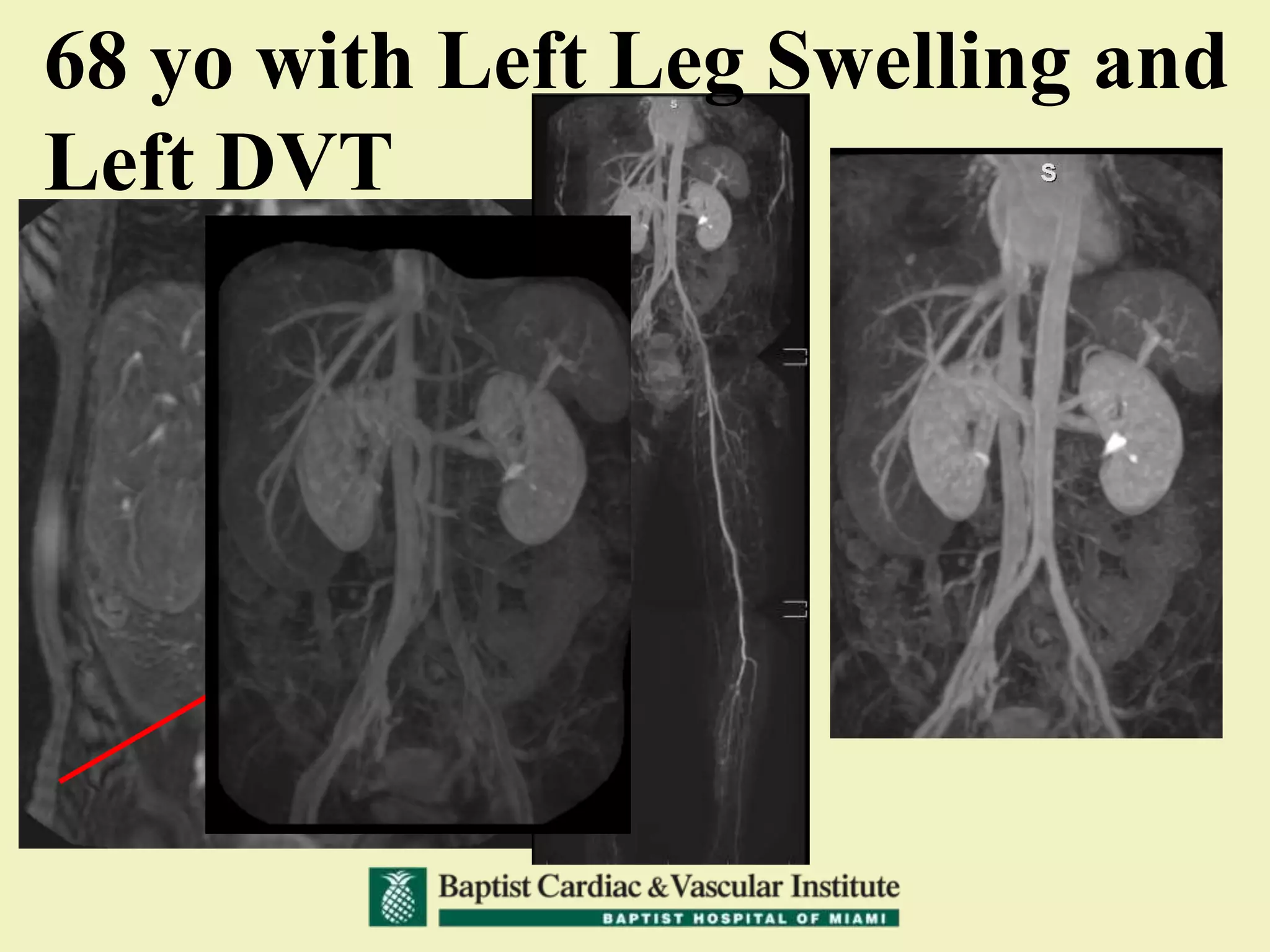
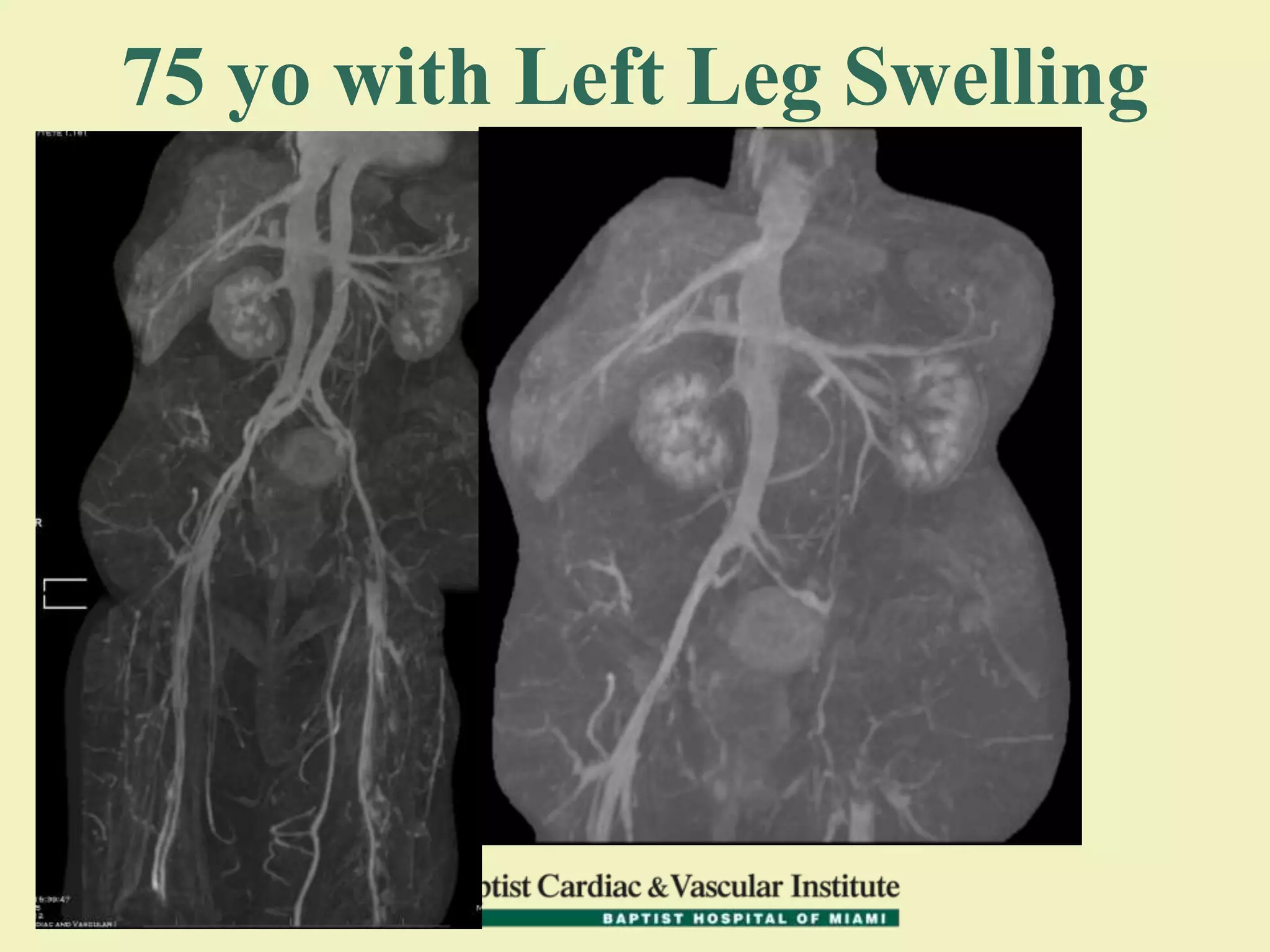
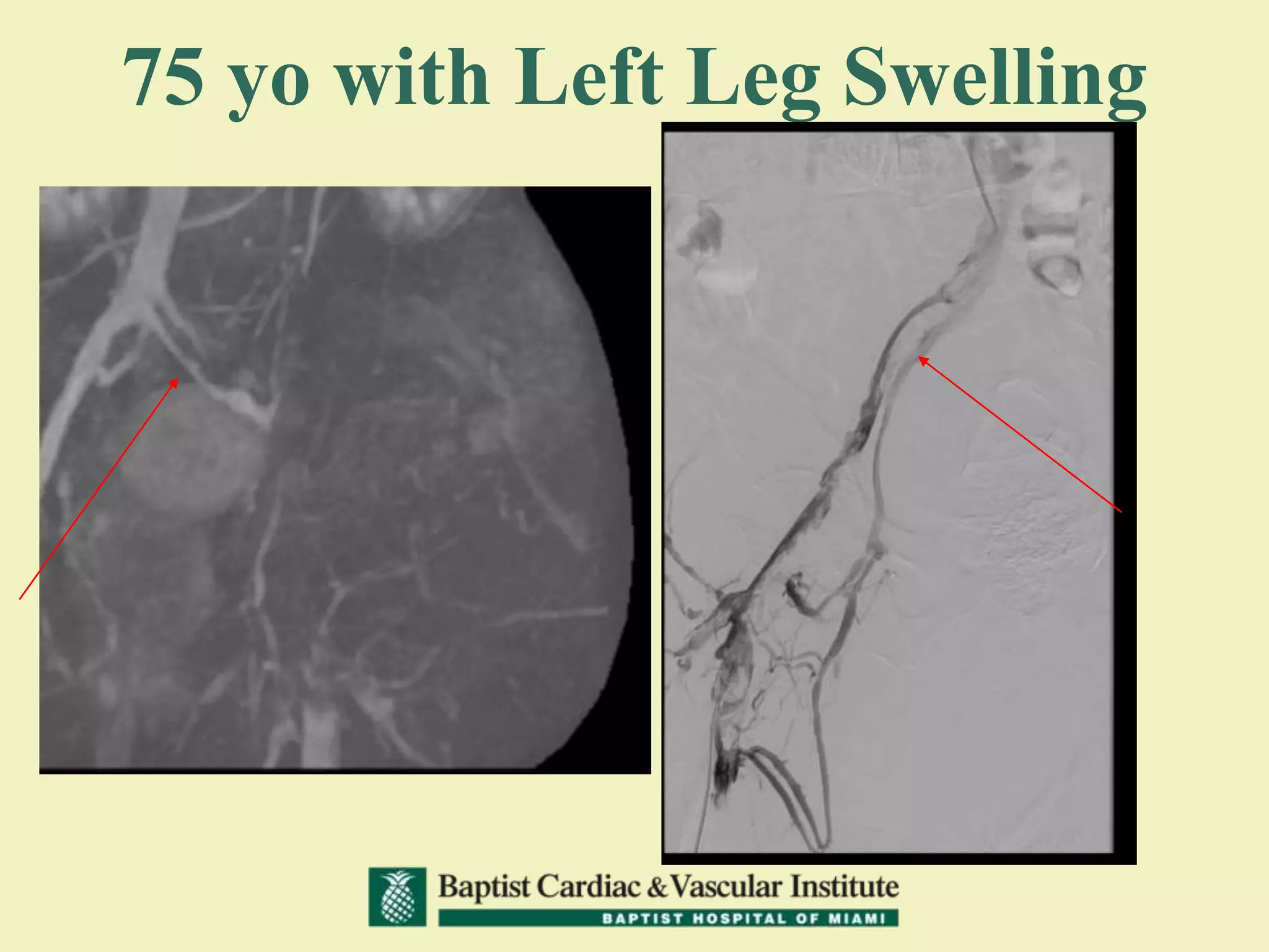
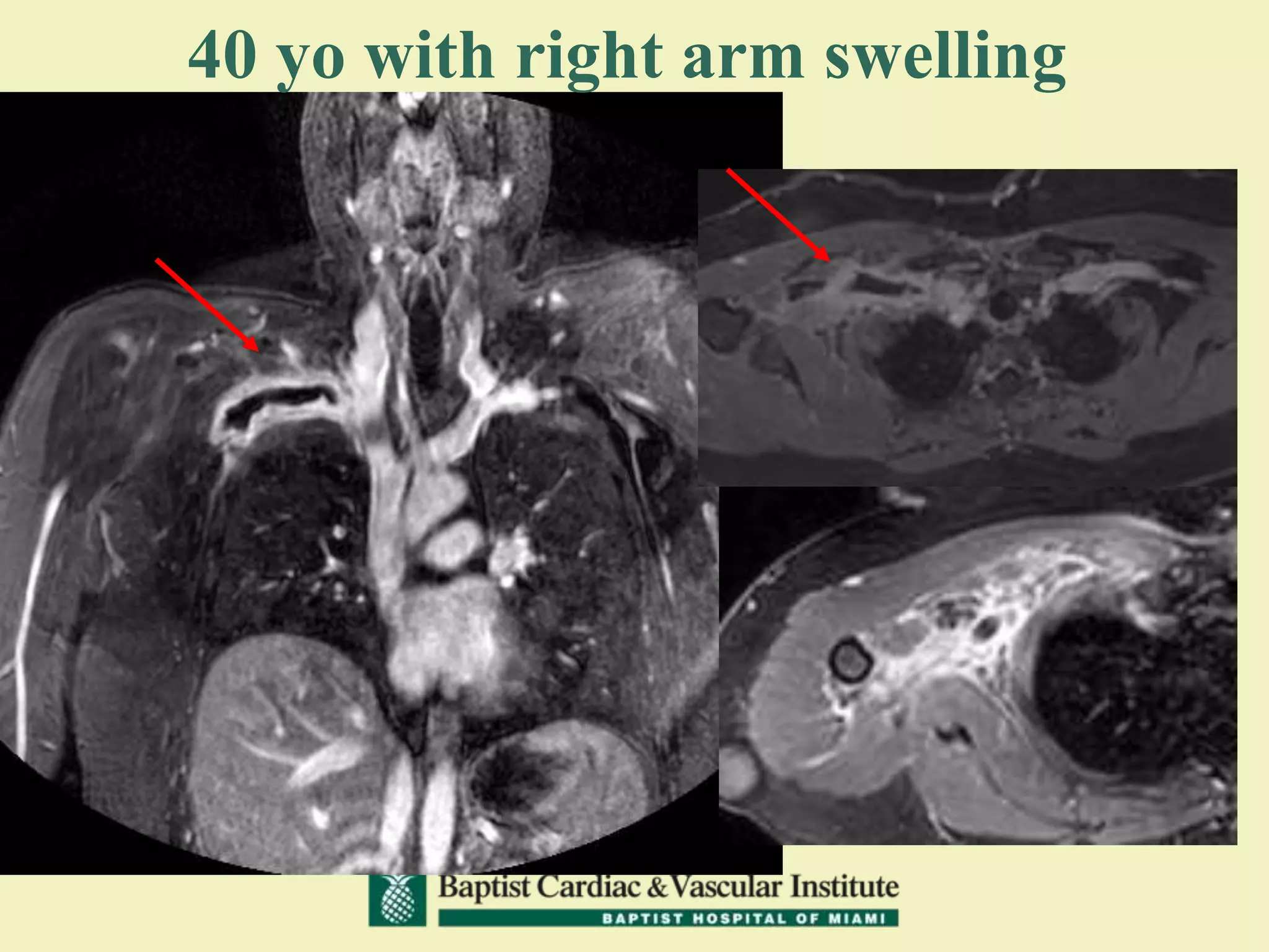
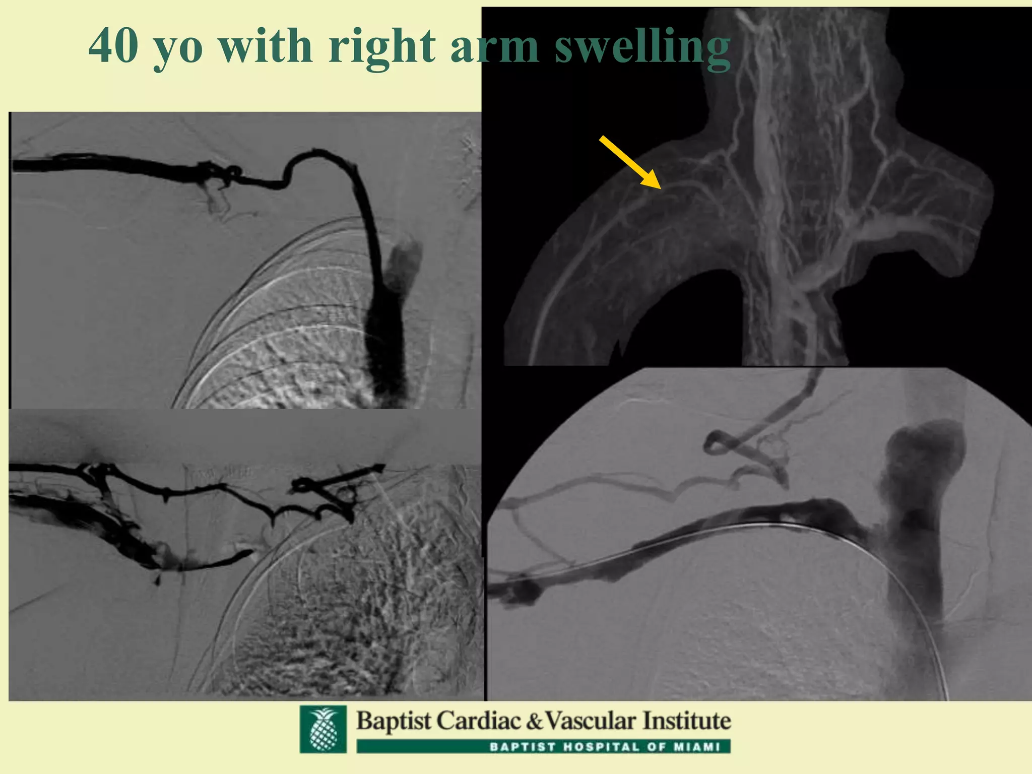
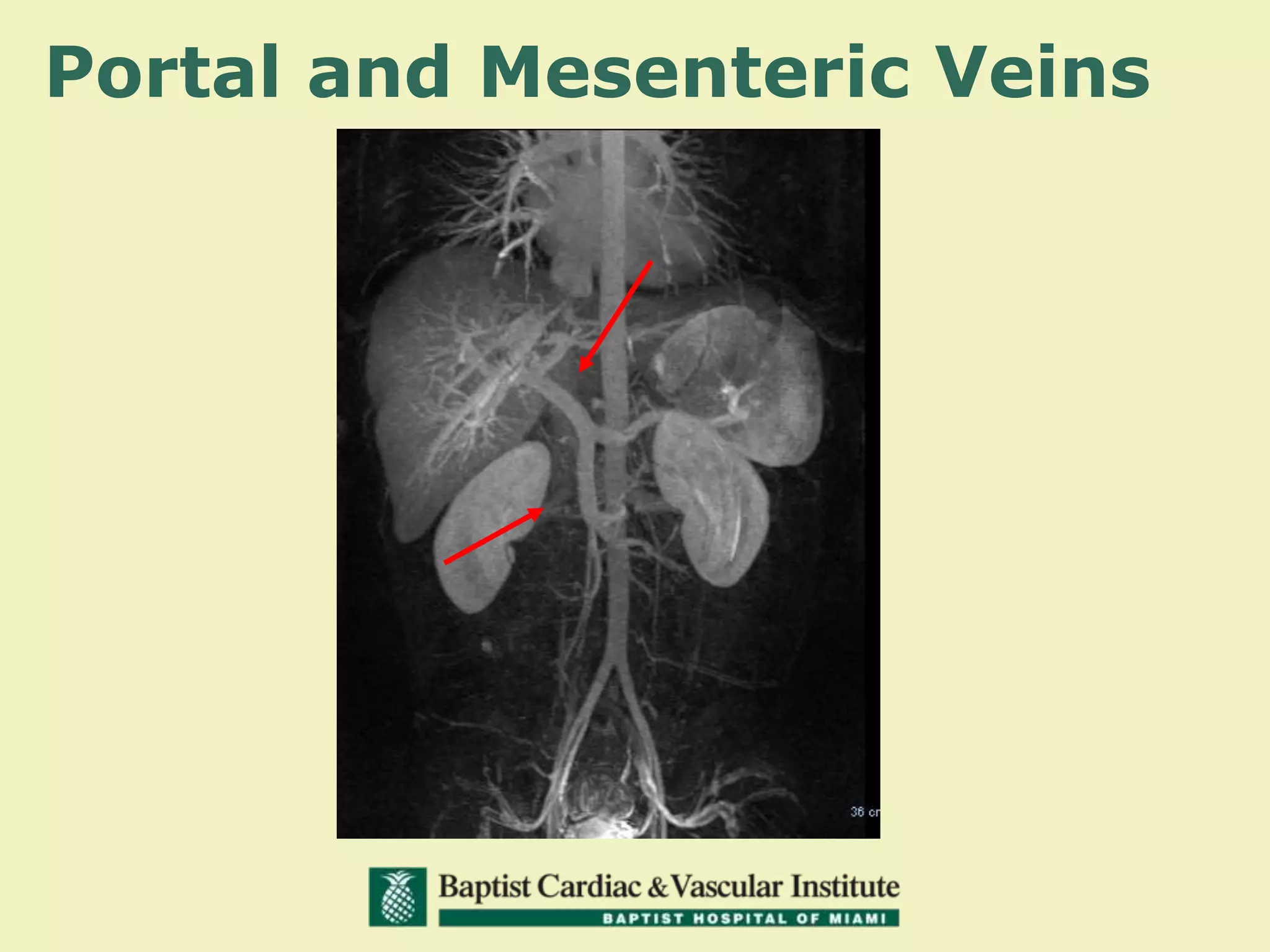
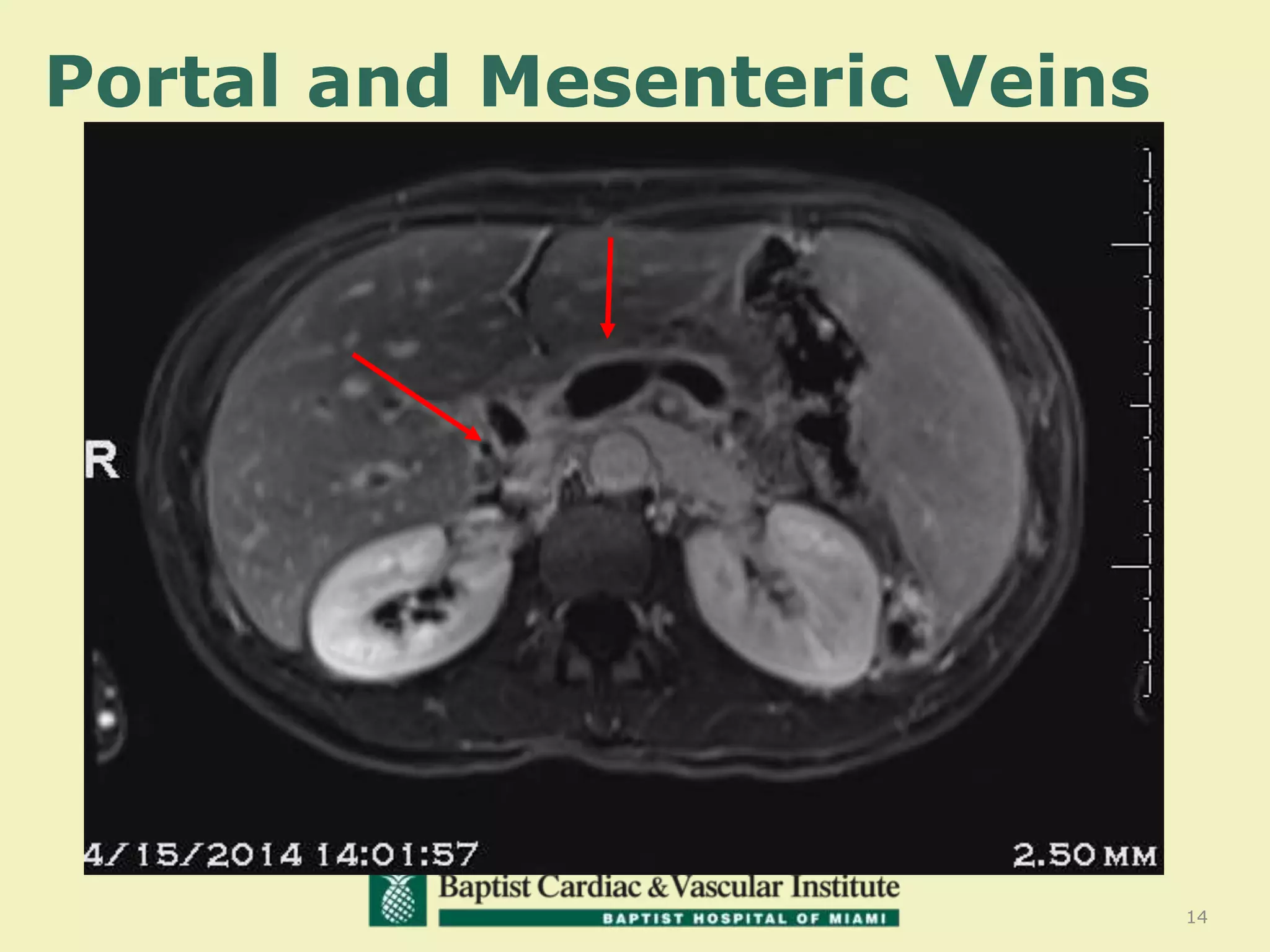
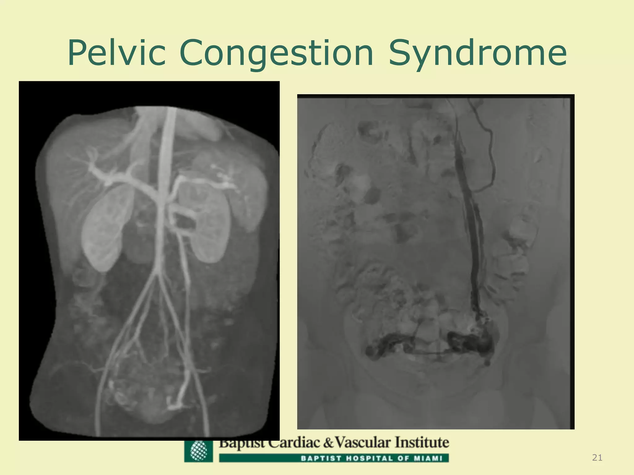
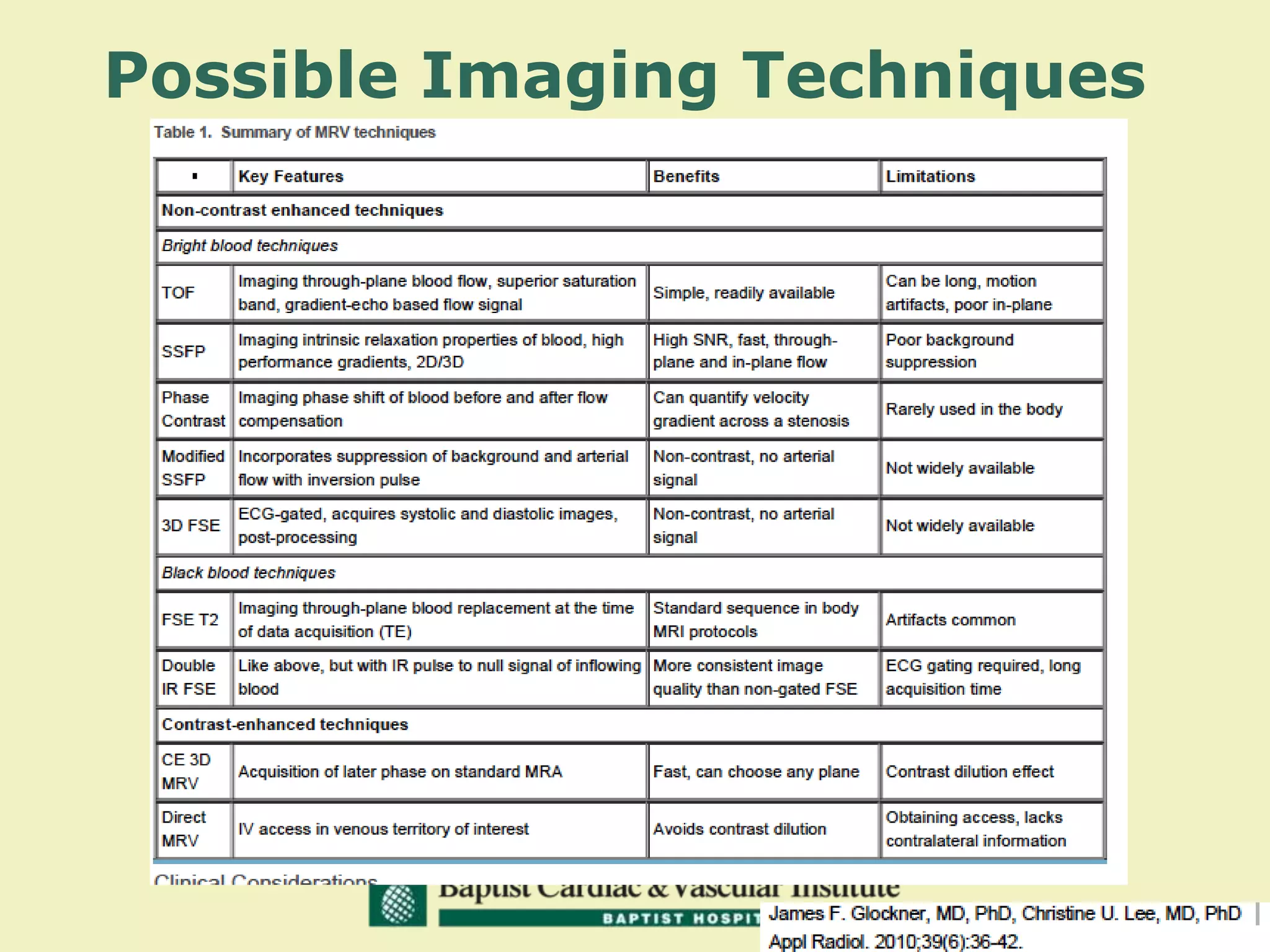
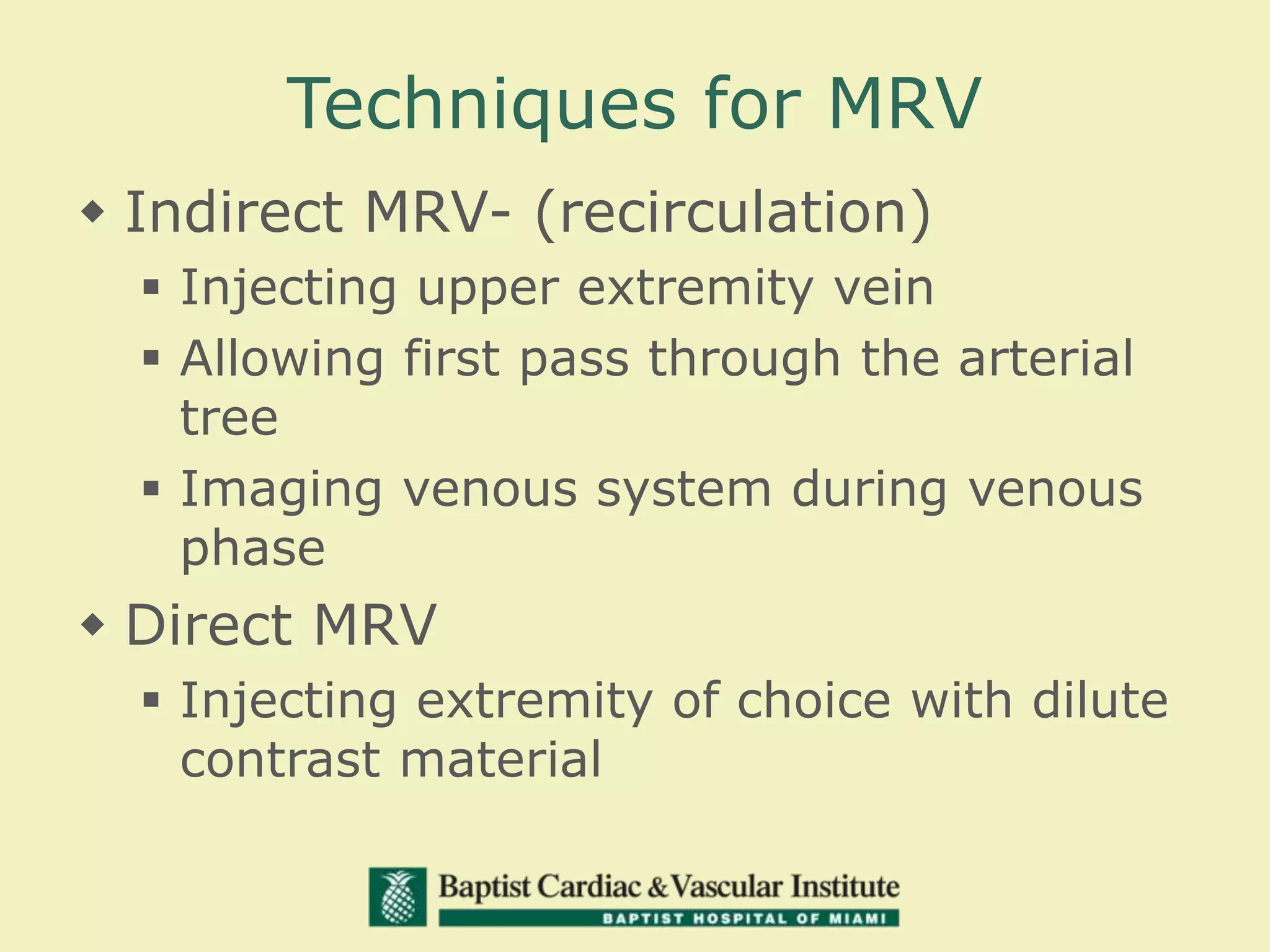
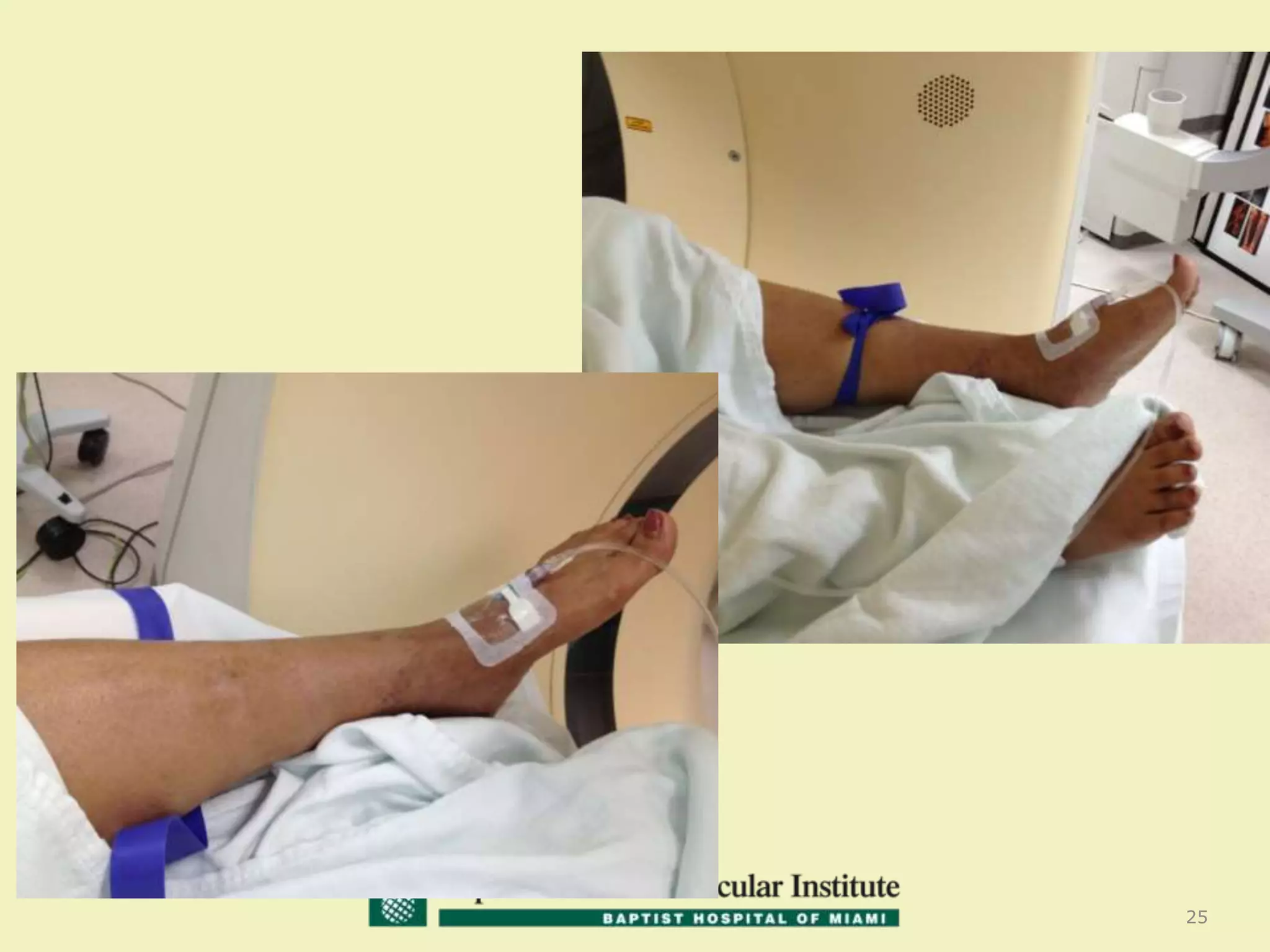
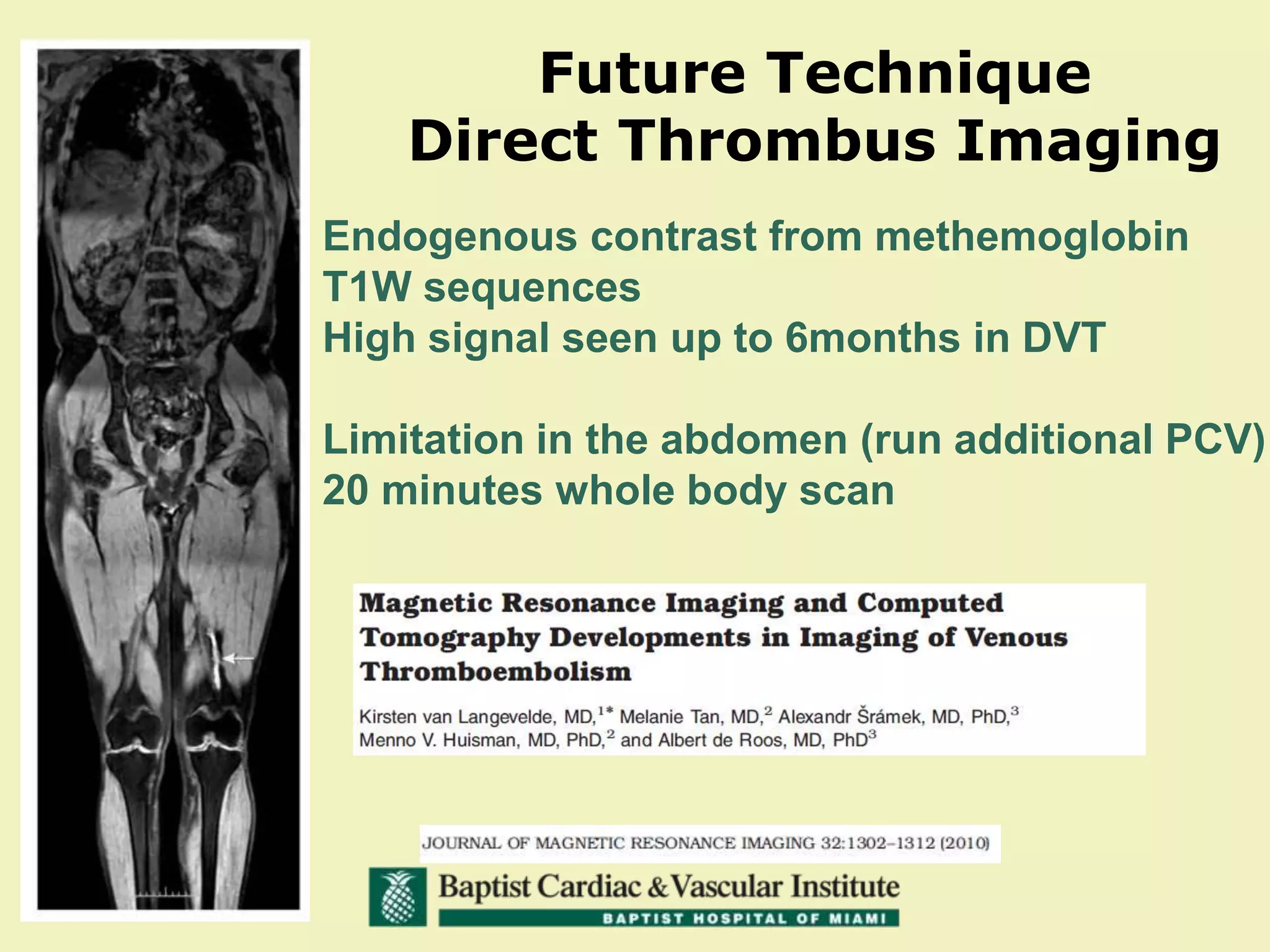
By: Constantino S.Peña
Visit VeinGlobal at http://www.veinglobal.com/ for more presentations and videos on this topic, or for more information on venous disease news, education and research.
By: Mark J. Garcia MD, MS, FSIR
Visit VeinGlobal at http://www.veinglobal.com/ for more presentations and videos on this topic, or for more information on venous disease news, education and research.
This document provides guidance on performing and interpreting coronary CT scans. It outlines steps for patient selection, protocols for calcium scoring and angiography images, and checklists for evaluating different anatomical areas. Key areas to examine include the coronary arteries, heart chambers, great vessels, lungs, mediastinum, abdomen, bones and skin/breasts. Specific pathological conditions are mentioned such as aneurysms, thrombi, stenoses, and valve abnormalities.
Cardiac CT uses X-rays and computer technology to image the heart and coronary arteries. It can detect calcium buildup in arteries, assess coronary arteries via angiography, and evaluate heart function. The test involves injecting contrast dye and potentially heart rate control drugs. Images are taken rapidly during breath-holds to freeze heart motion. Cardiac CT can diagnose heart disease by identifying plaque and blockages without the risks of angiography.
Cardiac CT provides a noninvasive way to evaluate the coronary arteries and cardiac structure. It has largely replaced invasive coronary angiography due to improvements in temporal and spatial resolution allowing for clear images of the heart. Cardiac CT is indicated to rule out coronary artery disease in low-moderate risk patients, assess anomalies, evaluate grafts and stents, and aid in surgical planning. It has limitations including irregular heart rates over 80 bpm, high calcium scores, small vessels, and radiation exposure. Proper patient selection and preparation are important to optimize results.
This document provides an overview of magnetic resonance angiography (MRA). It discusses the physics behind MRA, different techniques used including time-of-flight imaging and phase contrast imaging, as well as considerations for patient preparation and contraindications. Advantages of MRA include being non-invasive and avoiding risks associated with conventional angiography such as damage to arteries. Limitations include inability to depict small vessels or slow blood flow as well as conventional methods. Overall, the document provides a comprehensive introduction to MRA.
64 slice CT provides high resolution isotropic imaging that allows for improved diagnosis in cardiac, vascular, musculoskeletal and other applications. It provides advantages over older CT technologies such as reduced scan times allowing for single breathhold cardiac imaging. Cardiac applications include calcium scoring, assessment of coronary arteries and function. It is useful for evaluation of coronary artery disease in asymptomatic or symptomatic patients. Post processing techniques such as MIP and VRT allow sharp depiction of coronary arteries. While it has few disadvantages like artifacts from calcium and stents, 64 slice CT provides a non-invasive alternative to angiography for evaluation of many conditions.
This document discusses computed tomography angiography (CTA) and its applications in cardiology. CTA uses computed tomography to visualize blood vessels throughout the body, including coronary arteries. Coronary CTA can detect plaque buildup in coronary arteries without being invasive. Current multidetector CT systems can acquire high-resolution images of the heart within 20 seconds while the patient holds their breath. Coronary CTA provides diagnostic information but also exposes patients to radiation. It is most useful for evaluating cardiac symptoms in low-to-intermediate risk patients.
This document discusses cardiac imaging modalities such as CT, MRI, echocardiography, and nuclear scintigraphy used by cardiac surgeons. It provides information on how to perform good quality CT and MRI imaging, and what each modality can be used to visualize such as coronary artery anatomy, post-procedure evaluation, congenital heart disease, and cardiac function. The document also covers contraindications of CT and MRI, how to decide between the modalities, and risks of contrast-induced nephropathy.
Coronary CT angiography allows for noninvasive imaging of the heart and coronary arteries. It can be used to evaluate patients with chest pain, assess coronary arteries after revascularization, and detect congenital coronary anomalies. The scan involves a non-contrast scan for calcium scoring followed by a contrast-enhanced scan. Proper patient preparation including beta-blockers and nitroglycerin is important. Images are analyzed using techniques like multiplanar reformation, maximum intensity projection, volume rendering and curved reformation to evaluate coronary artery anatomy and detect any stenosis.
Non-invasive imaging plays an important role in the management of cardiovascular diseases. Different imaging modalities have advantages and limitations. Echocardiography is useful for assessing cardiac structure and function but limited for coronary artery disease evaluation. Nuclear imaging can evaluate perfusion and function but not coronary anatomy directly. CT and MRI can assess coronary anatomy in addition to function but CT involves radiation. The appropriate choice of imaging modality depends on the clinical question and no single test can replace all others for evaluating cardiovascular diseases. Integrating complementary information from different tests provides the most comprehensive assessment.
This document outlines the protocol for performing CT angiography (CTA) from the cerebral arteries to the lower limbs. It discusses indications for CTA including aneurysms, stenosis, dissections, and more. The preparation, positioning, and scanning protocols are provided for CTA of the head to lower limbs as well as the subclavian arteries. Pediatric protocols are also summarized. The document concludes with examples of CTA findings and references.
Cardiac CT and coronary CTA can play an important role in diagnosing cardiac patients. Coronary CTA is useful for ruling out obstructive coronary artery disease. Newer multislice CT scanners ranging from 4 to 320 slices allow for faster scanning times and better imaging of the heart. Proper patient preparation including beta blockers, fasting, and controlling heart rate are important for obtaining diagnostic images. Coronary CTA can detect obstructive coronary lesions, evaluate stents and grafts, and perform calcium scoring for risk stratification. It can also detect other cardiac abnormalities.
What is New in Cardiac CT? In Search of the Comprehensive and Conclusive Hear...Apollo Hospitals
Coronary CT Angiography (CT) with its noninvasive cross sectional information has seen remarkable growth in recent years. With the introduction of the new generation scanners, like the 320-slice CT, it has risen to a whole new level. Percent diameter stenosis determined with the use of 320-slice CT shows good correlation with Invasive catheter angiogram (ICA) without significant underestimation or overestimation. Plaque composition on CT regardless of lesion severity has emerged as a strong predictor of major cardiac events. The percentage stenosis mismatch between CT and ICA can be explained by the 2 dimensional nature of ICA and its interpretive inconsistencies. In the upcoming years, we need to evolve from focusing on lumen stenosis to a comprehensive assessment of CAD and its impact on patient outcome.
This document provides information on performing and interpreting CT angiography of the lower limbs. It discusses scanning techniques, protocols, contrast injection, and principles of timing acquisitions. Image post-processing includes MIP, VR, and MPR. Interpretation requires scrutinizing calcifications and stents to avoid overestimating stenosis. Peripheral CTA is useful for evaluating occlusive disease, aneurysms, trauma, infections, embolism, and postoperative surveillance. Examples demonstrate various vascular pathologies.
This document discusses the evolution and advances in coronary CT angiography (CCTA) technology and its role in the assessment of coronary artery disease (CAD). Key points include:
- CCTA has advanced from early CT scanners with 4-minute scan times to modern multi-detector scanners that can image the entire heart in a single heartbeat.
- CCTA provides information on coronary artery anatomy, plaque characteristics, and has prognostic value when assessing coronary artery calcium scoring.
- CCTA has good accuracy for detecting CAD compared to invasive coronary angiography, especially for ruling out disease, though its role in asymptomatic patients is still unclear.
- CCTA is useful for evaluating coronary anomalies, bypass grafts,
Cardiac CT-CCTA involves three main steps: patient preparation with beta blockade and nitroglycerine to lower heart rate, initial calcium scoring to identify atherosclerotic vessels, and coronary CTA scan using retrospective or prospective ECG gating. CCTA allows visualization of the coronary arteries and quantification of plaque type and stenosis. Normal coronary anatomy includes the left main artery bifurcating into the LAD and LCX, and the RCA originating from the right coronary cusp and dominantly supplying the posterior descending artery in most cases.
This document discusses the use of coronary CT angiography to detect and characterize coronary artery anatomy and exclude morphological abnormalities. It notes the technique involves a preliminary scout study followed by contrast-enhanced imaging of the coronary arteries. Reconstructions include curved multi-planar views. The quality was deemed excellent with no artifacts. The impression was a total calcium score of zero, no evidence of stenosis or plaque, and a CAD-RADS classification of 0, recommending reassurance.
Magnetic resonance venography & venous ultrasosnography for diagnosisng deep ...Prof. Shad Salim Akhtar
Method of detecting thrombosis in deep leg veins. Use of magnetic resonance venography in comparison to venous ultrasonography. A comparative blinded trial.
Dr. Muhammad Ayub discusses the technical considerations and applications of cardiac CT. He outlines the spatial and temporal resolution, contrast requirements, and radiation exposure of cardiac CT. He then discusses the advantages of cardiac CT including being noninvasive and providing 3D anatomical information. Potential limitations include contrast requirements, radiation exposure, and limited resolution. Applications covered include calcium scoring, CT angiography, assessment of coronary anomalies, cardiac masses, valves, grafts, aorta, pulmonary vessels, and congenital heart disease. Appropriateness criteria for various clinical indications are also provided.
The document provides an overview of coronary CT angiography (CCTA). It discusses recent advances in CCTA technology including perfusion imaging, spectral imaging, and fractional flow reserve CT (FFR-CT). The anatomy and physiology of the coronary arteries is described. The document outlines the equipment, indications, procedures, and post-processing techniques used in CCTA. It also discusses calcium scoring, artifacts, case studies, radiation dose, and limitations of CCTA.
1. Magnetic resonance angiography (MRA) is a non-invasive imaging technique that uses magnetic resonance imaging to visualize blood vessels and evaluate vascular anatomy and blood flow without using ionizing radiation or iodinated contrast material.
2. There are different MRA techniques including time-of-flight MRA, phase contrast MRA, and contrast-enhanced MRA. Time-of-flight MRA relies on differences in flowing and stationary blood signal while phase contrast MRA assesses velocity and direction of flow. Contrast-enhanced MRA uses gadolinium contrast to improve vessel depiction.
3. MRA has various clinical applications for evaluating carotid and intracranial arterial stenosis, aneurysms,
CT provides noninvasive evaluation of cardiac structure and function. It uses ionizing radiation and reconstruction algorithms to form images. Advancements include increasing detector rows for wider coverage and shorter scan times. CT can assess coronary calcium scoring, coronary angiography, bypass graft/stent patency, cardiac morphology/function, and pericardial diseases. It is useful for diagnosing chest pain, coronary anomalies, and evaluating surgical candidates. Risks include radiation exposure and contrast nephropathy, so appropriate patient selection is important.
Cardiac CT is a specialized type of CT scan used to image the heart. It works by rapidly taking multiple X-ray images as the patient is moved through the scanner, and uses techniques like ECG gating and beta blockers to slow the heart rate and improve images of the moving organ. Cardiac CT is useful for evaluating symptoms of chest pain or breathlessness when other tests are inconclusive, and can also check on surgical repairs like stents or bypass grafts. Good candidates are symptomatic or asymptomatic patients where heart disease needs ruling out, while arrhythmias, renal failure or an uncooperative patient are contraindications unless pre-treated.
Cardiac CT scan and echocardiography are non-invasive tests used to examine the heart. Cardiac CT scan uses x-rays to produce detailed images of the heart and can detect problems like calcium buildup, heart valve issues, or blood clots. Echocardiography uses ultrasound to create moving images of the heart and is useful for assessing heart valve function and detecting issues like coronary artery disease. Both tests provide information about heart size, shape, and pumping ability without radiation exposure for echocardiography.
Magnetic Resonance Angiography and techniquesAlwineAnto
This document discusses MR angiography techniques and vascular abnormalities. It begins by outlining the major vascular systems in the human body. It then describes various vascular abnormalities like stenosis, aneurysms, and arterial venous malformations. The document goes on to explain different MR angiography pulse sequences like TOF, CE MRA, and PC MRI. It provides details on TOF MRA principles and advantages/disadvantages. Common artifacts seen on TOF MRA like shine-through and susceptibility artifacts are also outlined. Finally, the document discusses CE MRA techniques including test bolus timing and advantages/disadvantages compared to TOF MRA.
Should C2 Disease Classification Be Broken Down Further? Who Progresses to C4?Vein Global
By: Nick Morrison, MD, FACS, FACPh, RPhS
Visit VeinGlobal at http://www.veinglobal.com/ for more presentations and videos on this topic, or for more information on venous disease news, education and research.
1. Despite prior endovenous thermal ablation of saphenous veins, varicose veins were present in 162 out of 197 limbs (82%).
2. Patients presented a median of 36 months after their initial endovenous ablation procedure. The majority were middle-aged females.
3. Recurrence of varicose veins was associated with recanalized saphenous veins in 36% of cases, perforator vein reflux in 31% of cases, and accessory vein reflux in 30% of cases.
This document discusses various treatment options for varicose veins, including conservative management using compression bandages and stockings, medications to improve lymph flow and protect veins, sclerotherapy to harden problematic veins, and surgical procedures such as vein ligation and stripping. More minimally invasive options like endovenous laser ablation and radiofrequency ablation are also covered, which involve inserting a laser or radiofrequency catheter into the vein to cause thermal damage and occlusion. All treatments have potential complications like pain, bruising, bleeding, or deep vein thrombosis.
Made by Ranjith R Thampi. A surgery powerpoint I made during internship for Management of Varicose Veins. Tried to cover as much as possible on the topic. Kindly comment before you download. Thanks!
This document discusses endovenous laser ablation (EVLA) for treating varicose veins. It begins with definitions of different types of abnormal veins like telangiectasias, reticular veins, and varicose veins. It then discusses patient assessment, which involves medical history, physical examination, and duplex ultrasound scan. For patients with superficial venous reflux and varicose veins, treatment options discussed include conservative management, sclerotherapy, and thermal ablation techniques like EVLA. EVLA involves using laser energy to close off diseased veins. The document provides details on patient selection, procedural technique, and outcomes of EVLA for varicose vein treatment.
Future of Non Thermal Ablation: Is the Future of Endovenous AblationVein Global
By: Steve Elias, MD, FACS
Visit VeinGlobal at http://www.veinglobal.com/ for more presentations and videos on this topic, or for more information on venous disease news, education and research.
How do Laser Wavelengths & Fibers Differ Clinically?Vein Global
By: Thomas M. Proebstle, M.D.
Visit VeinGlobal at http://www.veinglobal.com/ for more presentations and videos on this topic, or for more information on venous disease news, education and research.
Outcomes of Venous Interventions in C5-6 DiseaseVein Global
By: Mark H. Meissner, MD
Visit VeinGlobal at http://www.veinglobal.com/ for more presentations and videos on this topic, or for more information on venous disease news, education and research.
The Important Nerves During Venous AblationVein Global
By: John Mauriello, M.D.
Visit VeinGlobal at http://www.veinglobal.com/ for more presentations and videos on this topic, or for more information on venous disease news, education and research.
This document discusses varicose veins (VV), including their definition, anatomy, pathophysiology, etiology, clinical presentation, investigations, treatment options, and management approach. VV are dilated, tortuous superficial veins in the lower limbs. Treatment involves initial conservative measures for 3 months, including compression therapy and pharmacologic agents for symptomatic patients or those with reflux. Refractory patients may undergo ablative therapies like sclerotherapy, radiofrequency ablation, or laser ablation to treat refluxing veins. The goals of treatment are improved symptoms and appearance.
Radio Frequency Ablation (RFA Treatment ) -Modern Technology for management l...SafeMedTrip
Safemedtrip india's number one company of medical treatment, which provide low cost treatment package for international patient. if you have any problem Please scan and email your medical reports to us at hospitalindia@gmail.com or help@safemedtrip.com or call us at +91-9899993637 and we shall get you a Free, No Obligation Opinion from India's leading Specialist Doctors.
The document describes the venous drainage system of the lower extremity, including the long saphenous vein (LSV), short saphenous vein (SSV), deep veins, and perforating veins. It provides details on the anatomy and course of the LSV and SSV. Surgical procedures for varicose veins are discussed such as ligation and stripping, ligation of incompetent perforators, and newer minimally invasive techniques like foam sclerotherapy, endovenous laser ablation, and radiofrequency ablation. Post-operative care and potential complications are also summarized.
This document discusses varicose veins, including definitions, anatomy, causes, symptoms, examination techniques, and treatment options. Some key points:
- Varicose veins are dilated, tortuous veins, usually in the legs, caused by incompetent valves that allow blood to flow in the wrong direction.
- Annual incidence is about 2% and lifetime prevalence is around 40%, being more common in women.
- Symptoms can include pain, swelling, heaviness, and skin changes like pigmentation.
- Examination involves inspection, palpation, auscultation, and Doppler ultrasound to map veins and locate sites of reflux.
- Treatment options include conservative compression therapy, sclerotherapy
By: Steve Elias MD FACS
Visit VeinGlobal at http://www.veinglobal.com/ for more presentations and videos on this topic, or for more information on venous disease news, education and research.
Endovenous treatment for varicose veins – the first choice (laser, radiofre...Michał Molski
This document discusses the treatment of varicose veins, specifically endovenous ablation techniques like laser and radiofrequency ablation. It provides a brief history of varicose vein surgery techniques dating back to the early 1900s. More recent developments discussed include techniques like EVLA, RFA, sclerotherapy, steam vein sclerosis, and mechano-chemical ablation. The document also outlines the authors' hospital's experience with various treatment methods and provides their recommendations on when each technique is most appropriate based on factors like patient anatomy, vein diameter and location.
By: Mark Meissner, M.D.
Visit VeinGlobal at http://www.veinglobal.com/ for more presentations and videos on this topic, or for more information on venous disease news, education and research.
Who Needs More Testing Beyond Venous Duplex?Vein Global
By: William Marston, MD
Visit VeinGlobal at http://www.veinglobal.com/ for more presentations and videos on this topic, or for more information on venous disease news, education and research.
Venous Leg Ulcers: Wound Preparation & Adjuvants to HealingVein Global
By: William Marston, M.D.
Visit VeinGlobal at http://www.veinglobal.com/ for more presentations and videos on this topic, or for more information on venous disease news, education and research.
This document discusses varicose vein surgery. It provides an overview of the indications for varicose vein surgery, including pain, swelling, skin changes, ulceration, and appearance. It describes the importance of preoperative evaluation using duplex ultrasound mapping to identify refluxing veins and develop a surgical plan. Finally, it outlines procedural considerations for varicose vein treatment, including the goals of ablating reflux from deep to superficial veins and removing all branch varicosities.
Upper Extrem CT Venography_Hallett_2017_sm.pdfMonicaKamal5
CT venography is a technique used to assess venous anatomy and determine patency. It can be performed using direct or indirect methods. Direct CTV involves slowly infusing diluted contrast directly into veins of interest. Indirect CTV uses a larger bolus of full-strength contrast followed by a delayed scan to image recirculating contrast in the veins. CTV provides non-invasive evaluation of venous anatomy from the upper extremities to the pelvis with good spatial resolution and ability to combine with arterial imaging. It is useful for evaluating issues like deep vein thrombosis, thoracic outlet syndrome, and superior vena cava syndrome.
This document summarizes information on device therapy for congestive heart failure, including cardiac resynchronization therapy (CRT). It discusses:
1) The prevalence and mortality rates of heart failure in the US. Up to 30% of CHF patients have intraventricular conduction delays which increase mortality.
2) NYHA heart failure classifications and guidelines for CRT approval for classes III and IV.
3) Clinical trials that demonstrated the benefits of CRT including increased exercise capacity, quality of life, and decreased hospitalizations and mortality.
4) Anatomical challenges of CRT implantation via the coronary sinus and risks of the procedure. Proper lead placement is important to reduce asynchrony.
Angiography By Harvin Nelson.A Medical PhysicistHarvin Nelson
Angiography is an imaging test that uses x-rays and injection of dye to examine blood vessels. It can show blockages or abnormalities in arteries or veins. A small tube is inserted and dye injected to visualize the vessels. Angiography provides detailed images of blood vessels to diagnose issues and guide procedures like angioplasty. Potential risks include bleeding, pain, or allergic reaction to the dye.
[123doc] - bai-giang-sieu-am-tim-3d-trong-danh-gia-va-can-thiep-cac-benh-ly-v...Thọ Văn
3D echocardiography plays an important role in assessing and intervening in valve diseases. It can evaluate valve anatomy in detail, guide interventional procedures such as MitraClip and balloon valvuloplasty, and monitor outcomes. Real-time 3D TEE is especially useful for quantifying mitral regurgitation and measuring the mitral valve area during balloon valvuloplasty. 3D imaging also helps with patient selection and guidance for transcatheter aortic valve implantation.
Microvascular & Functional Ultrasound Imaging: Insights into Stroke and Neuro...InsideScientific
Professors Franck Lebrin and Denis Vivien discuss microvascular and functional ultrasound imaging techniques and their application to studying neurological disorders like stroke. They describe how ultrasound localization microscopy can image the brain vasculature down to small blood vessels, and how functional ultrasound imaging can detect hemodynamic changes with high spatial and temporal resolution. They provide examples of how these techniques have provided insights into diseases like hereditary hemorrhagic telangiectasia and allowed evaluation of potential treatments aimed at stabilizing the microvasculature.
This document discusses updates in venous thromboembolism (VTE), which includes deep vein thrombosis (DVT) and pulmonary embolism (PE). It describes the risk factors, signs, symptoms, and diagnostic testing for VTE including ultrasonography, CT pulmonary angiography (CTPA), ventilation-perfusion (V/Q) scanning, and magnetic resonance imaging (MRI). It also discusses specific considerations for diagnosing PE during pregnancy.
1) Neurovascular diseases like stroke are a major cause of death and disability worldwide. Advances in neuroimaging and endovascular neurointerventions now allow minimally invasive treatments for conditions like aneurysms and acute ischemic strokes.
2) Recent clinical trials have shown the benefits of endovascular therapies like stent retrievers for acute ischemic stroke. This represents a dramatic change in stroke management from older failed trials testing earlier devices.
3) A neurointerventionist, neurologist, neurosurgeon and radiologist working as a integrated team is key to utilizing the latest minimally invasive treatments for various neurovascular conditions like aneurysms, arteriovenous malformations and carotid artery stenosis.
3. VNVDAII - Venous diseases of lower extremitiesssuser787e5c1
This document discusses venous diseases of the lower extremities, including common conditions like varicose veins and deep vein thrombosis (DVT). It describes signs and symptoms, investigations like duplex venous scans, and treatment options for various conditions. Treatment may include compression stockings, sclerotherapy, surgery, laser ablation, radiofrequency ablation (RFA), mechanical occlusion and chemical ablation (MOCA). Endovascular options for DVT include catheter-directed thrombolysis, ultrasound-accelerated thrombolysis, percutaneous mechanical thrombectomy, and venoplasty or stenting. Preventing venous thromboembolism and reducing long-term morbidity from DVT involves considering endovascular treatments in addition to anticoagulation for
Transradial coil embolization of coronary artery fistulas (CAF) and left internal mammary artery (LIMA) side branches from radial approach. A case series - Zoltan Ruzsa
This document summarizes deep vein thrombosis (DVT) prophylaxis for orthopedic surgeries. It discusses that without prophylaxis, the risk of DVT is 50% for orthopedic surgeries and the risk of fatal pulmonary embolism is 2.0-2.5% for hip replacement and 2.5-7.5% for fractured hip. It reviews various risk assessment models and prophylaxis methods, including mechanical methods like compression stockings and intermittent pneumatic compression, and pharmacological methods like low molecular weight heparins, warfarin, and newer oral anticoagulants. It provides comparisons of effectiveness and safety between different prophylaxis options. National guidelines for
The document discusses assessment of mechanical dyssynchrony for cardiac resynchronization therapy. It defines electrical and mechanical dyssynchrony, and describes the deleterious hemodynamic effects of left ventricular dyssynchrony. It then summarizes various echocardiographic tools for assessing atrioventricular dyssynchrony, interventricular dyssynchrony, and intraventricular dyssynchrony, including M-mode, tissue Doppler imaging, and three-dimensional echocardiography. Measurement techniques for different dyssynchrony parameters such as septal-posterior wall motion delay, lateral wall postsystolic displacement, and time to peak systolic velocity are outlined.
This document discusses the use of multi-modality imaging in evaluating ischemic stroke. It compares CT and MRI for 5 objectives: 1) excluding hemorrhage, 2) eliminating other diagnoses, 3) detecting the infarct core, 4) evaluating salvageable tissue, and 5) assessing intracranial circulation. MRI is generally superior to CT for objectives 2-4 due to its increased sensitivity, while CT is comparable or better for objectives 1 and 5 due to its wider availability and faster scan times. The optimal imaging approach depends on the specific situation and capabilities of the facility.
Modern devices such as implantable defibrillators (ICDs) and cardiac resynchronization therapy (CRT) are playing an expanding role in treating heart failure. CRT aims to improve synchronization of ventricular contractions in patients with left bundle branch block and a widened QRS complex. It has been shown to improve symptoms, exercise capacity, and reduce hospitalizations and mortality in moderate to severe heart failure patients. ICDs provide protection against sudden cardiac death from arrhythmias in patients with reduced left ventricular ejection fraction. Guidelines recommend considering CRT for appropriate candidates with LVEF <35% and QRS >120ms and considering ICDs for those with LVEF <35% who are not in NYHA
Deep vein thrombosis (DVT) is a blood clot that forms inside a vein, usually in the leg veins. If not treated, the clots can break off and travel to other parts of the body. Risk factors include genetic factors, immobilization, surgery, cancer, and oral contraceptives. Symptoms may include leg swelling and pain. Treatment involves blood thinners to prevent clot growth and embolism. Proper prophylaxis including mechanical methods and anticoagulants depends on the type of surgery and patient risk factors. Care must be taken with neuraxial procedures and indwelling catheters.
Efek varicocelectomy tentang aliran arteri antara laaproskopik dengan mikroandilania
This study investigated the effects of laparoscopic varicocelectomy (LV) and microsurgical subinguinal varicocelectomy (MV) on testicular blood flow and semen quality. Color Doppler Flow Imaging was used to measure blood flow parameters in the testicular arteries before and after surgery. Both LV and MV significantly improved testicular blood flow and semen parameters. However, MV resulted in greater improvements in testicular blood flow earlier after surgery compared to LV. The study concludes that varicocelectomy improves testicular function by enhancing blood supply and microperfusion to the testes.
New and Emerging Advanced Vascular & Interventional Radiology ProceduresAllina Health
Radiology
1) The document introduces 6 new and emerging advanced vascular and interventional radiology procedures: radiation segmentectomy, radial artery access for visceral interventions, prostate artery embolization, advanced tumor ablation, balloon-occluded retrograde transvenous obliteration, and thrombolysis for venous thromboembolic disease.
2) The procedures provide minimally invasive alternatives to open surgery with benefits of shorter hospital stays, fewer complications, and improved quality of life.
3) Case examples are presented for each procedure to illustrate clinical applications and outcomes.
DVT is the formation of a blood clot in the deep veins, usually in the legs. Risk factors include age, immobilization, pregnancy, cancer, family history. Symptoms are leg swelling, pain, shortness of breath. Diagnosis involves a physical exam, Wells score, D-dimer test, ultrasound or CT scan. Treatment is blood thinners like heparin or warfarin to prevent clots from getting worse or causing pulmonary embolisms.
Similar to When is MR Venography Useful? What makes it so Operator Dependent? (20)
Polidocanol Endovenous Microfoam: Where Are We?Vein Global
By: Nick Morrison, MD, FACS, FACPh, RPhS
Visit VeinGlobal at http://www.veinglobal.com/ for more presentations and videos on this topic, or for more information on venous disease news, education and research.
2 Things New! 1290nm Laser & New Saphenous Vein Closure DeviceVein Global
By: Lowell S. Kabnick, MD
Visit VeinGlobal at http://www.veinglobal.com/ for more presentations and videos on this topic, or for more information on venous disease news, education and research.
Future of RF Ablation: Continuous or Segmental?Vein Global
By: Alan M. Dietzek, MD, RVT, RPVI, FACS
Visit VeinGlobal at http://www.veinglobal.com/ for more presentations and videos on this topic, or for more information on venous disease news, education and research.
Review of Randomized Controlled Trials Comparing Endovenous Thermal and Chemi...Vein Global
By: Edward G. Mackay, MD
Visit VeinGlobal at http://www.veinglobal.com/ for more presentations and videos on this topic, or for more information on venous disease news, education and research.
Choosing the Appropriate Truncal Vein Closure DeviceVein Global
By: Steve Elias, MD, FACS
Visit VeinGlobal at http://www.veinglobal.com/ for more presentations and videos on this topic, or for more information on venous disease news, education and research.
Does All Saphenous Reflux Need Ablation?Vein Global
By: Paul M. McNeill, MD, FACS
Visit VeinGlobal at http://www.veinglobal.com/ for more presentations and videos on this topic, or for more information on venous disease news, education and research.
By: Seshadri Raju, MD, FACS
Visit VeinGlobal at http://www.veinglobal.com/ for more presentations and videos on this topic, or for more information on venous disease news, education and research.
Thigh, Calf & Ankle Perforators: Are They Different?Vein Global
By: Nicos Labropoulos, PhD, RVT
Visit VeinGlobal at http://www.veinglobal.com/ for more presentations and videos on this topic, or for more information on venous disease news, education and research.
Diagnosis of Llio-caval Venous Obstruction: Causes of Venous ObstructionVein Global
By: William Marston, M.D.
Visit VeinGlobal at http://www.veinglobal.com/ for more presentations and videos on this topic, or for more information on venous disease news, education and research.
Detecting Deep Venous Disease with Duplex UltrasoundVein Global
By: Joseph Zygmunt, Jr., RVT, RPhS
Visit VeinGlobal at http://www.veinglobal.com/ for more presentations and videos on this topic, or for more information on venous disease news, education and research.
Deep Vein Pathophysiology: Reflux & ObstructionVein Global
By: Peter J. Pappas, M.D.
Visit VeinGlobal at http://www.veinglobal.com/ for more presentations and videos on this topic, or for more information on venous disease news, education and research.
By: Lowell S. Kabnick, MD, FACS, FACPh, RPhS
Visit VeinGlobal at http://www.veinglobal.com/ for more presentations and videos on this topic, or for more information on venous disease news, education and research.
1. The document discloses that Dr. Elias has financial relationships as a consultant for Covidien Inc. and Vascular Insights LLC.
2. It provides information on the anatomy of the small saphenous vein and discusses techniques for endovenous laser ablation of the small saphenous vein, including access points, positioning, and avoiding injury to nerves.
3. Potential complications of endovenous laser ablation of the small saphenous vein discussed include nerve injury and deep vein thrombosis.
By: Joseph Zygmunt, Jr., RVT, RPhS
Visit VeinGlobal at http://www.veinglobal.com/ for more presentations and videos on this topic, or for more information on venous disease news, education and research.
This document discusses thermal ablation as an alternative to surgery for treating great saphenous veins. It notes that randomized trials show thermal ablation is at least non-inferior to surgery for procedural success and clinical outcomes, with improved patient quality of life. The advantages of thermal ablation are outlined as being totally outpatient, avoiding sutures and reducing risks of infection, lymphatic damage, and limitations from anticoagulation. Key steps for performing thermal ablation are described, including patient selection, setting, ultrasound use, catheter placement skills, tumescent anesthesia, and ensuring sufficient heat delivery to the vein wall for durable success. Various technologies for heat delivery are mentioned, and segmental ablation is noted to allow faster patient recovery compared
By: Paul M. McNeill, M.D.
Visit VeinGlobal at http://www.veinglobal.com/ for more presentations and videos on this topic, or for more information on venous disease news, education and research.
Non-Thrombotic Iliac Vein Lesions: Permissive Role in CVD PathogenicityVein Global
By: Seshadri Raju, M.D.
Visit VeinGlobal at http://www.veinglobal.com/ for more presentations and videos on this topic, or for more information on venous disease news, education and research.
Future of Laser Ablation: New Wavelength or Fiber Tips?Vein Global
By: Thomas M. Proebstle, MD
Visit VeinGlobal at http://www.veinglobal.com/ for more presentations and videos on this topic, or for more information on venous disease news, education and research.
Does Multiple Axial Vein Incompetence Increase The Clinical Severity of Venou...Vein Global
By: Lowell S. Kabnick, MD, FACS
Visit VeinGlobal at http://www.veinglobal.com/ for more presentations and videos on this topic, or for more information on venous disease news, education and research.
One health condition that is becoming more common day by day is diabetes.
According to research conducted by the National Family Health Survey of India, diabetic cases show a projection which might increase to 10.4% by 2030.
Osteoporosis - Definition , Evaluation and Management .pdfJim Jacob Roy
Osteoporosis is an increasing cause of morbidity among the elderly.
In this document , a brief outline of osteoporosis is given , including the risk factors of osteoporosis fractures , the indications for testing bone mineral density and the management of osteoporosis
Cell Therapy Expansion and Challenges in Autoimmune DiseaseHealth Advances
There is increasing confidence that cell therapies will soon play a role in the treatment of autoimmune disorders, but the extent of this impact remains to be seen. Early readouts on autologous CAR-Ts in lupus are encouraging, but manufacturing and cost limitations are likely to restrict access to highly refractory patients. Allogeneic CAR-Ts have the potential to broaden access to earlier lines of treatment due to their inherent cost benefits, however they will need to demonstrate comparable or improved efficacy to established modalities.
In addition to infrastructure and capacity constraints, CAR-Ts face a very different risk-benefit dynamic in autoimmune compared to oncology, highlighting the need for tolerable therapies with low adverse event risk. CAR-NK and Treg-based therapies are also being developed in certain autoimmune disorders and may demonstrate favorable safety profiles. Several novel non-cell therapies such as bispecific antibodies, nanobodies, and RNAi drugs, may also offer future alternative competitive solutions with variable value propositions.
Widespread adoption of cell therapies will not only require strong efficacy and safety data, but also adapted pricing and access strategies. At oncology-based price points, CAR-Ts are unlikely to achieve broad market access in autoimmune disorders, with eligible patient populations that are potentially orders of magnitude greater than the number of currently addressable cancer patients. Developers have made strides towards reducing cell therapy COGS while improving manufacturing efficiency, but payors will inevitably restrict access until more sustainable pricing is achieved.
Despite these headwinds, industry leaders and investors remain confident that cell therapies are poised to address significant unmet need in patients suffering from autoimmune disorders. However, the extent of this impact on the treatment landscape remains to be seen, as the industry rapidly approaches an inflection point.
Integrating Ayurveda into Parkinson’s Management: A Holistic ApproachAyurveda ForAll
Explore the benefits of combining Ayurveda with conventional Parkinson's treatments. Learn how a holistic approach can manage symptoms, enhance well-being, and balance body energies. Discover the steps to safely integrate Ayurvedic practices into your Parkinson’s care plan, including expert guidance on diet, herbal remedies, and lifestyle modifications.
Histololgy of Female Reproductive System.pptxAyeshaZaid1
Dive into an in-depth exploration of the histological structure of female reproductive system with this comprehensive lecture. Presented by Dr. Ayesha Irfan, Assistant Professor of Anatomy, this presentation covers the Gross anatomy and functional histology of the female reproductive organs. Ideal for students, educators, and anyone interested in medical science, this lecture provides clear explanations, detailed diagrams, and valuable insights into female reproductive system. Enhance your knowledge and understanding of this essential aspect of human biology.
These lecture slides, by Dr Sidra Arshad, offer a quick overview of the physiological basis of a normal electrocardiogram.
Learning objectives:
1. Define an electrocardiogram (ECG) and electrocardiography
2. Describe how dipoles generated by the heart produce the waveforms of the ECG
3. Describe the components of a normal electrocardiogram of a typical bipolar lead (limb II)
4. Differentiate between intervals and segments
5. Enlist some common indications for obtaining an ECG
6. Describe the flow of current around the heart during the cardiac cycle
7. Discuss the placement and polarity of the leads of electrocardiograph
8. Describe the normal electrocardiograms recorded from the limb leads and explain the physiological basis of the different records that are obtained
9. Define mean electrical vector (axis) of the heart and give the normal range
10. Define the mean QRS vector
11. Describe the axes of leads (hexagonal reference system)
12. Comprehend the vectorial analysis of the normal ECG
13. Determine the mean electrical axis of the ventricular QRS and appreciate the mean axis deviation
14. Explain the concepts of current of injury, J point, and their significance
Study Resources:
1. Chapter 11, Guyton and Hall Textbook of Medical Physiology, 14th edition
2. Chapter 9, Human Physiology - From Cells to Systems, Lauralee Sherwood, 9th edition
3. Chapter 29, Ganong’s Review of Medical Physiology, 26th edition
4. Electrocardiogram, StatPearls - https://www.ncbi.nlm.nih.gov/books/NBK549803/
5. ECG in Medical Practice by ABM Abdullah, 4th edition
6. Chapter 3, Cardiology Explained, https://www.ncbi.nlm.nih.gov/books/NBK2214/
7. ECG Basics, http://www.nataliescasebook.com/tag/e-c-g-basics
8 Surprising Reasons To Meditate 40 Minutes A Day That Can Change Your Life.pptxHolistified Wellness
We’re talking about Vedic Meditation, a form of meditation that has been around for at least 5,000 years. Back then, the people who lived in the Indus Valley, now known as India and Pakistan, practised meditation as a fundamental part of daily life. This knowledge that has given us yoga and Ayurveda, was known as Veda, hence the name Vedic. And though there are some written records, the practice has been passed down verbally from generation to generation.
Our backs are like superheroes, holding us up and helping us move around. But sometimes, even superheroes can get hurt. That’s where slip discs come in.
When is MR Venography Useful? What makes it so Operator Dependent?
1. When is MR Venography Useful?
What makes it so Operator
Dependent ?
Constantino S.Peña
Interventional Radiologist
Medical Director, Vascular Imaging
Baptist Cardiac & Vascular Institute, Miami, Florida
Clinical Associate Professor of Medicine,
University of South Florida
And
Florida International University
Herbert Wertheim School of Medicine
2. Why MR Venography?
Evaluate Central Veins-
SVC, IVC, Pelvis
Patency and extrinsic compression
Allows for a 4 Dimensional Flow
evaluation- Time Resolved Imaging
Lack of Ionizing Radiation
Does not utilize Iodinated contrast
15. 4D Vascular MRA
Acquiring multiple 3D Volume datasets sequentially
Modified 3D Gradient sequence with parallel and/or
Keyhole imaging is used to achieve a high temporal
frame rate;
An initial mask is obtained
Morphology and time information acquired
Low dose of contrast agent used
22. Why is MR Venography
Operator Dependent?
What is the clinical question?
What is the best way to study?
What type of sequence and contrast
material?
24. Techniques for MRV
Indirect MRV- (recirculation)
Injecting upper extremity vein
Allowing first pass through the arterial
tree
Imaging venous system during venous
phase
Direct MRV
Injecting extremity of choice with dilute
contrast material
28. Future Technique
Direct Thrombus Imaging
Endogenous contrast from methemoglobin
T1W sequences
High signal seen up to 6months in DVT
Limitation in the abdomen (run additional PCV)
20 minutes whole body scan
29. When is CT Venography
better?
Evaluation of metal stent
Renal failure (Hemodialysis)
Pacemaker/AICD
30. Summary
MR Venography has a role in the diagnosis and
surveillance of “central venous” disease.
Indications include: SVC/IVC patency,
compressive syndromes, portal vein or visceral
vein patency. Usually performed when
treatment is being considered
MR Venography eliminates radiation risk
associated with CTV but is limited in terms of
heavy metal evaluation (bones and metal)
Off-label use of blood pool MR agents will
likely allow first pass as well as steady imaging
to improve resolution and evaluation of
compressive effects
Future MR imaging with Direct thrombus
imaging may deliver a manner to assess for
VTE without radiation or contrast agents