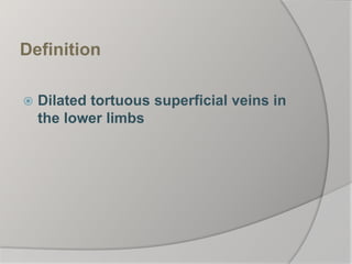
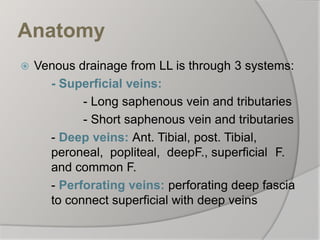
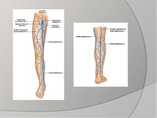
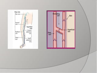
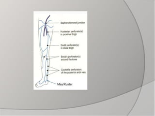
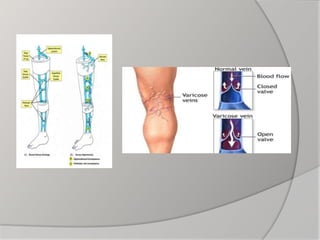
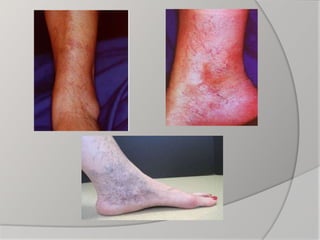
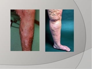
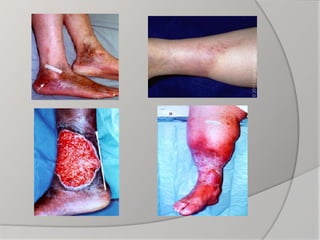
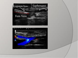
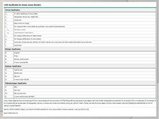
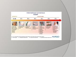
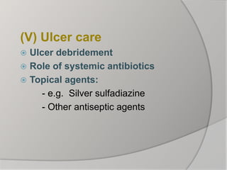
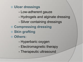
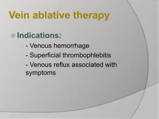
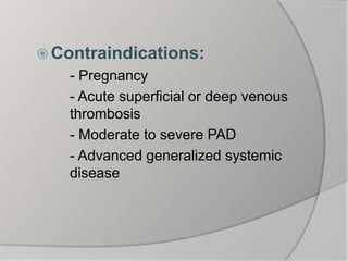
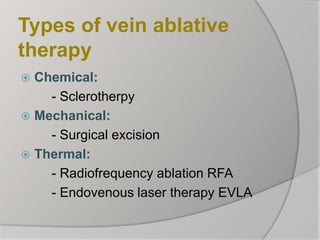
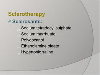
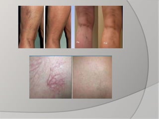
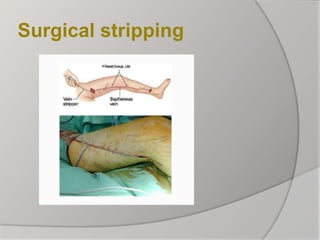
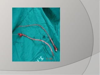
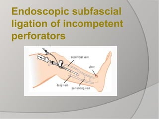
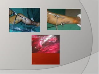
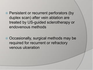
By
Mahmoud Zaghloul Raslan, MD
Consultant Surgeon, MGH
Madina
Definition
 Dilated tortuous superficial veins in
the lower limbs
Anatomy
 Venous drainage from LL is through 3 systems:
- Superficial veins:
- Long saphenous vein and tributaries
- Short saphenous vein and tributaries
- Deep veins: Ant. Tibial, post. Tibial,
peroneal, popliteal, deepF., superficial F.
and common F.
- Perforating veins: perforating deep fascia
to connect superficial with deep veins
Pathophysiology
Physiology:
_ Blood conductor:
- Muscular compression
- Negative ITP
- Calf Muscle pump
- Presence of valves
_ Blood reservoir:
- Dilatable
- Capacious
Etiology and Types
 Primary VV
_ High IVP: prolonged standing,
prolonged sitting with crossed
legs, chronic straining,
_ Weakness of vein walls:
- Obesity
- Pregnancy
- Hormonal
 Secondary VV:
- After previous DVT
- With pelvic tumours
Clinical Picture
 Thorough history taking:
- Occupation and prolonged standing
- Throbophlebitis or DVT
- Coaguation disorders
- Diabetes
- Results of previous treatment
- Pregnancy and contraceptive pills
 Symptoms:
- Pain: aching, throbbing, tingling
- Cramps, heaviness, tiredness of
legs, restless legs at night
- Of complications: Itching,
hyperpigmentation, skin ulceration
and bleeding
- Leg disfigurement
Trendlenberg test
Investigations
 General:
- Assess the general condition of the patient:
e.g. CBC,liver and kidney function tests
 Specific:
- Hand-held Doppler
- Duplex ultrasound: The best; gives both anatomical
and functional diagnosis
- Others: e.g. CT venography and MRI rarely needed
Treatment
Initial conservative
treatment
(I) General measures:
 Leg elevation
 Excerise
(II) Compression therapy:
(III) Pharmacologic therapy:
 Venoactive drugs
- Micronized purified flavonoid
MPFF e.g.Daflon
- Hydroxyethylrutoside
 Rheologic agents:
- Aspirin
- Stanozolol
- pentoxifylline
- prostacyclin analogues
(IV) Skin care:
 Skin cleansing e.g. Dove, Olay
 Emollients e.g. vaseline, cetaphil
 Barrier preparations e.g. zinc oxide
cream, Vaseline
 Topical corticosteroids
(V) Ulcer care
 Ulcer debridement
 Role of systemic antibiotics
 Topical agents:
- e.g. Silver sulfadiazine
- Other antiseptic agents
 Ulcer dressings
- Low-adherent gauze
- Hydrogels and alginate dressing
- Silver containing dressings
 Compressing dressing
 Skin grafting
 Others:
- Hyperbaric oxygen
- Electromagnetic therapy
- Therapeutic ultrasound
Vein ablative therapy
 Indications:
- Venous hemorrhage
- Superficial thrombophlebitis
- Venous reflux associated with
symptoms
 Contraindications:
- Pregnancy
- Acute superficial or deep venous
thrombosis
- Moderate to severe PAD
- Advanced generalized systemic
disease
Types of vein ablative
therapy
 Chemical:
- Sclerotherpy
 Mechanical:
- Surgical excision
 Thermal:
- Radiofrequency ablation RFA
- Endovenous laser therapy EVLA
Sclerotherapy
 Sclerosants:
_ Sodium tetradecyl sulphate
_ Sodium marrhuate
_ Polydocanol
_ Ethanolamine oleate
_ Hypertonic saline
Surgical stripping
Endoscopic subfascial
ligation of incompetent
perforators
Radiofrequency ablation
therapy
 By directing radiofrequency energy through a
vein, a narrow rim of tissue less than 1mm is
heated by an electrode.
 The amount of heating is modulated using a
microprocessor resulting in controlled
collagen contraction, thermocoagulation and
absorption of the vein.
 Endovenous laser ablation:
Initiates nonthrombotic occlusion by direct
thermal injury to vein wall causing
endothelial destruction, collagen
contraction and later fibrosis
Management by clinical category
 No visible or palpable signs of
venous dis. (CEAP category 0):
- If C/O venous symptoms:
Treated conservatively
 Telangiectasias and reticular veins
(CEAP category 1):
_ Asymptomatic patients with no
reflux: Treated by
- Sclerotherapy
- Laser light therapy
_ Symptomatic patients with reflux:
- Treat reflux first by surgical or
thermal ablation
 Varicose veins (CEAP category 2):
_ Isolated varicosities without trunkal
reflux are treated by:
- Sclerotherapy
- Surgical excision
Depending on size, location and
number of affected veins
_ With reflux: Treated by vein ablation
 Comparison of ablative therapies:
_ Recent studies reveal that minimally
invasive therapies are as effective as
surgical ablation with fewer
complications and rapid regain of daily
activities
 Advanced venous disease:
_ Patients with edema, skin changes or
ulceration (CEAP categories C3, C4,
C5, C6: Treated by initial conservative
measures
_ Refractory patients to conservative
measures: Treated by ablative therapy
Summary and conclusion
 Visibly dilated LL veins may indicate
underlying venous reflux, especially if
symptomatic, However, they can occur
in absence of symptoms or reflux
 Duplex US is essential to identify
superficial, deep or perforator vein reflux
and R/O DVT
 Patients with venous reflux and those
with complications should be referred to
venous or at least vascular specialist for
further evaluation and management
 Goals of treatment are improved
symptoms and appearance
 Most patients are treated by initial
conservative measures
 Patients refractory to conservative
measures for 3 months with documented
reflux (>0.5 second duration) are
candidates for vein ablative therapy
 Patients with telangiectasias, reticular
vein and VV with reflux should undergo
vein ablation before treating these
lesions
 Telangiectasias, reticular veins and
small VV large enough to admit 27 or 30
gauge needle are treated best by
sclerotherapy with good results
 Laser therapy is the only option for
telangiectasias too small to access,
allergy to sclerosants, patients afraid of
needles and after failed sclerotherapy
 Endovenous ablation techniques are
preferred for saphenous and other
trunkal veins over surgical ablation
 Persistent or recurrent perforators (by
duplex scan) after vein ablation are
treated by US-guided sclerotherapy or
endovenous methods
 Occasionally, surgical methods may be
required for recurrent or refractory
venous ulceration
Varicose veins

Varicose veins

  • 1.
    By Mahmoud Zaghloul Raslan,MD Consultant Surgeon, MGH Madina
  • 2.
    Definition  Dilated tortuoussuperficial veins in the lower limbs
  • 3.
    Anatomy  Venous drainagefrom LL is through 3 systems: - Superficial veins: - Long saphenous vein and tributaries - Short saphenous vein and tributaries - Deep veins: Ant. Tibial, post. Tibial, peroneal, popliteal, deepF., superficial F. and common F. - Perforating veins: perforating deep fascia to connect superficial with deep veins
  • 9.
    Pathophysiology Physiology: _ Blood conductor: -Muscular compression - Negative ITP - Calf Muscle pump - Presence of valves
  • 10.
    _ Blood reservoir: -Dilatable - Capacious
  • 11.
    Etiology and Types Primary VV _ High IVP: prolonged standing, prolonged sitting with crossed legs, chronic straining, _ Weakness of vein walls: - Obesity - Pregnancy - Hormonal
  • 12.
     Secondary VV: -After previous DVT - With pelvic tumours
  • 13.
  • 14.
     Thorough historytaking: - Occupation and prolonged standing - Throbophlebitis or DVT - Coaguation disorders - Diabetes - Results of previous treatment - Pregnancy and contraceptive pills
  • 15.
     Symptoms: - Pain:aching, throbbing, tingling - Cramps, heaviness, tiredness of legs, restless legs at night - Of complications: Itching, hyperpigmentation, skin ulceration and bleeding - Leg disfigurement
  • 19.
  • 20.
  • 21.
     General: - Assessthe general condition of the patient: e.g. CBC,liver and kidney function tests  Specific: - Hand-held Doppler - Duplex ultrasound: The best; gives both anatomical and functional diagnosis - Others: e.g. CT venography and MRI rarely needed
  • 27.
  • 28.
    Initial conservative treatment (I) Generalmeasures:  Leg elevation  Excerise (II) Compression therapy:
  • 29.
    (III) Pharmacologic therapy: Venoactive drugs - Micronized purified flavonoid MPFF e.g.Daflon - Hydroxyethylrutoside
  • 30.
     Rheologic agents: -Aspirin - Stanozolol - pentoxifylline - prostacyclin analogues
  • 31.
    (IV) Skin care: Skin cleansing e.g. Dove, Olay  Emollients e.g. vaseline, cetaphil  Barrier preparations e.g. zinc oxide cream, Vaseline  Topical corticosteroids
  • 32.
    (V) Ulcer care Ulcer debridement  Role of systemic antibiotics  Topical agents: - e.g. Silver sulfadiazine - Other antiseptic agents
  • 33.
     Ulcer dressings -Low-adherent gauze - Hydrogels and alginate dressing - Silver containing dressings  Compressing dressing  Skin grafting  Others: - Hyperbaric oxygen - Electromagnetic therapy - Therapeutic ultrasound
  • 34.
    Vein ablative therapy Indications: - Venous hemorrhage - Superficial thrombophlebitis - Venous reflux associated with symptoms
  • 35.
     Contraindications: - Pregnancy -Acute superficial or deep venous thrombosis - Moderate to severe PAD - Advanced generalized systemic disease
  • 36.
    Types of veinablative therapy  Chemical: - Sclerotherpy  Mechanical: - Surgical excision  Thermal: - Radiofrequency ablation RFA - Endovenous laser therapy EVLA
  • 37.
    Sclerotherapy  Sclerosants: _ Sodiumtetradecyl sulphate _ Sodium marrhuate _ Polydocanol _ Ethanolamine oleate _ Hypertonic saline
  • 40.
  • 42.
    Endoscopic subfascial ligation ofincompetent perforators
  • 44.
    Radiofrequency ablation therapy  Bydirecting radiofrequency energy through a vein, a narrow rim of tissue less than 1mm is heated by an electrode.  The amount of heating is modulated using a microprocessor resulting in controlled collagen contraction, thermocoagulation and absorption of the vein.
  • 49.
     Endovenous laserablation: Initiates nonthrombotic occlusion by direct thermal injury to vein wall causing endothelial destruction, collagen contraction and later fibrosis
  • 54.
    Management by clinicalcategory  No visible or palpable signs of venous dis. (CEAP category 0): - If C/O venous symptoms: Treated conservatively
  • 55.
     Telangiectasias andreticular veins (CEAP category 1): _ Asymptomatic patients with no reflux: Treated by - Sclerotherapy - Laser light therapy _ Symptomatic patients with reflux: - Treat reflux first by surgical or thermal ablation
  • 56.
     Varicose veins(CEAP category 2): _ Isolated varicosities without trunkal reflux are treated by: - Sclerotherapy - Surgical excision Depending on size, location and number of affected veins _ With reflux: Treated by vein ablation
  • 57.
     Comparison ofablative therapies: _ Recent studies reveal that minimally invasive therapies are as effective as surgical ablation with fewer complications and rapid regain of daily activities
  • 58.
     Advanced venousdisease: _ Patients with edema, skin changes or ulceration (CEAP categories C3, C4, C5, C6: Treated by initial conservative measures _ Refractory patients to conservative measures: Treated by ablative therapy
  • 59.
  • 60.
     Visibly dilatedLL veins may indicate underlying venous reflux, especially if symptomatic, However, they can occur in absence of symptoms or reflux  Duplex US is essential to identify superficial, deep or perforator vein reflux and R/O DVT
  • 61.
     Patients withvenous reflux and those with complications should be referred to venous or at least vascular specialist for further evaluation and management  Goals of treatment are improved symptoms and appearance
  • 62.
     Most patientsare treated by initial conservative measures  Patients refractory to conservative measures for 3 months with documented reflux (>0.5 second duration) are candidates for vein ablative therapy
  • 63.
     Patients withtelangiectasias, reticular vein and VV with reflux should undergo vein ablation before treating these lesions  Telangiectasias, reticular veins and small VV large enough to admit 27 or 30 gauge needle are treated best by sclerotherapy with good results
  • 64.
     Laser therapyis the only option for telangiectasias too small to access, allergy to sclerosants, patients afraid of needles and after failed sclerotherapy  Endovenous ablation techniques are preferred for saphenous and other trunkal veins over surgical ablation
  • 65.
     Persistent orrecurrent perforators (by duplex scan) after vein ablation are treated by US-guided sclerotherapy or endovenous methods  Occasionally, surgical methods may be required for recurrent or refractory venous ulceration