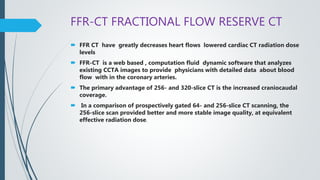
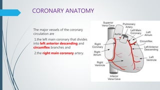
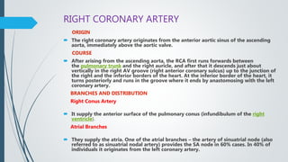
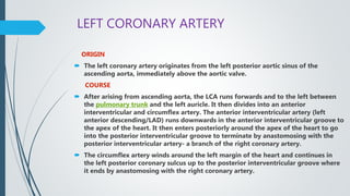
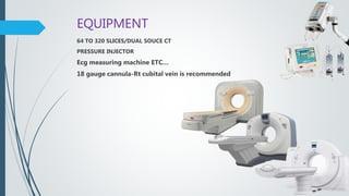
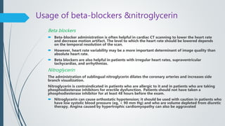
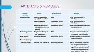
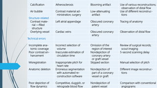
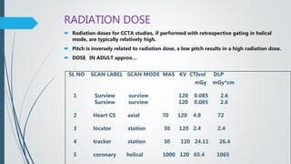
The document provides an overview of coronary CT angiography (CCTA). It discusses recent advances in CCTA technology including perfusion imaging, spectral imaging, and fractional flow reserve CT (FFR-CT). The anatomy and physiology of the coronary arteries is described. The document outlines the equipment, indications, procedures, and post-processing techniques used in CCTA. It also discusses calcium scoring, artifacts, case studies, radiation dose, and limitations of CCTA.
A detailed description of ct coronary angiography and calcium scoring with various aspects regarding the preparation, procedure, limitations and a short review regarding post CABG imaging.
CT coronary angiography uses ECG gating to synchronize data acquisition with the cardiac cycle in order to reduce motion artifacts. Data can be acquired retrospectively by continuously scanning over multiple heartbeats and reconstructing different cardiac phases, or prospectively by only scanning during a targeted phase like mid-diastole. Placement of the ROI for bolus tracking is important to ensure consistent coronary enhancement. High temporal resolution under 200ms can be achieved through techniques like partial scan or multisegment reconstruction.
A CT coronary angiogram (CTCA) uses computed tomography to non-invasively image the coronary arteries. It provides useful information about coronary artery disease. Specialists who interpret CTCAs must complete training requirements, including a minimum number of cases. CTCA is a low-risk, low-radiation exam that can accurately detect narrowings or anomalies in the coronary arteries. It may benefit those with suspected coronary artery disease, atypical chest pain, or to check grafts. Indications include chest pain with low-intermediate risk or family history. Preparation includes fasting and potentially taking a beta-blocker to lower the heart rate.
Coronary CT angiography allows for noninvasive imaging of the heart and coronary arteries. It can be used to evaluate patients with chest pain, assess coronary arteries after revascularization, and detect congenital coronary anomalies. The scan involves a non-contrast scan for calcium scoring followed by a contrast-enhanced scan. Proper patient preparation including beta-blockers and nitroglycerin is important. Images are analyzed using techniques like multiplanar reformation, maximum intensity projection, volume rendering and curved reformation to evaluate coronary artery anatomy and detect any stenosis.
This document discusses cardiac MRI (CMRI) and its clinical applications. CMRI provides anatomical and functional information to assess heart abnormalities through various sequences like ECG-gated bright and dark blood sequences. It is useful for evaluating congenital heart diseases, valvular heart diseases, ventricular function, coronary arteries, myocardial perfusion and viability, cardiac masses, and pericardial diseases. CMRI is more accurate than echocardiography for measuring ejection fraction, volumes, and assessing ventricular function and viability. It is useful for differentiating conditions like arrhythmogenic right ventricular dysplasia, restrictive vs constrictive cardiomyopathy, and determining feasibility of revascularization procedures.
- Coronary CT angiography uses x-rays and contrast material to examine the coronary arteries with high spatial and temporal resolution. It is non-invasive compared to traditional coronary angiography.
- Key factors for cardiac imaging include high temporal resolution (<250ms), spatial resolution (<0.75mm), and synchronization with the cardiac cycle using ECG gating. Prospective and retrospective gating, partial and multi-segment reconstruction, and low pitch values (<0.5) help achieve this.
- Advances in multi-detector CT scanners, faster gantry rotation times (<330ms), and improved reconstruction algorithms now allow temporal resolutions as low as 80ms for coronary CT angiography.
This document outlines the protocol for performing CT angiography (CTA) from the cerebral arteries to the lower limbs. It discusses indications for CTA including aneurysms, stenosis, dissections, and more. The preparation, positioning, and scanning protocols are provided for CTA of the head to lower limbs as well as the subclavian arteries. Pediatric protocols are also summarized. The document concludes with examples of CTA findings and references.
Coronary CT angiography is a noninvasive imaging modality used to evaluate coronary artery disease. It has a high sensitivity of 87-99% and specificity of 93-96% for detecting coronary artery stenosis. Coronary CT angiography is most useful in low- to intermediate-risk patients with chest pain to rule out coronary artery disease given its high negative predictive value of 93-100%. Coronary CT angiography involves acquiring images using ionizing radiation as the patient holds their breath and synchronizing the images with the patient's ECG signal.
This document discusses the use of coronary CT angiography to detect and characterize coronary artery anatomy and exclude morphological abnormalities. It notes the technique involves a preliminary scout study followed by contrast-enhanced imaging of the coronary arteries. Reconstructions include curved multi-planar views. The quality was deemed excellent with no artifacts. The impression was a total calcium score of zero, no evidence of stenosis or plaque, and a CAD-RADS classification of 0, recommending reassurance.
This document discusses the evolution and advances in coronary CT angiography (CCTA) technology and its role in the assessment of coronary artery disease (CAD). Key points include:
- CCTA has advanced from early CT scanners with 4-minute scan times to modern multi-detector scanners that can image the entire heart in a single heartbeat.
- CCTA provides information on coronary artery anatomy, plaque characteristics, and has prognostic value when assessing coronary artery calcium scoring.
- CCTA has good accuracy for detecting CAD compared to invasive coronary angiography, especially for ruling out disease, though its role in asymptomatic patients is still unclear.
- CCTA is useful for evaluating coronary anomalies, bypass grafts,
Presentation given at Arab Health congress on Jan. 29th 2013, with information about (dual source) Cardiac CT of the coronary arteries with technical & practical information and some clinical use cases
Multi detector ct cerebral angiographyEhab Elftouh
This document discusses techniques for computed tomography (CT) angiography. It covers advances in CT technology that have improved angiography, including faster scan speeds and thinner slices. Optimal CT angiography depends on scan technique factors like protocol and contrast injection, as well as image post-processing techniques. Newer multi-detector CT machines allow covering volumes more quickly and with higher resolution. Methods like multi-planar reformation and volume rendering help visualize vascular structures from CT image data.
This document discusses computed tomography angiography (CTA) and its applications in cardiology. CTA uses computed tomography to visualize blood vessels throughout the body, including coronary arteries. Coronary CTA can detect plaque buildup in coronary arteries without being invasive. Current multidetector CT systems can acquire high-resolution images of the heart within 20 seconds while the patient holds their breath. Coronary CTA provides diagnostic information but also exposes patients to radiation. It is most useful for evaluating cardiac symptoms in low-to-intermediate risk patients.
Copy of Mitsumori-CT angiography techniquesamin usmani
This document discusses techniques for CT angiography (CTA). It begins with an overview of contrast administration and achieving arterial enhancement through higher iodine concentration, injection flow rate, and duration. It describes using a timing bolus or bolus tracking to determine the optimal scan delay to image arteries during peak enhancement. Example CTA protocols are provided for pulmonary embolism, abdominal aorta, and thoracic aorta imaging.
CT calcium scoring can detect asymptomatic coronary artery disease by identifying coronary artery calcification. Calcium starts accumulating early in atherosclerosis and increases as the disease progresses. While a calcium score of zero does not rule out disease, it is associated with a very low risk of cardiac events. Higher calcium scores correlate with increased risk. CT calcium scoring provides individualized risk assessment and can guide aggressive risk factor modification in high-risk patients.
This document provides information on imaging of the carotid arteries and carotid angiography. It discusses various imaging modalities used to image the carotid arteries including ultrasound, CT, MRI, CT angiography, MR angiography, duplex ultrasound, and plain films. It then provides detailed information on carotid angiography including definitions, indications, complications, techniques, and how to avoid complications. Transcranial ultrasound in premature infants is also briefly discussed.
Normal Cardiac CT
This document summarizes the key aspects of performing and interpreting a normal cardiac CT scan. It discusses the technique, including protocols for ECG gating and contrast injection. It then reviews the anatomy of the coronary arteries and important post-processing techniques like MPR, MIP, and VR. Segmental models for describing coronary artery anatomy are presented. Metrics for normal coronary artery diameter and left atrial area are provided. Common cardiac imaging planes and structures like the left ventricle and valves are also depicted.
This document provides information on performing and interpreting CT angiography of the lower limbs. It discusses scanning techniques, protocols, contrast injection, and principles of timing acquisitions. Image post-processing includes MIP, VR, and MPR. Interpretation requires scrutinizing calcifications and stents to avoid overestimating stenosis. Peripheral CTA is useful for evaluating occlusive disease, aneurysms, trauma, infections, embolism, and postoperative surveillance. Examples demonstrate various vascular pathologies.
This document provides an overview of cardiac MRI techniques including gradient echo sequences which provide cine images of heart motion and white blood, spin echo sequences which produce static black blood images, and phase contrast imaging which uses Doppler to visualize blood flow direction and velocity. Delayed enhancement imaging identifies areas of scar or fibrosis by their contrast uptake several minutes after injection. Other techniques discussed are perfusion imaging, tissue tagging, and STIR imaging for edema detection. Common imaging planes and protocols are outlined along with common uses of cardiac MRI such as assessing function, cardiomyopathy, and viability.
Dual energy CT utilizes two different x-ray spectra to characterize tissues. It can help address challenges with single energy CT like lesion detection and image noise. Dual energy CT works by analyzing how materials attenuate x-rays differently at various energies, allowing differentiation of substances like iodine and calcium. There are several technical approaches to dual energy CT, including sequential acquisition with two scans, rapid voltage switching between two voltages, and dual-source CT with two tube-detector pairs. Post-processing involves material decomposition and differentiation using image-domain or projection-domain algorithms.
Role of ct angiography in diagnosis of coronary anomalies GhadaSheta
CT angiography plays an important role in diagnosing coronary artery anomalies. It provides detailed 3D images of the coronary arteries with high spatial and temporal resolution in a noninvasive manner. Proper patient preparation including beta blockers to lower heart rate and nitroglycerin to dilate arteries is important for optimal imaging. CT angiography can detect various types of anomalies such as anomalous coronary artery origins, fistulas, myocardial bridging, and duplication of arteries. It serves as a roadmap for cardiologists in guiding patient management.
This document provides information about dose reduction techniques in CT scanning. It discusses how CT scan technology has advanced but also leads to higher radiation doses compared to other modalities. Various techniques can help reduce dose like adjusting acquisition parameters such as tube current, voltage, and scan length. Equipment designs with features like iterative reconstruction and dual-layer detectors can also help lower dose. Selecting the appropriate scan protocol tailored to the clinical task is important to optimize image quality while keeping radiation exposure as low as reasonably achievable.
Computerized tomography (CT) was pioneered by Godfrey Hounsfield and Allan Cormack in the 1970s. CT uses X-rays and computer processing to create cross-sectional images of the body. The first CT scanners used a translate-rotate design, while later generations used multiple detectors and spiral scanning for faster, more detailed imaging. Image reconstruction uses back projection to convert attenuation measurements into pixel values and display slices. CT provides excellent anatomical detail and is widely used for diagnosing conditions of the brain, blood vessels, lungs and other organs.
Cardiac CT-CCTA involves three main steps: patient preparation with beta blockade and nitroglycerine to lower heart rate, initial calcium scoring to identify atherosclerotic vessels, and coronary CTA scan using retrospective or prospective ECG gating. CCTA allows visualization of the coronary arteries and quantification of plaque type and stenosis. Normal coronary anatomy includes the left main artery bifurcating into the LAD and LCX, and the RCA originating from the right coronary cusp and dominantly supplying the posterior descending artery in most cases.
This document discusses the basics of CT scanning, including its history and key components. It describes how CT scanning works, from the x-ray tube emitting radiation that is detected after passing through the body, to the computer using this data to reconstruct cross-sectional images. It outlines the main parts of a CT system, including the gantry, detector, and control console. It also explains different scanning methods and how image quality is determined.
This document discusses digital subtraction angiography (DSA), including its history, equipment, and applications. DSA involves acquiring digital fluoroscopic images before and after injecting contrast material, and using computer subtraction to remove bone structures and leave an image of blood vessels. It originated in the 1970s and allows for real-time angiography with improved vessel contrast compared to conventional techniques. Key components of DSA systems include an x-ray unit, image intensifier, computer, and software for image processing functions like subtraction, enhancement, and roadmapping.
Post processing of computed tomography images allows radiologists to view images in different planes and highlight key anatomical structures. Techniques like multiplanar reconstruction generate coronal and sagittal views from axial scans, while maximum intensity projection highlights contrast-filled vessels. Together, these techniques provide additional diagnostic information beyond the original axial images.
coronary angiography, LV angiogram and coronary anomaliesSalman Ahmed
Coronary angiography is a procedure that uses dye and x-rays to visualize blood flow through the coronary arteries. It remains the gold standard for detecting significant coronary artery disease. The procedure involves passing a catheter into the heart and injecting contrast dye to see blockages. Coronary angiography is indicated when objective demonstration of the coronary anatomy could help resolve a problem, with competent staff and facilities. It provides information on stenosis levels and collateral circulation. Different views are used to visualize different coronary artery segments. Cannulation techniques depend on arterial origins and graft types.
The document discusses Triple Rule Out CT (TRO-CT), which is a CT exam that evaluates the coronary arteries, pulmonary arteries, and aorta to diagnose causes of acute chest pain. TRO-CT is appropriate when there is suspicion of acute coronary syndrome along with pulmonary embolism or acute aortic syndrome. It describes the anatomy of the aorta, pulmonary arteries, and coronary arteries. It also discusses common causes of chest pain related to the heart, lungs, chest wall, and digestion. The criteria and exclusion criteria for TRO-CT are provided.
Triple rule-out CT (TRO-CT) is an imaging exam that evaluates the coronary arteries, pulmonary arteries, and aorta in patients presenting with acute chest pain. It aims to diagnose the cause of chest pain, which could be due to conditions in any of the three vascular territories. The exam involves a contrast-enhanced CT scan of the chest during a single breath-hold. It allows for simultaneous assessment of the entire thorax, improving diagnosis and reducing the need for multiple tests. TRO-CT can help identify life-threatening causes of chest pain such as pulmonary embolism, aortic dissection and coronary artery disease.
This document provides an overview of carotid Doppler ultrasound. It begins with the anatomy of the carotid arteries and their branches. It then discusses the technique of carotid Doppler ultrasound, including instrumentation, examination protocol, and interpretation of ultrasound findings. It provides details on evaluating the internal carotid, external carotid, and vertebral arteries for stenosis or occlusion. The document also covers characterizing carotid plaques and differentiating true findings from artifacts.
Coronary angiography is a procedure that uses dye and x-rays to see how blood flows through the coronary arteries of the heart. It is the gold standard for evaluating coronary artery disease and can identify the location and severity of any blockages. A coronary angiogram involves inserting a catheter into the heart and injecting dye so that blockages are highlighted on x-ray images. Potential complications are rare but can include heart attack, stroke, or kidney injury from the dye. The results of the angiogram are used to determine if further procedures like angioplasty or bypass surgery are needed.
Coronary angiography is a procedure that uses dye and x-rays to see how blood flows through the coronary arteries of the heart. It is the gold standard for evaluating coronary artery disease and can identify the location and severity of any blockages. A coronary angiogram involves inserting a catheter into the heart and injecting dye so that blockages are highlighted on x-ray images. Potential complications are usually minor but can include heart attack, stroke, or kidney injury from the dye. The results of the angiogram are used to determine if further procedures like angioplasty or bypass surgery are needed.
The document discusses coronary artery anatomy and various imaging techniques used to assess myocardial viability. It provides details on:
1. The origins, branches and distributions of the right and left coronary arteries.
2. Imaging modalities for evaluating viable myocardium including dobutamine stress echocardiography, nuclear techniques like SPECT and PET, and cardiac MRI.
3. Cardiac MRI's use of late gadolinium enhancement to identify infarcted versus viable myocardium based on the degree and transmurality of enhancement.
The document discusses coronary artery anatomy and techniques for assessing myocardial viability. It provides details on:
1. The origins, branches and distributions of the right and left coronary arteries.
2. Imaging modalities for evaluating myocardial viability including dobutamine stress echocardiography, nuclear techniques using thallium/technetium and FDG PET, and cardiac MRI with late gadolinium enhancement.
3. The interpretation of these tests to determine viability, with areas of uptake on nuclear imaging over 50% or absence of late gadolinium enhancement on MRI suggesting viable myocardium.
This document provides information on renal artery anatomy and Doppler ultrasound evaluation of the renal arteries. It describes:
1. The typical origin and course of the right and left renal arteries. Approximately 30% of individuals have variant anatomy with more than one renal artery on each side.
2. How Doppler ultrasound is used to image the renal arteries from different approaches and measure parameters like peak systolic velocity to evaluate for renal artery stenosis.
3. The normal Doppler waveforms expected in the main renal artery and intrarenal arteries, as well as normal values for measured parameters.
4. How a bilateral renal Doppler examination is performed, including evaluating each kidney, the renal arteries and veins, and measuring parameters to identify
This document provides guidance on performing and interpreting coronary angiograms. It discusses techniques such as catheter selection, standard angiographic views, contrast injection settings, and complications. It also covers evaluating angiograms by quantifying lesions, classifying them using ACC/AHA criteria, and assessing TIMI flow. Interpretation involves a systematic analysis of the coronary anatomy and any areas of stenosis.
This document discusses various radiological techniques for examining the cardiovascular system, including fluoroscopy, roentgenography, fluorography, tomography, angiography, computed tomography, ultrasonography, and magnetic resonance imaging. It describes how to evaluate the heart shape and position using these techniques. Common cardiovascular abnormalities are outlined, including mitral valve disease, aortic valve disease, diffuse myocardial disease, and limited vascular abnormalities. The document also discusses analyzing coronary arteries, congenital heart diseases, and evaluating cardiac function and plaque types using multidetector computed tomography.
Raja Lahiri provides an overview of coronary angiography. Key points include:
- Coronary angiography is the current gold standard for visualizing the coronary arteries through X-ray imaging with contrast injection.
- The history of coronary angiography began in the 1920s-1940s with early experiments in cerebral and cardiac catheterization.
- Modern techniques involve accessing arteries typically through the femoral or radial arteries to insert a catheter for contrast injection into the coronary arteries under X-ray imaging.
- Multiple angiographic views are needed to visualize different segments of the left and right coronary arteries. Coronary angiography is used to evaluate coronary artery disease, graft patency, and left ventricular function.
Radiological diagnostics of Cardiovascular SystemEneutron
This document discusses various radiological techniques for examining the cardiovascular system, including fluoroscopy, roentgenography, fluorography, tomography, angiography, computed tomography, ultrasonography, and magnetic resonance imaging. It provides details on evaluating heart size and shape, positioning, cardiac contours and arcs seen in different views. Common cardiovascular abnormalities are described based on their radiological presentations, such as mitral and aortic heart shapes. Congenital heart diseases are categorized into three groups based on blood flow abnormalities. Methods for examining coronary arteries both invasively and non-invasively are also outlined.
Here are the key points about the different types of blood vessels:
- Arteries carry oxygenated blood away from the heart to tissues and organs. They have an outer
tunica externa layer of connective tissue, a middle tunica media layer of smooth muscle, and an
inner tunica intima layer of endothelium. Larger elastic arteries near the heart have more elastic
tissue.
- Capillaries are the microscopic vessels that connect arterioles and venules. They allow for the
exchange of water, oxygen, nutrients, hormones, carbon dioxide and waste between blood and
tissues. Capillaries have a single layer of endothelial cells and connective tissue.
-
The document describes the anatomy and blood supply of the heart. It discusses the right and left coronary arteries, their branches, and the areas they supply. It also covers the conducting system of the heart, including the sinoatrial node, atrioventricular node, bundle of His, and Purkinje fibers. Additionally, it summarizes the venous drainage of the heart through coronary sinus and its tributaries.
This document provides an overview of echocardiography, including its definition, history, uses, types, parameters, and measurements. Echocardiography involves using ultrasound to create real-time images of the heart. Key developments include the discovery of piezoelectricity in 1880 and the first clinical echocardiogram in the 1950s. Echocardiography can evaluate both cardiac structure and function, and provides important information on systolic and diastolic performance through measurements like ejection fraction, fractional shortening, and mitral inflow velocities. Standard views and protocols aim to comprehensively assess cardiac health.
Coronary angiography remains the gold standard for detecting coronary artery disease. The technique was first performed in 1958 and is used to visualize the coronary arteries and assess for stenosis. It can determine treatment options and prognosis. Complications are rare but include vascular injury and contrast reactions. Proper angiographic views are important for evaluating different coronary artery segments.
The document discusses ruptured aneurysms of the aorta, specifically focusing on ruptured abdominal aortic aneurysms (RAAAs). It describes the typical presentation of RAAAs, which includes abdominal or back pain, hypotension, and the potential presence of a pulsatile abdominal mass. It notes that RAAAs have a high mortality rate if not treated emergently through open repair or potentially endovascular aneurysm repair (EVAR). Unusual presentations of RAAAs are also discussed, which can include symptoms like leg paralysis or groin/testicular pain that mimic other conditions and delay diagnosis.
share - Lions, tigers, AI and health misinformation, oh my!.pptxTina Purnat
• Pitfalls and pivots needed to use AI effectively in public health
• Evidence-based strategies to address health misinformation effectively
• Building trust with communities online and offline
• Equipping health professionals to address questions, concerns and health misinformation
• Assessing risk and mitigating harm from adverse health narratives in communities, health workforce and health system
TEST BANK For Basic and Clinical Pharmacology, 14th Edition by Bertram G. Kat...rightmanforbloodline
TEST BANK For Basic and Clinical Pharmacology, 14th Edition by Bertram G. Katzung, Verified Chapters 1 - 66, Complete Newest Version.
Integrating Ayurveda into Parkinson’s Management: A Holistic ApproachAyurveda ForAll
Explore the benefits of combining Ayurveda with conventional Parkinson's treatments. Learn how a holistic approach can manage symptoms, enhance well-being, and balance body energies. Discover the steps to safely integrate Ayurvedic practices into your Parkinson’s care plan, including expert guidance on diet, herbal remedies, and lifestyle modifications.
Adhd Medication Shortage Uk - trinexpharmacy.comreignlana06
The UK is currently facing a Adhd Medication Shortage Uk, which has left many patients and their families grappling with uncertainty and frustration. ADHD, or Attention Deficit Hyperactivity Disorder, is a chronic condition that requires consistent medication to manage effectively. This shortage has highlighted the critical role these medications play in the daily lives of those affected by ADHD. Contact : +1 (747) 209 – 3649 E-mail : sales@trinexpharmacy.com
These lecture slides, by Dr Sidra Arshad, offer a quick overview of the physiological basis of a normal electrocardiogram.
Learning objectives:
1. Define an electrocardiogram (ECG) and electrocardiography
2. Describe how dipoles generated by the heart produce the waveforms of the ECG
3. Describe the components of a normal electrocardiogram of a typical bipolar lead (limb II)
4. Differentiate between intervals and segments
5. Enlist some common indications for obtaining an ECG
6. Describe the flow of current around the heart during the cardiac cycle
7. Discuss the placement and polarity of the leads of electrocardiograph
8. Describe the normal electrocardiograms recorded from the limb leads and explain the physiological basis of the different records that are obtained
9. Define mean electrical vector (axis) of the heart and give the normal range
10. Define the mean QRS vector
11. Describe the axes of leads (hexagonal reference system)
12. Comprehend the vectorial analysis of the normal ECG
13. Determine the mean electrical axis of the ventricular QRS and appreciate the mean axis deviation
14. Explain the concepts of current of injury, J point, and their significance
Study Resources:
1. Chapter 11, Guyton and Hall Textbook of Medical Physiology, 14th edition
2. Chapter 9, Human Physiology - From Cells to Systems, Lauralee Sherwood, 9th edition
3. Chapter 29, Ganong’s Review of Medical Physiology, 26th edition
4. Electrocardiogram, StatPearls - https://www.ncbi.nlm.nih.gov/books/NBK549803/
5. ECG in Medical Practice by ABM Abdullah, 4th edition
6. Chapter 3, Cardiology Explained, https://www.ncbi.nlm.nih.gov/books/NBK2214/
7. ECG Basics, http://www.nataliescasebook.com/tag/e-c-g-basics
Histololgy of Female Reproductive System.pptxAyeshaZaid1
Dive into an in-depth exploration of the histological structure of female reproductive system with this comprehensive lecture. Presented by Dr. Ayesha Irfan, Assistant Professor of Anatomy, this presentation covers the Gross anatomy and functional histology of the female reproductive organs. Ideal for students, educators, and anyone interested in medical science, this lecture provides clear explanations, detailed diagrams, and valuable insights into female reproductive system. Enhance your knowledge and understanding of this essential aspect of human biology.
8 Surprising Reasons To Meditate 40 Minutes A Day That Can Change Your Life.pptxHolistified Wellness
We’re talking about Vedic Meditation, a form of meditation that has been around for at least 5,000 years. Back then, the people who lived in the Indus Valley, now known as India and Pakistan, practised meditation as a fundamental part of daily life. This knowledge that has given us yoga and Ayurveda, was known as Veda, hence the name Vedic. And though there are some written records, the practice has been passed down verbally from generation to generation.
Rasamanikya is a excellent preparation in the field of Rasashastra, it is used in various Kushtha Roga, Shwasa, Vicharchika, Bhagandara, Vatarakta, and Phiranga Roga. In this article Preparation& Comparative analytical profile for both Formulationon i.e Rasamanikya prepared by Kushmanda swarasa & Churnodhaka Shodita Haratala. The study aims to provide insights into the comparative efficacy and analytical aspects of these formulations for enhanced therapeutic outcomes.
Does Over-Masturbation Contribute to Chronic Prostatitis.pptxwalterHu5
In some case, your chronic prostatitis may be related to over-masturbation. Generally, natural medicine Diuretic and Anti-inflammatory Pill can help mee get a cure.
Cell Therapy Expansion and Challenges in Autoimmune DiseaseHealth Advances
There is increasing confidence that cell therapies will soon play a role in the treatment of autoimmune disorders, but the extent of this impact remains to be seen. Early readouts on autologous CAR-Ts in lupus are encouraging, but manufacturing and cost limitations are likely to restrict access to highly refractory patients. Allogeneic CAR-Ts have the potential to broaden access to earlier lines of treatment due to their inherent cost benefits, however they will need to demonstrate comparable or improved efficacy to established modalities.
In addition to infrastructure and capacity constraints, CAR-Ts face a very different risk-benefit dynamic in autoimmune compared to oncology, highlighting the need for tolerable therapies with low adverse event risk. CAR-NK and Treg-based therapies are also being developed in certain autoimmune disorders and may demonstrate favorable safety profiles. Several novel non-cell therapies such as bispecific antibodies, nanobodies, and RNAi drugs, may also offer future alternative competitive solutions with variable value propositions.
Widespread adoption of cell therapies will not only require strong efficacy and safety data, but also adapted pricing and access strategies. At oncology-based price points, CAR-Ts are unlikely to achieve broad market access in autoimmune disorders, with eligible patient populations that are potentially orders of magnitude greater than the number of currently addressable cancer patients. Developers have made strides towards reducing cell therapy COGS while improving manufacturing efficiency, but payors will inevitably restrict access until more sustainable pricing is achieved.
Despite these headwinds, industry leaders and investors remain confident that cell therapies are poised to address significant unmet need in patients suffering from autoimmune disorders. However, the extent of this impact on the treatment landscape remains to be seen, as the industry rapidly approaches an inflection point.
2. OVERVIEW
Recent Advances in Coronary CT
Anatomy and Physiology
Equipment and Physics
Indications
Coronary procedure
In detail Calcium Score
Techniques and post processing
Artefacts
Case studies
Contraindications
Radiation Dose
Limitations
Summary and References.
3. RECENT ADVANCES IN CORONARY CT
New technology supports CT as prime cardiac imaging
modality.
Perfusion imaging
Spectral imaging and
Non invasive Fractional flow reserve dose levels FFR-CT
PERFUSION IMAGING
CT perfusion imaging can accurately assess functional
blood flow in the heart by Software like TERARECON
and VITAL IMAGES etc… without the need for a
nuclear exam/MRI.
4. SPECTRAL IMAGING
Spectral imaging allow to differentiate anatomical features
which are enhanced and easier to see at different levels
It can highlight /eliminate specific chemical compounds
based on their atomic numbers including iodine and
calcium
Enables differentiation between calcified coronary lesion
and iodine contrast in the blood vessel.
5. FFR-CT FRACTIONAL FLOW RESERVE CT
FFR CT have greatly decreases heart flows lowered cardiac CT radiation dose
levels
FFR-CT is a web based , computation fluid dynamic software that analyzes
existing CCTA images to provide physicians with detailed data about blood
flow with in the coronary arteries.
The primary advantage of 256- and 320-slice CT is the increased craniocaudal
coverage.
In a comparison of prospectively gated 64- and 256-slice CT scanning, the
256-slice scan provided better and more stable image quality, at equivalent
effective radiation dose.
6. CORONARY ANATOMY
The major vessels of the coronary
circulation are
1.the left main coronary that divides
into left anterior descending and
circumflex branches and
2.the right main coronary artery.
7. RIGHT CORONARY ARTERY
ORIGIN
The right coronary artery originates from the anterior aortic sinus of the ascending
aorta, immediately above the aortic valve.
COURSE
After arising from the ascending aorta, the RCA first runs forwards between
the pulmonary trunk and the right auricle, and after that it descends just about
vertically in the right AV groove (right anterior coronary sulcus) up to the junction of
the right and the inferior borders of the heart. At the inferior border of the heart, it
turns posteriorly and runs in the groove where it ends by anastomosing with the left
coronary artery.
BRANCHES AND DISTRIBUTION
Right Conus Artery
It supply the anterior surface of the pulmonary conus (infundibulum of the right
ventricle).
Atrial Branches
They supply the atria. One of the atrial branches – the artery of sinuatrial node (also
referred to as sinuatrial nodal artery) provides the SA node in 60% cases. In 40% of
individuals it originates from the left coronary artery.
8. Anterior Ventricular Branches
They’re 2 or 3 and supply the anterior surface of the right ventricle.
The marginal branch is the largest and runs along the lower margin of the sternocostal
surface to make it to the apex.
Posterior Ventricular Branches
They may be generally 2 and supply the diaphragmatic surface of the right ventricle.
Posterior Interventricular Artery
It runs in the posterior interventricular groove up to the apex. It supplies the posterior
part of the interventricular septum, atrioventricular node (AV node) in 60% of the cases,
and right and left ventricles.
In 10% individuals, the posterior interventricular artery originates from the left coronary
artery.
9. LEFT CORONARY ARTERY
ORIGIN
The left coronary artery originates from the left posterior aortic sinus of the
ascending aorta, immediately above the aortic valve.
COURSE
After arising from ascending aorta, the LCA runs forwards and to the left between
the pulmonary trunk and the left auricle. It then divides into an anterior
interventricular and circumflex artery. The anterior interventricular artery (left
anterior descending/LAD) runs downwards in the anterior interventricular groove to
the apex of the heart. It then enters posteriorly around the apex of the heart to go
into the posterior interventricular groove to terminate by anastomosing with the
posterior interventricular artery- a branch of the right coronary artery.
The circumflex artery winds around the left margin of the heart and continues in
the left posterior coronary sulcus up to the posterior interventricular groove where
it ends by anastomosing with the right coronary artery.
10. BRANCHES AND DISTRIBUTION
1. Anterior interventricular artery/left anterior descending (LAD) artery: It provides (a)
anterior part of interventricular septum, (b) greater part of the left ventricle and part of
right ventricle, and (c) a part of left bundle branch (of His) posterior atrioventricular groove
(right posterior coronary sulcus) up to the posterior interventricular.
2. Circumflex artery: It supplies a left marginal artery that provides the left margin of the
left ventricle up to the apex of the heart.
3. Diagonal artery: It may originate directly from the trunk of the left coronary artery.
4. Conus artery: It supplies the pulmonary conus.
5. Atrial branches: They supply the left atrium.
Anatomic Region of Heart Coronary Artery (most likely associated)
Inferior Right coronary
Anteroseptal Left anterior descending
Anteroapical Left anterior descending (distal)
Anterolateral Circumflex
Posterior Right coronary artery
11. PHYSIOLOGY
Extravascular compression (shown
to the right) during systole
markedly affects coronary flow;
therefore, most of the coronary
flow occurs during diastole.
Because of extravascular
compression, the endocardium is
more susceptible to ischemia
especially at lower perfusion
pressures.
13. INDICATIONS
1. Ruling out significant luminal stenosis in stable patients with suspected coronary
stenosis, but intermediate pretest likelihood of disease
2. Ruling out coronary artery disease in acute chest pain
3. Coronary anomalies
4. Ruling out stenosis before non coronary cardiac surgery
5. Determine patency of coronary artery bypass grafts
6. Using CT as an alternative when cardiac catheterization is impossible or carries a
high risk
7. Clarifying unclear findings after invasive angiography
8. Providing pre-interventional information for percutaneous coronary
intervention
9. Assessing coronary artery stents
10. Determining the presence and extent of coronary atherosclerotic plaque
14. CORONARY PROCEDURE
.
Give an appointment with prescribed beta-blockers in higher heart rate
patients
Serum creatine <1.50
instruct to avoid eating solid food 4 hours before the study and to increase
fluid intake prior to the exam.
Standard precautions with regard to contrast allergy are taken.
Instruct to avoid smoking ,drug chewing and other medication intake.
Administration of beta blockers in adults
Heart rate beta blockers
1. <55 none
2. 70<HR<80 propranolol 40 mg orally 15-45 min berfore the scan
3. >80HR>90 100 mg metaprolol orally 1 hour before the scan
15. Usage of beta-blockers &nitroglycerin
Beta blockers
Beta-blocker administration is often helpful in cardiac CT scanning to lower the heart rate
and decrease motion artifact. The level to which the heart rate should be lowered depends
on the temporal resolution of the scan.
However, heart rate variability may be a more important determinant of image quality than
absolute heart rate.
Beta blockers are also helpful in patients with irregular heart rates, supraventricular
tachycardias, and arrhythmias.
Nitroglycerin
The administration of sublingual nitroglycerin dilates the coronary arteries and increases side
branch visualization.
Nitroglycerin is contraindicated in patients who are allergic to it and in patients who are taking
phosphodiesterase inhibitors for erectile dysfunction. Patients should not have taken a
phosphodiesterase inhibitor for at least 48 hours before the exam.
Nitroglycerin can cause orthostatic hypotension; it should be used with caution in patients who
have low systolic blood pressure (eg, < 90 mm Hg) and who are volume depleted from diuretic
therapy. Angina caused by hypertrophic cardiomyopathy can also be aggravated
16. CALCIUM SCORE
CALCIUM SCORES
The amount of calcium in the coronary arteries can be quantitated. Various
methods have been proposed for this purpose.
The most attractive and the most commonly used is the Agatston score.
Other methods described include calcium volume and mass (mineral) score.
AGATSTON SCORES
To measure total calcium scores based on the number, areas and peak HU CT
numbers of the calcific lesions detected.
Subsequent studies have confirmed the high correlation between calcium
scores and histopathologic coronary disease and also that absence of
calcification was highly indicative of absence of CAD .
Inter reader variability of the Agatston score is about 3%, intra reader
variability is less than 1% and inter scan variability is thought to be about
15%
17. Method of calculation
The calculation is based on the weighted density score given to the highest
attenuation value (HU) multiplied by area of the calcification speck.
Density factor
130-199 HU: 1
200-299 HU: 2
300-399 HU: 3
400+ HU: 4
For example, if a calcified speck has maximum attenuation value of 400 HU and
occupies 8 sq mm area, then its calcium score will be 32.
The score of every calcified speck is summed up to give the total calcium score.
18. VOLUME SCORE
The calcium volume can be calculated by multiplying the number of voxels (Vn) with the voxel
volume (Vv ) using a technique of isotropic interpolation as mentioned by Callister.
The main limitations of this technique, are that a third spatial dimension of the plaque is not
taken into account, and that, there is introduction of an arbitrary attenuation scaling factor .
MASS SCORE
The mass score is calculated as the product of the calcium concentration and calcified plaque
volume .
19. TECHNIQUES
Patient Preparation
Instruct the patient about procedure and Consent/health history forms
1. Feet first scanning is recommended
2. Position patient on couch, feet first supine (with cushion under knees)
3. Ensure proper skin prep,
4.Place ECG electrodes on patient and connect ECG leads)
5.Verify the ECG wave display on gantry
6.Offset patient on the couch so the patient’s heart is in the middle of
the field of view.
7.Have the patient assume the posture for the scan; raise the arms above the
head.
8. Ask the patient to simulate a breath hold with arms above their head.
observe the ECG signal during the breath hold.
20. .
Select one Coronary CTA protocols.
Verify the Surview scan parameters.
It is recommended to do a Dual Surview
Scan the Surview from the manubrial notch to the mid abdomen Perform
the Surview with a short inspiration
Plan on Surview
Verify the Locator scan parameters.
Position the Locator line one disc level below the carina.
Coronary Note:
CTA start point is placed 1 to 2 cm above the first slice where a coronary artery can be
seen (slightly below the level of the carina).
The end point is 1 to 2 cm below the apex of the left ventricle
21. CONTRAST ADMINISTRATION & CORONARY AQUISION
Verify the Tracker scan parameters.
Verify the Coronary CTA scan parameters.
Use the contrast injection parameters per your site’s requirements
Bolus Tracking is 150 with a 5 – 8 second post threshold delay
If desired, define the coronaries and functional phases using the Edit
phase option under the Cardiac tab
Verify the patient’s heart rate on the ECG viewer
If needed, adjust
1 rotation time 0.33 and
2 pitch 0.1 based on the patient’s heart rate
22. Perform the Locator scan
Place the ROI in the descending Aorta, using the Auto ROI feature.
Perform the Tracker scan
Follow the on-screen instructions and perform and complete Coronary CTA
scan with a short inspiration
Adjust the images and edit the ECG as needed.
Acquisition Mode:
For imaging the rapidly moving heart, projection data must be acquired as fast as
possible in order to freeze the heart motion. This is achieved in multiple-row
detector CT either by prospective ECG triggering or by retrospective ECG gating
25. POST PROCEDURE
Patient observation and instruction after the scan
1. Have patients stand up slowly.
2. Help them walk to a chair and sit with continued IV hydration and observation for
15 min.
3. If oral beta-blockers were given, let them remain at the center for 1 h.
4. Utilize a teaching sheet to remind patients about post-hydration, when they may
eat and when to restart their routine medications (including metformin).
5. Remove the iv cannula.
26. Coronary artery stenosis detection
High-grade stenosis of the mid-right
coronary artery in a 55-year-old man
with atypical chest pain.
(A) A maximum intensity projection,
with a high-grade luminal reduction
distal to a calcified segment.
(B) A curved multiplanar reconstruction.
(C) A three-dimensional rendering of
the heart and right coronary artery.
(D) shows the corresponding coronary
angiogram.
CASE STUDIES
27. ARTEFACTS & REMEDIES
Problem Cause Manifestation Remedy
Artifact
Cardiac motion Heart rate exceeded Blur Prior administration of
speed of acquisition -blockers
Heart rate varied Stepladder artifact Prior administration of
-blockers
Inappropriate recon- Stepladder artifact Selection of appropriate recon-
struction window was struction window
selected
Pulmonary motion Respiration during im- Blur Oxygen supplementation; in-
age acquisition struction in breath holding
Body motion Voluntary motion Stepladder artifact Minimization of anatomic cov-
erage; instruction
Beam hardening
Metallic object Surgical clip, marker, or Blooming artifact Use of nonmetallic materials
and image reconstruction
algorithms; optimization of
the reconstruction window;
observation of distal flow
28. Calcification Atherosclerosis Blooming artifact Use of various reconstructions;
observation of distal flow
Air bubble Contrast material ad- Low-attenuating Use of different reconstruc-
ministration; surgery artifact tions
Structure-related
Contrast mate- Left atrial appendage Obscured coronary Tracing of anatomy
rial —filled artery
structure
Overlying vessel Cardiac veins Obscured coronary Observation of distal flow
artery
Technical errors
Incomplete ana- Incorrect selection of Omission of the Review of surgical records;
tomic coverage volume region of interest scout imaging
Poor contrast en- Inaccurate estimation of Nondepiction of 5-second scanning delay
hancement circulation time coronary artery
or graft vessel
Misregistration Inappropriate pitch for Skipped section Manual selection of pitch
heart rate
Anatomic deletion Erroneous segmentation Nondepiction of Different image reconstruc-
with automated re- part of a coronary tions
construction software vessel or graft
Poor depiction of Competitive, sluggish, or Nondepiction of Comparison with conventional
flow dynamics retrograde blood flow patent vessel angiograms
30. RADIATION DOSE
Radiation doses for CCTA studies, if performed with retrospective gating in helical
mode, are typically relatively high.
Pitch is inversely related to radiation dose, a low pitch results in a high radiation dose.
DOSE IN ADULT approx…
SL NO SCAN LABEL SCAN MODE MAS KV CTIvol DLP
mGy mGy*cm
1 Surview surview 120 0.085 2.6
Surview surview 120 0.085 2.6
2 Heart CS axial 70 120 4.8 72
3 locator station 30 120 2.4 2.4
4 tracker station 30 120 24.11 26.4
5 coronary helical 1000 120 65.4 1065
31. CONTRAINDICATIONS
Adverse effects include contrast-induced nephropathy
Extravasation of contrast
Initial treatment: Elevate extremity. Ice pack recommended three times per day and
may be alternated with warm soaks
Contrast reactions are as follows:
1. Moderate-to-severe itching/flushing/rash.
2. Nausea.
3. Mild respiratory distress such as wheezing.
4. Signs of anaphylaxis.
5.Morbid obesity
6.Asthmatic patients
7.Low blood pressure
8.Anaphylactic shock
9.Cardiac Arrest
10.Other relative contraindications include: the presence of arrhythmias, high coronary
calcification scores
33. SUMMARY
The most recent MDCT scanner generations allow for robust morphological and functional
imaging of the heart. Clinically, the main focus of cardiac CT is coronary artery imaging. The
assessment of coronary anomalies by coronary CT angiography is straightforward and CT is
indicated for that purpose. Under certain prerequisites, which include a low and regular heart
rate, a carefully performed coronary CT angiography investigation allows for the accurate
detection of coronary artery stenoses. On the basis of clinical considerations and initial
clinical trials, this may be of particular utility in situations that require to reliably rule out CAD
even though the pre-test likelihood for disease is not high, such as in patients with atypical
chest pain, patients with equivocal stress test results, patients with acute chest pain in the
absence of ECG changes or enzyme elevations, or patients before non-coronary cardiac
surgery. In these situations, the rationale for using CT is to achieve more rapid and definitive
stratification and to avoid invasive coronary angiography if CT demonstrates the absence of
stenoses. In patients with a high pre-test likelihood of disease, however, the use of CT
angiography will most likely not result in a ‘negative’ scan that would help to avoid invasive
angiography and is therefore not recommendable.
34. Besides the detection of coronary stenoses, cardiac CT has the potential to visualize earlier stages
of coronary atherosclerosis. Coronary calcium, a surrogate marker for the presence and amount of
coronary atherosclerotic plaque, can be detected and quantified by non-contrast CT. Coronary
calcium allows to stratify asymptomatic individuals concerning their future cardiovascular risk with
a predictive power that is stronger than and independent of traditional cardiovascular risk factors.
Coronary calcium measurements by CT may be useful in patients who, based on prior assessment
of standard risk factors, seem to be at intermediate risk for future CAD events and may be
appropriate in order to facilitate a decision concerning lipid-lowering therapy or other risk factor
modification.
Although clinical application of cardiac CT is possible today in the situations outlined earlier, it
can be expected that technology will continue to evolve rapidly. Spatial and temporal resolution
will increase further, current indications as well as cost-effectiveness will be more firmly
established by large clinical trials, and new applications will be developed. In addition, it will be
necessary to establish adequate training programmes for cardiac CT, and to develop
reimbursement structures which, tied to stringent guidelines on specific clinical situations for
which cardiac CT is considered appropriate, will be necessary to allow more widespread use of CT
in the diagnostic workup of patients with cardiac disease.