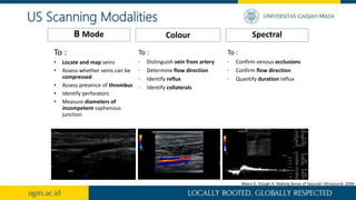
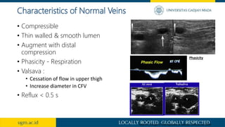
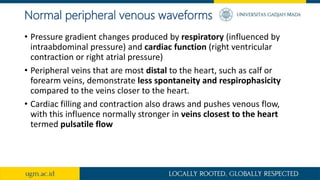
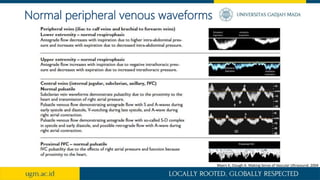
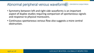
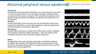
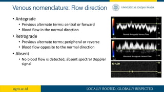
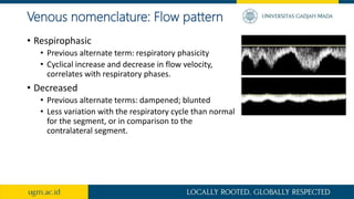
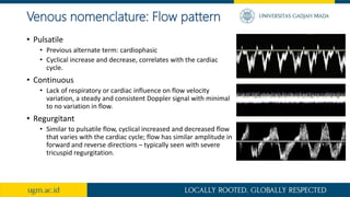
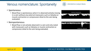
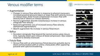
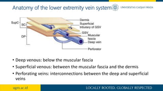
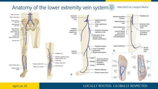
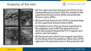
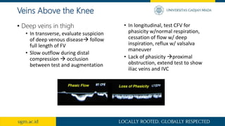
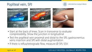
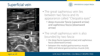
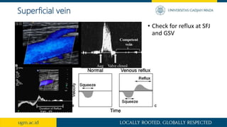
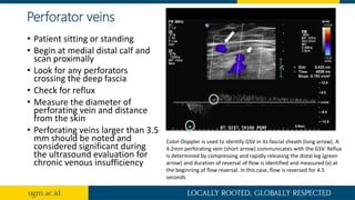
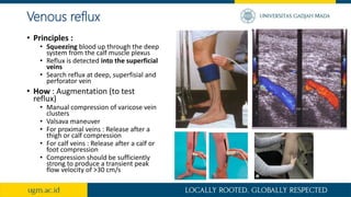
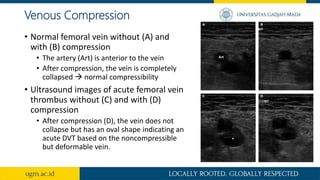
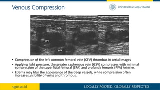
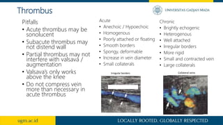
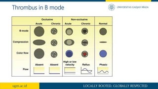
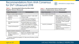
Interpretation of peripheral venous
Doppler in lower extrimity
Effika Nurningtyas Putri
Supervisors:
Dr. dr. Hariadi Hariawan, SpPD, SpJP(K)
dr. M. Taufik Ismail, SpJP (K)
Introduction
• Venous ultrasound is the standard imaging test for patients suspected
of having acute deep venous thrombosis (DVT).
• 200 million people diagnosed with peripheral artery disease (PAD)
worldwide and >600,000 hospital admissions yearly for venous
thromboembolies (VTE)
• 1/3 of DVT complicate with a clot in the lungs
• Recurrence at 5 years : 28%
• Case-fatality rate (recurrence) : 3% -6%
• The lack of a single protocol has led to misunderstandings between
ordering clinicians and facilities providing the service. This may result
in underdiagnosis, unnecessary testing, and insufficient imaging.
Indication for Venous Ultrasound
Indication
• Ass. VTE or venous obstruction in
symptomatic or high-risk asymptomatic
individuals
• Ass. venous insufficiency, reflux, and
varicosities
• Venous mapping before surgery
• Post procedural assessment of venous
ablation or other interventions
• Ass. of dialysis access.
• Evaluation of veins before venous
access.
• Follow-up
• Venous malformations
Limitations
• Oedematous tissue
• Extensive ulceration
• Obesity
• Operator dependent
Indication for Venous Ultrasound
To :
• Locate and map veins
• Assess whether veins can be
compressed
• Assess presence of thrombus
• Identify perforators
• Measure diameters of
incompetent saphenous
junction
B Mode Spectral
Colour
To :
• Distinguish vein from artery
• Determine flow direction
• Identify reflux
• Identify collaterals
To :
• Confirm venous occlusions
• Confirm flow direction
• Quantify duration reflux
Myers K, Clough A. Making Sense of Vascular Ultrasound. 2004
US Scanning Modalities
• Compressible
• Thin walled & smooth lumen
• Augment with distal
compression
• Phasicity - Respiration
• Valsava :
• Cessation of flow in upper thigh
• Increase diameter in CFV
• Reflux < 0.5 s
Characteristics of Normal Veins
Myers K, Clough A. Making Sense of Vascular Ultrasound. 2004
Phasicity
Normal peripheral venous waveforms
• Pressure gradient changes produced by respiratory (influenced by
intraabdominal pressure) and cardiac function (right ventricular
contraction or right atrial pressure)
• Peripheral veins that are most distal to the heart, such as calf or
forearm veins, demonstrate less spontaneity and respirophasicity
compared to the veins closer to the heart.
• Cardiac filling and contraction also draws and pushes venous flow,
with this influence normally stronger in veins closest to the heart
termed pulsatile flow
Normal peripheral venous waveforms
Myers K, Clough A. Making Sense of Vascular Ultrasound. 2004
Abnormal peripheral venous waveforms
• Symmetry between left and right side waveforms is an important
aspect of duplex studies requiring comparison of spontaneous signals
and response to physical maneuvers.
• Continuous spontaneous venous flow also suggests a more central
obstruction.
Abnormal peripheral venous waveforms
Venous
• Key major descriptors
• Flow direction (antegrade, retrograde, absent)
• Flow pattern (respirophasic, decreased, pulsatile, continuous, regurgitant)
• Spontaneity (spontaneous, nonspontaneous)
• Additional modifier terms
• Augmentation (normal, reduced, absent)
• Reflux
• Fistula flow
Summary of Nomenclature from Society for
Vascular Medicine 2020
Venous nomenclature: Flow direction
• Antegrade
• Previous alternate terms: central or forward
• Blood flow in the normal direction
• Retrograde
• Previous alternate terms: peripheral or reverse
• Blood flow opposite to the normal direction
• Absent
• No blood flow is detected, absent spectral Doppler
signal
Venous nomenclature: Flow pattern
• Respirophasic
• Previous alternate term: respiratory phasicity
• Cyclical increase and decrease in flow velocity,
correlates with respiratory phases.
• Decreased
• Previous alternate terms: dampened; blunted
• Less variation with the respiratory cycle than normal
for the segment, or in comparison to the
contralateral segment.
Venous nomenclature: Flow pattern
• Pulsatile
• Previous alternate term: cardiophasic
• Cyclical increase and decrease, correlates with the cardiac
cycle.
• Continuous
• Lack of respiratory or cardiac influence on flow velocity
variation, a steady and consistent Doppler signal with minimal
to no variation in flow.
• Regurgitant
• Similar to pulsatile flow, cyclical increased and decreased flow
that varies with the cardiac cycle; flow has similar amplitude in
forward and reverse directions – typically seen with severe
tricuspid regurgitation.
Venous nomenclature: Spontaneity
• Spontaneous
• Blood flow is spontaneous when it is observed actively moving
in a vein without any external maneuvers such as Valsalva or
muscle contraction or compression distal to the vein being
evaluated.
• Nonspontaneous
• Blood flow is not actively observed in a vein and only noted
with maneuvers such as Valsalva or muscle contraction or
compression distal to the vein being evaluated.
Venous modifier terms
• Augmentation
• Changes in venous flow velocity in response to physical maneuvers:
increased proximal pressure lowers or stops flow with velocity increase
on release (top); distal muscle compression increases flow with
velocity low or absent on release (bottom).
• Normal augmented: Discrete instantaneous increase in venous
antegrade flow velocity
• Reduced augmented: Loss of increased venous flow velocity
(dampened augmentation)
• Absent augmented: No increase in venous flow/return
• Reflux
• Persistent retrograde flow beyond the normal venous valve closure
time, typically noted upon release of distal compression. Also noted in
response to Valsalva maneuver.
• Fistula flow
• Venous flow with an arteriovenous fistula becomes pulsatile due to
direct communication with an artery; sharp peaks often appearing
pulsatile with spectral broadening.
Anatomy of the lower extremity vein system
• Deep venous: below the muscular fascia
• Superficial venous: between the muscular fascia and the dermis
• Perforating veins: interconnections between the deep and superficial
veins
Anatomy of the lower extremity vein system
Anatomy of the vein
• (A) The right common femoral vein (CFV) at the
saphenofemoral junction (SFJ) lies medial to the
superficial femoral artery (SFA) and profunda
femoris artery (PFA)
• (B) Superficial femoral vein (SFV) is located deep
to the superficial femoral artery (SFA)
• (C) Popliteal veins (PV) are found superficial to
the popliteal artery (PA).The gastrocnemius
veins (GV) project toward the PV in typical vein–
artery–vein formation.
• (D) Peroneal veins (PerV) are imaged superficial
to the fibula (Fib).The posterior tibial veins (PTV)
approach the peroneal veins in the proximal calf.
• “Mickey Mouse Image”
• Determine source of reflux colour or
spectral doppler, can be elicited
using Valsalva manouvre/
compression
• In transverse, determine destination
of reflux (GSV or major thigh
tributaries)
• If GSV Ø ↑ , suspect a source of reflux
• Follow full length of GSV and
tributaries to the knee. Test
compressibility and reflux every few
cm
• Measure Ø at junction and along
GSV
Veins Above the Knee
Myers K, Clough A. Making Sense of Vascular Ultrasound. 2004
• Deep veins in thigh
• In transverse, evaluate suspicion
of deep venous disease follow
full length of FV
• Slow outflow during distal
compression  occlusion
between test and augmentation
• In longitudinal, test CFV for
phasicity w/normal respiration,
cessation of flow w/ deep
inspiration, reflux w/ valsalva
maneuver
• Lack of phasicity proximal
obstruction, extend test to show
iliac veins and IVC
Veins Above the Knee
• Start at the back of knee, Scan in transverse to evaluate
compressibility, Show the junction in longitudinal
• Test the popliteal vein proximal and distal to the SPJ, gastrocnemius
veins insertion and SPJ with distal augmentation
• If there is reflux/retrograde flow, measure Ø SPJ, SSV
Popliteal vein, SPJ
• Crural veins
• Reflux in PTV best reflects clinical
features
• Scan in longitudinal, Examine PTV w/
medial or posteromedial view,
peroneal vein w/ posteromedial or
posterior view, ATV w/ anterolateral
view (Scanning ATV is optional)
Veins Below the Knee
Taken from : Myers K, Clough A. Making Sense of Vascular Ultrasound. 2004
• The great saphenous vein lies
between two fascia and its
appearance called “Cleopatra eyes”
• deep muscular fascia (upward arrow)
and saphenous fascia (downward
arrow)
Superficial vein
• The small saphenous vein is also
bounded by two fascia
• the deep fascia (upward arrow) and saphenous
fascia (downward arrow)
• between the medial gastrocnemius muscle
(MG) and lateral gastrocnemius muscle (LG)
Superficial vein
• Check for reflux at SFJ
and GSV
Perforator veins
• Patient sitting or standing
• Begin at medial distal calf and
scan proximally
• Look for any perforators
crossing the deep fascia
• Check for reflux
• Measure the diameter of
perforating vein and distance
from the skin
• Perforating veins larger than 3.5
mm should be noted and
considered significant during
the ultrasound evaluation for
chronic venous insufficiency
Color-Doppler is used to identify GSV in its fascial sheath (long arrow). A
4.2mm perforating vein (short arrow) communicates with the GSV. Reflux
is determined by compressing and rapidly releasing the distal leg (green
arrow) and duration of reversal of flow is identified and measured (x) at
the beginning of flow reversal. In this case, flow is reversed for 4.5
seconds.
Venous reflux
• Principles :
• Squeezing blood up through the deep
system from the calf muscle plexus
• Reflux is detected into the superficial
veins
• Search reflux at deep, superfisial and
perforator vein
• How : Augmentation (to test
reflux)
• Manual compression of varicose vein
clusters
• Valsava maneuver
• For proximal veins : Release after a
thigh or calf compression
• For calf veins : Release after a calf or
foot compression
• Compression should be sufficiently
strong to produce a transient peak
flow velocity of >30 cm/s
Venous Compression
• Normal femoral vein without (A) and
with (B) compression
• The artery (Art) is anterior to the vein
• After compression, the vein is completely
collapsed  normal compressibility
• Ultrasound images of acute femoral vein
thrombus without (C) and with (D)
compression
• After compression (D), the vein does not
collapse but has an oval shape indicating an
acute DVT based on the noncompressible
but deformable vein.
Venous Compression
• Compression of the left common femoral vein (CFV) thrombus in serial images
• Applying light pressure, the greater saphenous vein (GSV) compresses with minimal
compression of the superficial femoral (SFA) and profunda femoris (PFA) Arteries
• Edema may blur the appearance of the deep vessels, while compression often
increases,visibility of veins and thrombus.
Acute
• Anechoic / Hypoechoic
• Homogenous
• Poorly attached or floating
• Smooth borders
• Spongy, deformable
• Increase in vein diameter
• Small collaterals
Chronic
• Brightly echogenic
• Heterogenous
• Well attached
• Irregular borders
• More rigid
• Small and contracted vein
• Large collaterals
Thrombus
Irregular borders Collateral veins
Pitfalls
• Acute thrombus may be
sonolucent
• Subacute thrombus may
not distend wall
• Partial thrombus may not
interfere with valsava /
augmentation
• Valsava’s only works
above the knee
• Do not compress vein
more than necessary in
acute thrombus
Thrombus in B mode
Recommendations from ESVS Chronic
Venous Disease 2022
• Morphological examination (patency, reflux, compressibility, lumen
echogenicity, wall irregularity) of deep veins and evaluation of normal
spontaneity and phasicity flow in the CFV may be performed in the
supine position (CFV, FV, and POPV)
• Evaluation of the presence or absence of reflux and size of vessel, the
upright position with the knee of the investigated leg slightly bent.
(GSV, AASV, SSV, SFJ and SPJ)
• Reflux must be provoked, either from above with dependency testing or a
Valsalva manoeuvre, or from below, using an automatic pneumatic pressure
cuff or manual compression of the thigh, calf, or foot.
• Reflux cut off values
• 1 second for reflux in the CFV, FV and POPV
• 0.5 seconds in superficial veins and PVs
Recommendations from AHA Consensus
for DVT Ultrasound 2018
• Complete duplex ultrasound (CDUS) is the preferred venous
ultrasound test for the diagnosis of acute DVT.
• compression of the deep veins from the inguinal ligament to the ankle
(including posterior tibial and peroneal veins in the calf) - performed at 2cm
intervals
• right and left common femoral vein spectral Doppler waveforms (to evaluate
symmetry)
• popliteal spectral Doppler
• color Doppler images
Recommendations from AHA Consensus
for DVT Ultrasound 2018
• 2-CUS (2-region compression ultrasound)
compression ultrasound including the femoral veins
1 to 2 cm above and below the saphenofemoral
junction and the popliteal veins up to the calf veins
confluence
• ECUS (extended compression ultrasound), the
compression ultrasound from common femoral
vein through the popliteal vein up to the calf veins
confluence
• CCUS (complete compression ultrasound),
compression ultrasound from common femoral
vein to the ankle
• CDUS (complete duplex ultrasound), compression
ultrasound from the common femoral vein to the
ankle, color and spectral Doppler of the common
femoral (or iliac veins) on both sides, color and
spectral Doppler of the popliteal vein on the
symptomatic side
Recommendations from AHA Consensus
for DVT Ultrasound 2018
Abnormalities should be classified into acute venous thrombosis,
chronic postthrombotic change, or indeterminate (equivocal).
• Acute venous thrombosis
• vein noncompressibility, but the thrombus is soft and deformable with probe
pressure
• the surface of the thrombus is smooth and the vein is larger than normal
• loosely adherent or free-floating edge may be seen but is less common
• Chronic postthrombotic
• noncompressible, but the intraluminal material is rigid and nondeformable
with probe pressure
• the material may retract and produce thin webs (synechiae) or thicker flat
bands
• recanalization may produce regular or irregular wall thickening
Recommendations from AHA Consensus
for DVT Ultrasound 2018
Thank You

Book reading doppler vena.pptx

  • 1.
    Interpretation of peripheralvenous Doppler in lower extrimity Effika Nurningtyas Putri Supervisors: Dr. dr. Hariadi Hariawan, SpPD, SpJP(K) dr. M. Taufik Ismail, SpJP (K)
  • 2.
    Introduction • Venous ultrasoundis the standard imaging test for patients suspected of having acute deep venous thrombosis (DVT). • 200 million people diagnosed with peripheral artery disease (PAD) worldwide and >600,000 hospital admissions yearly for venous thromboembolies (VTE) • 1/3 of DVT complicate with a clot in the lungs • Recurrence at 5 years : 28% • Case-fatality rate (recurrence) : 3% -6% • The lack of a single protocol has led to misunderstandings between ordering clinicians and facilities providing the service. This may result in underdiagnosis, unnecessary testing, and insufficient imaging.
  • 3.
  • 4.
    Indication • Ass. VTEor venous obstruction in symptomatic or high-risk asymptomatic individuals • Ass. venous insufficiency, reflux, and varicosities • Venous mapping before surgery • Post procedural assessment of venous ablation or other interventions • Ass. of dialysis access. • Evaluation of veins before venous access. • Follow-up • Venous malformations Limitations • Oedematous tissue • Extensive ulceration • Obesity • Operator dependent Indication for Venous Ultrasound
  • 5.
    To : • Locateand map veins • Assess whether veins can be compressed • Assess presence of thrombus • Identify perforators • Measure diameters of incompetent saphenous junction B Mode Spectral Colour To : • Distinguish vein from artery • Determine flow direction • Identify reflux • Identify collaterals To : • Confirm venous occlusions • Confirm flow direction • Quantify duration reflux Myers K, Clough A. Making Sense of Vascular Ultrasound. 2004 US Scanning Modalities
  • 6.
    • Compressible • Thinwalled & smooth lumen • Augment with distal compression • Phasicity - Respiration • Valsava : • Cessation of flow in upper thigh • Increase diameter in CFV • Reflux < 0.5 s Characteristics of Normal Veins Myers K, Clough A. Making Sense of Vascular Ultrasound. 2004 Phasicity
  • 7.
    Normal peripheral venouswaveforms • Pressure gradient changes produced by respiratory (influenced by intraabdominal pressure) and cardiac function (right ventricular contraction or right atrial pressure) • Peripheral veins that are most distal to the heart, such as calf or forearm veins, demonstrate less spontaneity and respirophasicity compared to the veins closer to the heart. • Cardiac filling and contraction also draws and pushes venous flow, with this influence normally stronger in veins closest to the heart termed pulsatile flow
  • 8.
    Normal peripheral venouswaveforms Myers K, Clough A. Making Sense of Vascular Ultrasound. 2004
  • 9.
    Abnormal peripheral venouswaveforms • Symmetry between left and right side waveforms is an important aspect of duplex studies requiring comparison of spontaneous signals and response to physical maneuvers. • Continuous spontaneous venous flow also suggests a more central obstruction.
  • 10.
  • 11.
    Venous • Key majordescriptors • Flow direction (antegrade, retrograde, absent) • Flow pattern (respirophasic, decreased, pulsatile, continuous, regurgitant) • Spontaneity (spontaneous, nonspontaneous) • Additional modifier terms • Augmentation (normal, reduced, absent) • Reflux • Fistula flow Summary of Nomenclature from Society for Vascular Medicine 2020
  • 12.
    Venous nomenclature: Flowdirection • Antegrade • Previous alternate terms: central or forward • Blood flow in the normal direction • Retrograde • Previous alternate terms: peripheral or reverse • Blood flow opposite to the normal direction • Absent • No blood flow is detected, absent spectral Doppler signal
  • 13.
    Venous nomenclature: Flowpattern • Respirophasic • Previous alternate term: respiratory phasicity • Cyclical increase and decrease in flow velocity, correlates with respiratory phases. • Decreased • Previous alternate terms: dampened; blunted • Less variation with the respiratory cycle than normal for the segment, or in comparison to the contralateral segment.
  • 14.
    Venous nomenclature: Flowpattern • Pulsatile • Previous alternate term: cardiophasic • Cyclical increase and decrease, correlates with the cardiac cycle. • Continuous • Lack of respiratory or cardiac influence on flow velocity variation, a steady and consistent Doppler signal with minimal to no variation in flow. • Regurgitant • Similar to pulsatile flow, cyclical increased and decreased flow that varies with the cardiac cycle; flow has similar amplitude in forward and reverse directions – typically seen with severe tricuspid regurgitation.
  • 15.
    Venous nomenclature: Spontaneity •Spontaneous • Blood flow is spontaneous when it is observed actively moving in a vein without any external maneuvers such as Valsalva or muscle contraction or compression distal to the vein being evaluated. • Nonspontaneous • Blood flow is not actively observed in a vein and only noted with maneuvers such as Valsalva or muscle contraction or compression distal to the vein being evaluated.
  • 16.
    Venous modifier terms •Augmentation • Changes in venous flow velocity in response to physical maneuvers: increased proximal pressure lowers or stops flow with velocity increase on release (top); distal muscle compression increases flow with velocity low or absent on release (bottom). • Normal augmented: Discrete instantaneous increase in venous antegrade flow velocity • Reduced augmented: Loss of increased venous flow velocity (dampened augmentation) • Absent augmented: No increase in venous flow/return • Reflux • Persistent retrograde flow beyond the normal venous valve closure time, typically noted upon release of distal compression. Also noted in response to Valsalva maneuver. • Fistula flow • Venous flow with an arteriovenous fistula becomes pulsatile due to direct communication with an artery; sharp peaks often appearing pulsatile with spectral broadening.
  • 17.
    Anatomy of thelower extremity vein system • Deep venous: below the muscular fascia • Superficial venous: between the muscular fascia and the dermis • Perforating veins: interconnections between the deep and superficial veins
  • 18.
    Anatomy of thelower extremity vein system
  • 19.
    Anatomy of thevein • (A) The right common femoral vein (CFV) at the saphenofemoral junction (SFJ) lies medial to the superficial femoral artery (SFA) and profunda femoris artery (PFA) • (B) Superficial femoral vein (SFV) is located deep to the superficial femoral artery (SFA) • (C) Popliteal veins (PV) are found superficial to the popliteal artery (PA).The gastrocnemius veins (GV) project toward the PV in typical vein– artery–vein formation. • (D) Peroneal veins (PerV) are imaged superficial to the fibula (Fib).The posterior tibial veins (PTV) approach the peroneal veins in the proximal calf.
  • 20.
    • “Mickey MouseImage” • Determine source of reflux colour or spectral doppler, can be elicited using Valsalva manouvre/ compression • In transverse, determine destination of reflux (GSV or major thigh tributaries) • If GSV Ø ↑ , suspect a source of reflux • Follow full length of GSV and tributaries to the knee. Test compressibility and reflux every few cm • Measure Ø at junction and along GSV Veins Above the Knee Myers K, Clough A. Making Sense of Vascular Ultrasound. 2004
  • 21.
    • Deep veinsin thigh • In transverse, evaluate suspicion of deep venous disease follow full length of FV • Slow outflow during distal compression  occlusion between test and augmentation • In longitudinal, test CFV for phasicity w/normal respiration, cessation of flow w/ deep inspiration, reflux w/ valsalva maneuver • Lack of phasicity proximal obstruction, extend test to show iliac veins and IVC Veins Above the Knee
  • 22.
    • Start atthe back of knee, Scan in transverse to evaluate compressibility, Show the junction in longitudinal • Test the popliteal vein proximal and distal to the SPJ, gastrocnemius veins insertion and SPJ with distal augmentation • If there is reflux/retrograde flow, measure Ø SPJ, SSV Popliteal vein, SPJ
  • 23.
    • Crural veins •Reflux in PTV best reflects clinical features • Scan in longitudinal, Examine PTV w/ medial or posteromedial view, peroneal vein w/ posteromedial or posterior view, ATV w/ anterolateral view (Scanning ATV is optional) Veins Below the Knee Taken from : Myers K, Clough A. Making Sense of Vascular Ultrasound. 2004
  • 24.
    • The greatsaphenous vein lies between two fascia and its appearance called “Cleopatra eyes” • deep muscular fascia (upward arrow) and saphenous fascia (downward arrow) Superficial vein • The small saphenous vein is also bounded by two fascia • the deep fascia (upward arrow) and saphenous fascia (downward arrow) • between the medial gastrocnemius muscle (MG) and lateral gastrocnemius muscle (LG)
  • 25.
    Superficial vein • Checkfor reflux at SFJ and GSV
  • 26.
    Perforator veins • Patientsitting or standing • Begin at medial distal calf and scan proximally • Look for any perforators crossing the deep fascia • Check for reflux • Measure the diameter of perforating vein and distance from the skin • Perforating veins larger than 3.5 mm should be noted and considered significant during the ultrasound evaluation for chronic venous insufficiency Color-Doppler is used to identify GSV in its fascial sheath (long arrow). A 4.2mm perforating vein (short arrow) communicates with the GSV. Reflux is determined by compressing and rapidly releasing the distal leg (green arrow) and duration of reversal of flow is identified and measured (x) at the beginning of flow reversal. In this case, flow is reversed for 4.5 seconds.
  • 27.
    Venous reflux • Principles: • Squeezing blood up through the deep system from the calf muscle plexus • Reflux is detected into the superficial veins • Search reflux at deep, superfisial and perforator vein • How : Augmentation (to test reflux) • Manual compression of varicose vein clusters • Valsava maneuver • For proximal veins : Release after a thigh or calf compression • For calf veins : Release after a calf or foot compression • Compression should be sufficiently strong to produce a transient peak flow velocity of >30 cm/s
  • 28.
    Venous Compression • Normalfemoral vein without (A) and with (B) compression • The artery (Art) is anterior to the vein • After compression, the vein is completely collapsed  normal compressibility • Ultrasound images of acute femoral vein thrombus without (C) and with (D) compression • After compression (D), the vein does not collapse but has an oval shape indicating an acute DVT based on the noncompressible but deformable vein.
  • 29.
    Venous Compression • Compressionof the left common femoral vein (CFV) thrombus in serial images • Applying light pressure, the greater saphenous vein (GSV) compresses with minimal compression of the superficial femoral (SFA) and profunda femoris (PFA) Arteries • Edema may blur the appearance of the deep vessels, while compression often increases,visibility of veins and thrombus.
  • 30.
    Acute • Anechoic /Hypoechoic • Homogenous • Poorly attached or floating • Smooth borders • Spongy, deformable • Increase in vein diameter • Small collaterals Chronic • Brightly echogenic • Heterogenous • Well attached • Irregular borders • More rigid • Small and contracted vein • Large collaterals Thrombus Irregular borders Collateral veins Pitfalls • Acute thrombus may be sonolucent • Subacute thrombus may not distend wall • Partial thrombus may not interfere with valsava / augmentation • Valsava’s only works above the knee • Do not compress vein more than necessary in acute thrombus
  • 31.
  • 32.
    Recommendations from ESVSChronic Venous Disease 2022 • Morphological examination (patency, reflux, compressibility, lumen echogenicity, wall irregularity) of deep veins and evaluation of normal spontaneity and phasicity flow in the CFV may be performed in the supine position (CFV, FV, and POPV) • Evaluation of the presence or absence of reflux and size of vessel, the upright position with the knee of the investigated leg slightly bent. (GSV, AASV, SSV, SFJ and SPJ) • Reflux must be provoked, either from above with dependency testing or a Valsalva manoeuvre, or from below, using an automatic pneumatic pressure cuff or manual compression of the thigh, calf, or foot. • Reflux cut off values • 1 second for reflux in the CFV, FV and POPV • 0.5 seconds in superficial veins and PVs
  • 33.
    Recommendations from AHAConsensus for DVT Ultrasound 2018 • Complete duplex ultrasound (CDUS) is the preferred venous ultrasound test for the diagnosis of acute DVT. • compression of the deep veins from the inguinal ligament to the ankle (including posterior tibial and peroneal veins in the calf) - performed at 2cm intervals • right and left common femoral vein spectral Doppler waveforms (to evaluate symmetry) • popliteal spectral Doppler • color Doppler images
  • 34.
    Recommendations from AHAConsensus for DVT Ultrasound 2018 • 2-CUS (2-region compression ultrasound) compression ultrasound including the femoral veins 1 to 2 cm above and below the saphenofemoral junction and the popliteal veins up to the calf veins confluence • ECUS (extended compression ultrasound), the compression ultrasound from common femoral vein through the popliteal vein up to the calf veins confluence • CCUS (complete compression ultrasound), compression ultrasound from common femoral vein to the ankle • CDUS (complete duplex ultrasound), compression ultrasound from the common femoral vein to the ankle, color and spectral Doppler of the common femoral (or iliac veins) on both sides, color and spectral Doppler of the popliteal vein on the symptomatic side
  • 35.
    Recommendations from AHAConsensus for DVT Ultrasound 2018 Abnormalities should be classified into acute venous thrombosis, chronic postthrombotic change, or indeterminate (equivocal). • Acute venous thrombosis • vein noncompressibility, but the thrombus is soft and deformable with probe pressure • the surface of the thrombus is smooth and the vein is larger than normal • loosely adherent or free-floating edge may be seen but is less common • Chronic postthrombotic • noncompressible, but the intraluminal material is rigid and nondeformable with probe pressure • the material may retract and produce thin webs (synechiae) or thicker flat bands • recanalization may produce regular or irregular wall thickening
  • 36.
    Recommendations from AHAConsensus for DVT Ultrasound 2018
  • 37.