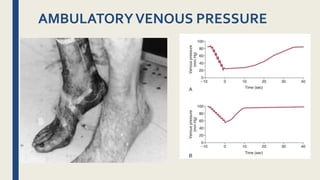
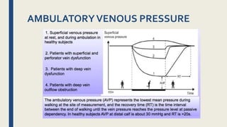
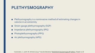
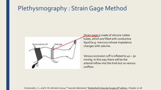
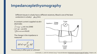
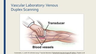
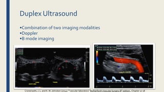
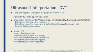
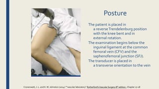
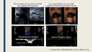
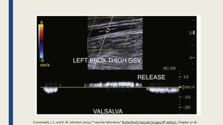
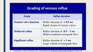
VENOUS
PHYSIOLOGY
ASSESSMENT
F1 PARACH SIRISRIRO
3OTH April 2018
REFERENCE
Outline
■ Vascular Laboratory:Venous Physiologic Assessment
– AMBULATORY VENOUS PRESSURE
– PLETHYSMOGRAPHY
■ Vascular Laboratory:Venous Duplex Scanning
– Acute deep vein thrombosis
– Venous Incompetency
Function ofVeins:
• Return blood to heart for re-oxygenation and recirculation
• Storage of blood
• 60-80% of resting blood volume in the venous system
• Maintain cardiovascular stability through changes in
capacitance
•Through sympathetic mediated smooth muscle tone
Cronenwett, J. L. and K.W. Johnston (2014).””vascular laboratory” Rutherford'sVascular Surgery 8th edition , Chapter 17-18
AMBULATORYVENOUS PRESSURE
■ The sum of the hemodynamic factors involved in venous
insufficiency
■ AVP is defined as the venous pressure in a dorsal foot vein
after 10 tiptoe maneuvers in a standing position.
■ Most physicians consider AVP the “gold standard”
measure of venous hemodynamics.
Cronenwett, J. L. and K.W. Johnston (2014).””vascular laboratory” Rutherford'sVascular Surgery 8th edition , Chapter 17-18
AMBULATORYVENOUS PRESSURE
Limitation
■ Pressure at the pedal vein may not accurately reflect changes in venous
pressure within the leg, particularly in the deep venous system
■ invasive and time-consuming procedure that may be difficult to perform
in many patients,
– active ulcers
– Severe edema or dermal thickening
Cronenwett, J. L. and K.W. Johnston (2014).””vascular laboratory” Rutherford'sVascular Surgery 8th edition , Chapter 17-18
PLETHYSMOGRAPHY
■ Plethysmography is a noninvasive method of estimating changes in
volume in an extremity
■ Strain-gauge plethysmography (SGP)
■ Impedance plethysmography (IPG)
■ Photoplethysmography (PPG)
■ Air plethysmography (APG)
Cronenwett, J. L. and K.W. Johnston (2014).””vascular laboratory” Rutherford'sVascular Surgery 8th edition , Chapter 17-18
Plethysmography : Strain Gage Method
Strain gage is made of silicone rubber
tubes, which are filled with conductive
liquid (e.g. mercury) whose impedance
changes with volume.
Venous occlusion cuff is inflated to 40 – 50
mmHg. In this way there will be the
arterial inflow into the limb but no venous
outflow.
Cronenwett, J. L. and K.W. Johnston (2014).””vascular laboratory” Rutherford'sVascular Surgery 8th edition , Chapter 17-18
Photoplethysmography
PPG uses light absorbance by hemoglobin as a reflection of blood volume in
the venous and capillary networks in the skin to estimate the degree of venous
stasis
Cronenwett, J. L. and K.W. Johnston (2014).””vascular laboratory” Rutherford'sVascular Surgery 8th edition , Chapter 17-18
Interpretation
■ In normal individuals,VRT tends be more than 20 seconds and can
extend to as long as 60 seconds.
■ Significant venous reflux results in aVRT of less than 20 seconds, and
reducing
times generally reflect increasing severity of reflux
■ In the presence of abnormal findings, the test can be repeated with a
tourniquet inflated (50 mm Hg) alternately above and below the knee to
identify the source of reflux
Cronenwett, J. L. and K.W. Johnston (2014).””vascular laboratory” Rutherford'sVascular Surgery 8th edition , Chapter 17-18
Airplethysmography
Cronenwett, J. L. and K.W. Johnston (2014).””vascular laboratory” Rutherford'sVascular Surgery 8th edition , Chapter 17-18
Interpretation
Cronenwett, J. L. and K.W. Johnston (2014).””vascular laboratory” Rutherford'sVascular Surgery 8th edition , Chapter 17-18
Impedanceplethysmography
Different tissues in a body have a different resistivity. Blood is one of the best
conductors in a body ( = 1,5 Ωm)
A constant current is applied via skin
electrodes
The change in the impedance is
measured
The accuracy is often poor or unknown
I = 0,5 – 4 mA rms (SNR)
f = 50 – 100 kHz
(Zskin-electrode+shock)
Z
Z
L
Vol  2
0
2

Cronenwett, J. L. and K.W. Johnston (2014).””vascular laboratory” Rutherford'sVascular Surgery 8th edition , Chapter 17-18
■ Duplex examination : to identify the sites of reflux, evaluate the
deep and perforator systems, and guide intervention.
■ APG serve improvement and predict long-term outcome after intervention
■ PPG is relatively rapid to perform and a reasonable choice to detect venous
disease without details concerning anatomy or the severity of CVI
■ Physiologic assessment with PPG or APG can provide evidence of global
venous dysfunction
SELECTIVE USE OFVENOUS
PHYSIOLOGICTESTING
Cronenwett, J. L. and K.W. Johnston (2014).””vascular laboratory” Rutherford'sVascular Surgery 8th edition , Chapter 17-18
Vascular Laboratory:Venous
Duplex Scanning
Cronenwett, J. L. and K.W. Johnston (2014).””vascular laboratory” Rutherford'sVascular Surgery 8th edition , Chapter 17-18
Duplex Ultrasound
• Several important roles in phlebology
• Assist in the evaluation of
Deep venous disease
Superficial venous disease
Mapping prior to intervention
Peri‐procedural imaging
Post‐procedural imaging (success, complications,
failure)
Cronenwett, J. L. and K.W. Johnston (2014).””vascular laboratory” Rutherford'sVascular Surgery 8th edition , Chapter 17-18
Duplex Ultrasound
•Combination of two imaging modalities
•Doppler
•B mode imaging
Cronenwett, J. L. and K.W. Johnston (2014).””vascular laboratory” Rutherford'sVascular Surgery 8th edition , Chapter 17-18
■ Ultrasound Imaging
Pulses of sound waves are
transmitted into the body
and the returning “echoes”
from various structures are
detected
by the probe and
converted into
images on a screen
Duplex
Ultrasound
Cronenwett, J. L. and K.W. Johnston (2014).””vascular laboratory” Rutherford'sVascular Surgery 8th edition , Chapter 17-18
Ultrasound Interpretation ‐ DVT
■ DUS is the test of choice for diagnosis of proximal DVT
• Sensitivity >95%, Specificity >95%
■ Diagnostic components :Visualization, compressibility, flow, and augmentation
•Transducer compression maneuvers
• Doppler evaluation (color and spectral Doppler waveform analysis)
• Augmentation maneuvers
■ Acute DVT
• Loss of compression
• Dilated vein (diameter > artery)
• Intraluminal echoes from thrombus
• Abnormal/absent color Doppler
• Abnormal/absent PW spectral
Cronenwett, J. L. and K.W. Johnston (2014).””vascular laboratory” Rutherford'sVascular Surgery 8th edition , Chapter 17-18
Posture
The patient is placed in
a reverseTrendelenburg position
with the knee bent and in
external rotation.
The examination begins below the
inguinal ligament at the common
femoral vein (CFV) and the
saphenofemoral junction (SFJ).
The transducer is placed in
a transverse orientation to the vein
Cronenwett, J. L. and K.W. Johnston (2014).””vascular laboratory” Rutherford'sVascular Surgery 8th edition , Chapter 17-18
1. N Engl J Med. 1989;320(6):342. 2. Bruit. 1982;7:41–42.
Ultrasound
Interpretation ‐ DVT
■ • Acuity of thrombus
• Acute (<2 weeks)
• Subacute (2 weeks – 6 months)
• Chronic (>6 months)
• Ultrasound parameters
• B‐mode appearance (hypoechoic,
isoechoic, hyeprechoic)
•Vein lumen size
•Vein wall appearance
•Venous compressibility
• Function of venous valves
• Presence of collaterals
Cronenwett, J. L. and K.W. Johnston (2014).””vascular laboratory” Rutherford'sVascular Surgery 8th edition , Chapter 17-18
RecurrentThrombosis
■ Recurrent thrombosis is defined as a repeated thrombotic
event. It may occur in the contralateral or ipsilateral limb
■ Three DUS criteria can be used to diagnose recurrent DVT:
■ extension of the thrombus more than 9 cm,
■ noncompressibility of a vein segment that had previously been
compressible or had previously recanalized,
■ increase in thrombus thickness by 4 mm.
Cronenwett, J. L. and K.W. Johnston (2014).””vascular laboratory” Rutherford'sVascular Surgery 8th edition , Chapter 17-18
RecurrentThrombosis
Cronenwett, J. L. and K.W. Johnston (2014).””vascular laboratory” Rutherford'sVascular Surgery 8th edition , Chapter 17-18
CentralVein
Stenosis
■ The best sonographic
criterion for detecting
significant vein stenosis
was found to be aV2/V1
ratio of
greater than 2.5 (with
only two false-positive
and false negative test
results reported)
Cronenwett, J. L. and K.W. Johnston (2014).””vascular laboratory” Rutherford'sVascular Surgery 8th edition , Chapter 17-18
SuperficialVeinThrombosis
Contiguous extension
of the thrombotic process from the
superficial to the deep veins can occur
in three ways,
- most commonly from the great
saphenous vein (GSV) to the femoral
vein.
- Less often, the thrombus extends
from the small saphenous vein (SSV)
to the popliteal vein.
- Extension through perforator veins to
deep veins can also occur.
Cronenwett, J. L. and K.W. Johnston (2014).””vascular laboratory” Rutherford'sVascular Surgery 8th edition , Chapter 17-18
Ultrasound –Venous Incompetency
Cronenwett, J. L. and K.W. Johnston (2014).””vascular laboratory” Rutherford'sVascular Surgery 8th edition , Chapter 17-18
■ Venous reflux: reversal of flow in the veins of the lower extremity
•Physiologic: fraction of a second it takes for valve leaflets to appose, time varies by
location
•Pathologic: reflux of ≥0.5 seconds in superficial veins
■ Reflux can be elicited by
•Valsalva maneuver
•Augmentation: compression and release distal to point of examination
■ Best results obtained with patient
•Standing with weight on contralateral limb
•Sitting with torso elevated >45 degrees
•ReverseTrendelenberg
Ultrasound –Venous Incompetency
Cronenwett, J. L. and K.W. Johnston (2014).””vascular laboratory” Rutherford'sVascular Surgery 8th edition , Chapter 17-18
Examination Protocol
■ Deep system:
•CFV, FV, Popliteal vein
•Reflux >1.0 sec
■ Superficial System: (vein diameter msmtsincluded)
•Entire length of GSV +/‐entire length of SSV
•Reflux >0.5 sec
■ Perforators
– •Examine entire calf
– •Focus on areas of ulcerations
– •Reflux ≥0.35 sec
– •Diameter >3.5 mm likely competent
Ultrasound –Venous Incompetency
Cronenwett, J. L. and K.W. Johnston (2014).””vascular laboratory” Rutherford'sVascular Surgery 8th edition , Chapter 17-18
VALVES AND REFLUX
■ There are essentially four ways to check for reflux:
■ 1. Direct visualization of valve closure
■ 2. Doppler waveform evaluation
■ 3. Color flow evaluation
■ 4. Gray-scale evaluation
Cronenwett, J. L. and K.W. Johnston (2014).””vascular laboratory” Rutherford'sVascular Surgery 8th edition , Chapter 17-18
Direct visualization of valve closure
Cronenwett, J. L. and K.W. Johnston (2014).””vascular laboratory” Rutherford'sVascular Surgery 8th edition , Chapter 17-18
Doppler waveform evaluation
Cronenwett, J. L. and K.W. Johnston (2014).””vascular laboratory” Rutherford'sVascular Surgery 8th edition , Chapter 17-18
SUMMARY OF GUIDELINES FOR
MANAGEMENT OF PATIENTS WITHVARICOSE
VEINS AND
ASSOCIATED CHRONICVENOUS DISEASE■ Duplex :
SUMMARY OF GUIDELINES FOR
MANAGEMENT OF PATIENTS WITHVARICOSE
VEINS AND
ASSOCIATED CHRONICVENOUS DISEASE■ Duplex :
SUMMARY OF GUIDELINES FOR
MANAGEMENT OF PATIENTS WITH
VARICOSEVEINS AND
ASSOCIATED CHRONICVENOUS DISEASE

Venous physiology assessment

  • 1.
  • 2.
  • 3.
    Outline ■ Vascular Laboratory:VenousPhysiologic Assessment – AMBULATORY VENOUS PRESSURE – PLETHYSMOGRAPHY ■ Vascular Laboratory:Venous Duplex Scanning – Acute deep vein thrombosis – Venous Incompetency
  • 4.
    Function ofVeins: • Returnblood to heart for re-oxygenation and recirculation • Storage of blood • 60-80% of resting blood volume in the venous system • Maintain cardiovascular stability through changes in capacitance •Through sympathetic mediated smooth muscle tone Cronenwett, J. L. and K.W. Johnston (2014).””vascular laboratory” Rutherford'sVascular Surgery 8th edition , Chapter 17-18
  • 5.
    AMBULATORYVENOUS PRESSURE ■ Thesum of the hemodynamic factors involved in venous insufficiency ■ AVP is defined as the venous pressure in a dorsal foot vein after 10 tiptoe maneuvers in a standing position. ■ Most physicians consider AVP the “gold standard” measure of venous hemodynamics. Cronenwett, J. L. and K.W. Johnston (2014).””vascular laboratory” Rutherford'sVascular Surgery 8th edition , Chapter 17-18
  • 6.
  • 7.
  • 8.
    Limitation ■ Pressure atthe pedal vein may not accurately reflect changes in venous pressure within the leg, particularly in the deep venous system ■ invasive and time-consuming procedure that may be difficult to perform in many patients, – active ulcers – Severe edema or dermal thickening Cronenwett, J. L. and K.W. Johnston (2014).””vascular laboratory” Rutherford'sVascular Surgery 8th edition , Chapter 17-18
  • 9.
    PLETHYSMOGRAPHY ■ Plethysmography isa noninvasive method of estimating changes in volume in an extremity ■ Strain-gauge plethysmography (SGP) ■ Impedance plethysmography (IPG) ■ Photoplethysmography (PPG) ■ Air plethysmography (APG) Cronenwett, J. L. and K.W. Johnston (2014).””vascular laboratory” Rutherford'sVascular Surgery 8th edition , Chapter 17-18
  • 10.
    Plethysmography : StrainGage Method Strain gage is made of silicone rubber tubes, which are filled with conductive liquid (e.g. mercury) whose impedance changes with volume. Venous occlusion cuff is inflated to 40 – 50 mmHg. In this way there will be the arterial inflow into the limb but no venous outflow. Cronenwett, J. L. and K.W. Johnston (2014).””vascular laboratory” Rutherford'sVascular Surgery 8th edition , Chapter 17-18
  • 11.
    Photoplethysmography PPG uses lightabsorbance by hemoglobin as a reflection of blood volume in the venous and capillary networks in the skin to estimate the degree of venous stasis Cronenwett, J. L. and K.W. Johnston (2014).””vascular laboratory” Rutherford'sVascular Surgery 8th edition , Chapter 17-18
  • 12.
    Interpretation ■ In normalindividuals,VRT tends be more than 20 seconds and can extend to as long as 60 seconds. ■ Significant venous reflux results in aVRT of less than 20 seconds, and reducing times generally reflect increasing severity of reflux ■ In the presence of abnormal findings, the test can be repeated with a tourniquet inflated (50 mm Hg) alternately above and below the knee to identify the source of reflux Cronenwett, J. L. and K.W. Johnston (2014).””vascular laboratory” Rutherford'sVascular Surgery 8th edition , Chapter 17-18
  • 13.
    Airplethysmography Cronenwett, J. L.and K.W. Johnston (2014).””vascular laboratory” Rutherford'sVascular Surgery 8th edition , Chapter 17-18
  • 14.
    Interpretation Cronenwett, J. L.and K.W. Johnston (2014).””vascular laboratory” Rutherford'sVascular Surgery 8th edition , Chapter 17-18
  • 15.
    Impedanceplethysmography Different tissues ina body have a different resistivity. Blood is one of the best conductors in a body ( = 1,5 Ωm) A constant current is applied via skin electrodes The change in the impedance is measured The accuracy is often poor or unknown I = 0,5 – 4 mA rms (SNR) f = 50 – 100 kHz (Zskin-electrode+shock) Z Z L Vol  2 0 2  Cronenwett, J. L. and K.W. Johnston (2014).””vascular laboratory” Rutherford'sVascular Surgery 8th edition , Chapter 17-18
  • 16.
    ■ Duplex examination: to identify the sites of reflux, evaluate the deep and perforator systems, and guide intervention. ■ APG serve improvement and predict long-term outcome after intervention ■ PPG is relatively rapid to perform and a reasonable choice to detect venous disease without details concerning anatomy or the severity of CVI ■ Physiologic assessment with PPG or APG can provide evidence of global venous dysfunction SELECTIVE USE OFVENOUS PHYSIOLOGICTESTING Cronenwett, J. L. and K.W. Johnston (2014).””vascular laboratory” Rutherford'sVascular Surgery 8th edition , Chapter 17-18
  • 17.
    Vascular Laboratory:Venous Duplex Scanning Cronenwett,J. L. and K.W. Johnston (2014).””vascular laboratory” Rutherford'sVascular Surgery 8th edition , Chapter 17-18
  • 18.
    Duplex Ultrasound • Severalimportant roles in phlebology • Assist in the evaluation of Deep venous disease Superficial venous disease Mapping prior to intervention Peri‐procedural imaging Post‐procedural imaging (success, complications, failure) Cronenwett, J. L. and K.W. Johnston (2014).””vascular laboratory” Rutherford'sVascular Surgery 8th edition , Chapter 17-18
  • 19.
    Duplex Ultrasound •Combination oftwo imaging modalities •Doppler •B mode imaging Cronenwett, J. L. and K.W. Johnston (2014).””vascular laboratory” Rutherford'sVascular Surgery 8th edition , Chapter 17-18
  • 20.
    ■ Ultrasound Imaging Pulsesof sound waves are transmitted into the body and the returning “echoes” from various structures are detected by the probe and converted into images on a screen Duplex Ultrasound Cronenwett, J. L. and K.W. Johnston (2014).””vascular laboratory” Rutherford'sVascular Surgery 8th edition , Chapter 17-18
  • 21.
    Ultrasound Interpretation ‐DVT ■ DUS is the test of choice for diagnosis of proximal DVT • Sensitivity >95%, Specificity >95% ■ Diagnostic components :Visualization, compressibility, flow, and augmentation •Transducer compression maneuvers • Doppler evaluation (color and spectral Doppler waveform analysis) • Augmentation maneuvers ■ Acute DVT • Loss of compression • Dilated vein (diameter > artery) • Intraluminal echoes from thrombus • Abnormal/absent color Doppler • Abnormal/absent PW spectral Cronenwett, J. L. and K.W. Johnston (2014).””vascular laboratory” Rutherford'sVascular Surgery 8th edition , Chapter 17-18
  • 22.
    Posture The patient isplaced in a reverseTrendelenburg position with the knee bent and in external rotation. The examination begins below the inguinal ligament at the common femoral vein (CFV) and the saphenofemoral junction (SFJ). The transducer is placed in a transverse orientation to the vein Cronenwett, J. L. and K.W. Johnston (2014).””vascular laboratory” Rutherford'sVascular Surgery 8th edition , Chapter 17-18
  • 23.
    1. N EnglJ Med. 1989;320(6):342. 2. Bruit. 1982;7:41–42.
  • 24.
    Ultrasound Interpretation ‐ DVT ■• Acuity of thrombus • Acute (<2 weeks) • Subacute (2 weeks – 6 months) • Chronic (>6 months) • Ultrasound parameters • B‐mode appearance (hypoechoic, isoechoic, hyeprechoic) •Vein lumen size •Vein wall appearance •Venous compressibility • Function of venous valves • Presence of collaterals Cronenwett, J. L. and K.W. Johnston (2014).””vascular laboratory” Rutherford'sVascular Surgery 8th edition , Chapter 17-18
  • 25.
    RecurrentThrombosis ■ Recurrent thrombosisis defined as a repeated thrombotic event. It may occur in the contralateral or ipsilateral limb ■ Three DUS criteria can be used to diagnose recurrent DVT: ■ extension of the thrombus more than 9 cm, ■ noncompressibility of a vein segment that had previously been compressible or had previously recanalized, ■ increase in thrombus thickness by 4 mm. Cronenwett, J. L. and K.W. Johnston (2014).””vascular laboratory” Rutherford'sVascular Surgery 8th edition , Chapter 17-18
  • 26.
    RecurrentThrombosis Cronenwett, J. L.and K.W. Johnston (2014).””vascular laboratory” Rutherford'sVascular Surgery 8th edition , Chapter 17-18
  • 27.
    CentralVein Stenosis ■ The bestsonographic criterion for detecting significant vein stenosis was found to be aV2/V1 ratio of greater than 2.5 (with only two false-positive and false negative test results reported) Cronenwett, J. L. and K.W. Johnston (2014).””vascular laboratory” Rutherford'sVascular Surgery 8th edition , Chapter 17-18
  • 28.
    SuperficialVeinThrombosis Contiguous extension of thethrombotic process from the superficial to the deep veins can occur in three ways, - most commonly from the great saphenous vein (GSV) to the femoral vein. - Less often, the thrombus extends from the small saphenous vein (SSV) to the popliteal vein. - Extension through perforator veins to deep veins can also occur. Cronenwett, J. L. and K.W. Johnston (2014).””vascular laboratory” Rutherford'sVascular Surgery 8th edition , Chapter 17-18
  • 29.
    Ultrasound –Venous Incompetency Cronenwett,J. L. and K.W. Johnston (2014).””vascular laboratory” Rutherford'sVascular Surgery 8th edition , Chapter 17-18
  • 30.
    ■ Venous reflux:reversal of flow in the veins of the lower extremity •Physiologic: fraction of a second it takes for valve leaflets to appose, time varies by location •Pathologic: reflux of ≥0.5 seconds in superficial veins ■ Reflux can be elicited by •Valsalva maneuver •Augmentation: compression and release distal to point of examination ■ Best results obtained with patient •Standing with weight on contralateral limb •Sitting with torso elevated >45 degrees •ReverseTrendelenberg Ultrasound –Venous Incompetency Cronenwett, J. L. and K.W. Johnston (2014).””vascular laboratory” Rutherford'sVascular Surgery 8th edition , Chapter 17-18
  • 31.
    Examination Protocol ■ Deepsystem: •CFV, FV, Popliteal vein •Reflux >1.0 sec ■ Superficial System: (vein diameter msmtsincluded) •Entire length of GSV +/‐entire length of SSV •Reflux >0.5 sec ■ Perforators – •Examine entire calf – •Focus on areas of ulcerations – •Reflux ≥0.35 sec – •Diameter >3.5 mm likely competent Ultrasound –Venous Incompetency Cronenwett, J. L. and K.W. Johnston (2014).””vascular laboratory” Rutherford'sVascular Surgery 8th edition , Chapter 17-18
  • 32.
    VALVES AND REFLUX ■There are essentially four ways to check for reflux: ■ 1. Direct visualization of valve closure ■ 2. Doppler waveform evaluation ■ 3. Color flow evaluation ■ 4. Gray-scale evaluation Cronenwett, J. L. and K.W. Johnston (2014).””vascular laboratory” Rutherford'sVascular Surgery 8th edition , Chapter 17-18
  • 33.
    Direct visualization ofvalve closure Cronenwett, J. L. and K.W. Johnston (2014).””vascular laboratory” Rutherford'sVascular Surgery 8th edition , Chapter 17-18
  • 34.
    Doppler waveform evaluation Cronenwett,J. L. and K.W. Johnston (2014).””vascular laboratory” Rutherford'sVascular Surgery 8th edition , Chapter 17-18
  • 35.
    Cronenwett, J. L.and K.W. Johnston (2014).””vascular laboratory” Rutherford'sVascular Surgery 8th edition , Chapter 17-18
  • 36.
    Cronenwett, J. L.and K.W. Johnston (2014).””vascular laboratory” Rutherford'sVascular Surgery 8th edition , Chapter 17-18
  • 37.
    Cronenwett, J. L.and K.W. Johnston (2014).””vascular laboratory” Rutherford'sVascular Surgery 8th edition , Chapter 17-18
  • 38.
    SUMMARY OF GUIDELINESFOR MANAGEMENT OF PATIENTS WITHVARICOSE VEINS AND ASSOCIATED CHRONICVENOUS DISEASE■ Duplex :
  • 39.
    SUMMARY OF GUIDELINESFOR MANAGEMENT OF PATIENTS WITHVARICOSE VEINS AND ASSOCIATED CHRONICVENOUS DISEASE■ Duplex :
  • 40.
    SUMMARY OF GUIDELINESFOR MANAGEMENT OF PATIENTS WITH VARICOSEVEINS AND ASSOCIATED CHRONICVENOUS DISEASE

Editor's Notes

  • #7 By placing a small needle into one of the veins on the back of the foot and connecting the needle to a blood pressure measurement machine. The test has three parts Standing venous pressure is around 90 mmHg During exercise this should fall to around 30 mmHg.
  • #8 Note the rapid fall in pressure during 10 calf contractions, indicative of good venous outflow and calf muscle pump function. Also note the slow rise to baseline pressure on completion of the calf contractions, indicative of absence of venous reflux. B, Tracing of AVP in a patient with chronic venous insuffciency. Note the higher baseline pressure, slow fall in pressure with tiptoe maneuvers, and high pressure on completion of the contractions, indicative of venous outflow obstruction and poor calf muscle pump function. Also note the rapid rise to baseline on completion of the calf contractions, indicative of severe venous reflux.
  • #11 SGP is designed to detect changes in calf volume after inflation and then rapid deflation of a thigh occlusion cuff SGP provides an estimate of VO obstruction and has primarily been used in the past for the diagnosis of deep venous thrombosis (DVT).
  • #12 A, Standard position for photoplethysmography (PPG), with the phototransducer located above the medial malleolus. The transducer is applied to the leg and a baseline recording is obtained. The patient is then asked to perform five consecutive ankle flexion and extension maneuvers. The tracing drops as the calf muscles empty the veins. The time taken to recover to 90% of the original baseline tracing after the exertion is completed is recorded as the venous refill time (VRT)
  • #13 Superfcial venous reflux can be differentiated from deep venous reflux by application of tourniquets to compress the great and small saphenous veins. A tourniquet (latex tubing or blood pressure cuff inflated to 45 mm Hg) is initially placed above the knee. The test is repeated as described previously. If the venous refll time normalizes to longer than 20 seconds, the superfcial venous system is implicated as the source of incompetence. If the refll time improves but does not normalize, the data imply that both the deep and superfcial systems are incompetent
  • #14 measures changes in size in a large sample area of the lower part of the leg by using an air-filled cuff snuggly applied to the extremity A low-pressure, air-filled cuff measuring 30 to 40 cm in length is applied to the lower part of the leg from the knee to the ankle. The air-filled cuff is connected to a plethysmograph and serves as the sensor for changes in volume
  • #15 A VFI of less than 2 mL/s is associated with clinically normal limbs. The VFI may be increased to 30 mL/s in severe reflux, and increasing VFI values indicate more severe reflux, more symptoms, and higher grades of CVI
  • #16 Impedance plethysmography measures the change in venous capacitance and rate of emptying of the venous volume on temporary occlusion and release of the occlusion of the venous system. A cuff is inflated around the upper thigh until the electrical signal has plateaued. When the cuff is deflated, there is usually rapid outflow and reduction of volume. With a venous thrombosis, one notes a prolongation of the outflow wave. It is not useful clinically for the detection of calf venous thrombosis and of patients with prior venous thrombosis.
  • #17 APG better measure of the global venous function of the limb than PPG that APG accurately separated normal limbs from those with CVI, and with parameters that significantly differentiated the two groups were VFI, VV, EF, and RVF
  • #18 the best screening, perioperative, and follow-up tool available for the evaluation of vascular disease. In the assessment of venous disease, DUS is used to detect acute deep venous thrombosis (DVT) and venous reflux and to evaluate chronic venous obstruction
  • #19 An angle of insonation of 45 to 60 degrees between the transducer and the vein should be used to achieve the optimum Doppler waveform.
  • #23 The four components that should be examined : Visualization, compressibility, flow, and augmentation
  • #27 Recurrent thrombosis in the common femoral vein in a patient with limb swelling and chest pain. The patient previously had a femoropopliteal thrombosis documented in our hospital. The vein contains both echogenic (downward arrow) and echolucent (upward arrow) material (left panel). It is dilated (significantly larger than the common femoral artery, arrow, right panel) and noncompressible.
  • #36 Direct visualization of valve closur
  • #37 Using color Doppler, the examiner views the blood flow in the vein moving away from the transducer  (back  toward  the  heart),  displayed  as  blue  (A). During  the  Valsalva  maneuver,  the  direction  of  blood flow reverses to red, suggesting the presence of venous reflux  (B). If the valves were competent, no flow would be seen during the Valsalva maneuver.
  • #38 Color flow images demonstrating valvular incompetence.  A, Venous flow is antegrade through an  incompetent  valve  when  manual  compression  is applied distal to the valve.  B, Blood flow direction is retrograde  (reflux)  through  the  incompetent  valve  when manual  compression  is  released.  (Color  assignment  is red  for  flow  toward  the  transducer  and  blue  for  flow away  from  the  transducer.)
  • #39 Reflux  displayed  using  Doppler  spectral analysis.  Note  how  flow  is  below  the  base  line  at  rest  and switches to above the line with Valsalva. If the valves were competent, no flow would be seen during the Valsalva maneuver.