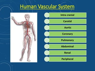
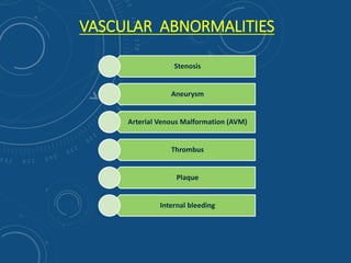
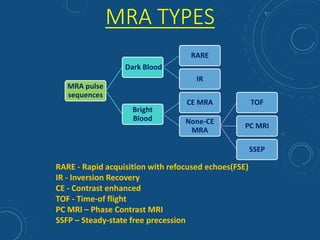
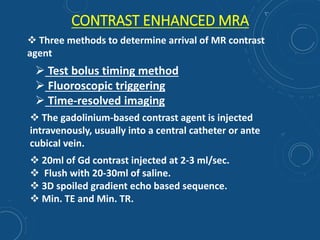
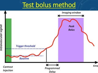
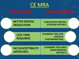
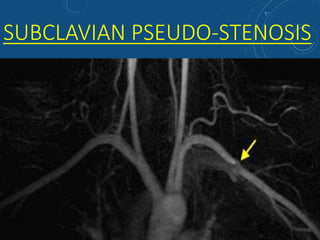
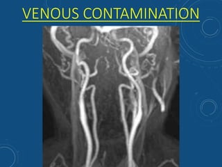
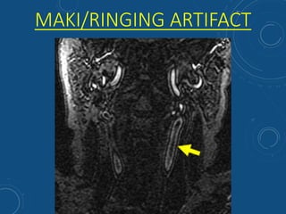
ALWINE ANTO
2ND Year BSc RADIOGRAPHY STUDENT
DEPT OF NIIR , NIMHANS, BENGALURU-29
MR ANGIOGRAPHY & TECHNIQUES
Human Vascular System
Intra cranial
Carotid
Aortic
Coronary
Pulmonary
Abdominal
Renal
Peripheral
VASCULAR ABNORMALITIES
Stenosis
Aneurysm
Arterial Venous Malformation (AVM)
Thrombus
Plaque
Internal bleeding
MRA TYPES
MRA pulse
sequences
Dark Blood
RARE
IR
Bright
Blood
CE MRA
None-CE
MRA
TOF
PC MRI
SSEP
RARE - Rapid acquisition with refocused echoes(FSE)
IR - Inversion Recovery
CE - Contrast enhanced
TOF - Time-of flight
PC MRI – Phase Contrast MRI
SSFP – Steady-state free precession
TOF MRA
Based on inflow effect with use of GRE sequence keeping
* TR 40msec (to saturate stationary tissues)
* TE 5msec (to reduce spin dephasing)
Methods:
-2D multislice
-3D singleslab
-3D multislice
PRINCIPLE OF TOF
- TOF is based on the effect of unsaturated spins inflowing into a region of
saturated stationary spins
2D vs 3D TOF MRA
2D TOF 3D TOF
↓ saturation effect ↑ saturation effect
Slow as well as fast flow Fast flow
Low spatial resolution High spatial resoultion
Good for large FOVs Good for small FOVs
Cerebral venography, abdominal and
peripheral angiography
Cerebral angiography
TOF MRA
ADVANTAGES
DISADVANTAGES
Overestimation of stenosis in turbulent or very slow flow
MethHbin thrombosed vessel may mimic blood flow.
CSF pulsation artifact simulating vessel lesions.
Susceptibility artifacts.
Can be used in renal failure patients
Can be used in patients allergic to gadolinium.
ARTIFACTS IN TOF MRA
• SHINE THROUGH ARTIFACTS
• FLOW REVERSAL ARTIFACT
• VENETIAN BLOOD ARTIFACT(3D MRA ONLY)
• SUSPECTIBILITY ARTIFACTS
• IN-PLANE SATURATION ARTIFACT
• STAIR STEP ARTIFACT
SHINE-THROUGH ARTIFACTS
3D TOF MRA showing shine through of high signal from hematoma (H) and fat (F)
at skull base
FLOW-REVERSAL ARTIFACT
2D TOF MRA shows only right
vertebral artery. No flow related
signal in the left vertebral artery is
seen.
Contrast-enhanced MRA shows
retrograde filling of left vertebral
artery (subclavian steal
phenomenon)
VENETIAN BLIND ARTIFACT (3D MRA ONLY)
SUSCEPTIBILITY ARTIFACTS
Source image shows
susceptibility field distortion
due to aneurysm clip
MRA shows spurious loss of flow
in entire right middle cerebral
artery due to susceptibility
artifact from clip
IN-PLANE SATURATION ARTIFACT
3D-TOF MRA showing
artifactual in-plane signal loss
within both middle cerebral
arteries
2D-TOF MRA shows
artifactual in-plane signal
loss in horizontal portions of
both anterior tibial arteries
STAIR-STEP ARTIFACT (2D TOF ONLY)
Mild "stair-step"
artifact due to non-
isotropic voxels
Mild motion artifact
causing horizontal
banding on this 2D-TOF
MRA of the aorta
Severe artifacts with
jagged edges due to
gross motion for carotid
TOF MRA study
CONTRAST ENHANCED MRA
 Test bolus timing method
 Fluoroscopic triggering
 Time-resolved imaging
 Three methods to determine arrival of MR contrast
agent
 The gadolinium-based contrast agent is injected
intravenously, usually into a central catheter or ante
cubical vein.
 20ml of Gd contrast injected at 2-3 ml/sec.
 Flush with 20-30ml of saline.
 3D spoiled gradient echo based sequence.
 Min. TE and Min. TR.
Test bolus method
CE MRA
ADVANTAGES DISADVANTAGES
BETTER SPATIAL
RESOLUTION
LESS TIME
REQUIRED
NO SUSCEPTIBILITY
ARTEFCATS.
SUBCLAVIAN PSEUDO-
STENOSIS ARTIFACT
SCANNING TOO LATE
(VENOUS
CONTAMINATION)
SCANNING TOO EARLY
(MAKI/RINGING
ARTIFACT)
SUBCLAVIAN PSEUDO-STENOSIS
VENOUS CONTAMINATION
MAKI/RINGING ARTIFACT
Magnetic Resonance Angiography and techniques

Magnetic Resonance Angiography and techniques

  • 1.
    ALWINE ANTO 2ND YearBSc RADIOGRAPHY STUDENT DEPT OF NIIR , NIMHANS, BENGALURU-29 MR ANGIOGRAPHY & TECHNIQUES
  • 2.
    Human Vascular System Intracranial Carotid Aortic Coronary Pulmonary Abdominal Renal Peripheral
  • 3.
    VASCULAR ABNORMALITIES Stenosis Aneurysm Arterial VenousMalformation (AVM) Thrombus Plaque Internal bleeding
  • 4.
    MRA TYPES MRA pulse sequences DarkBlood RARE IR Bright Blood CE MRA None-CE MRA TOF PC MRI SSEP RARE - Rapid acquisition with refocused echoes(FSE) IR - Inversion Recovery CE - Contrast enhanced TOF - Time-of flight PC MRI – Phase Contrast MRI SSFP – Steady-state free precession
  • 5.
    TOF MRA Based oninflow effect with use of GRE sequence keeping * TR 40msec (to saturate stationary tissues) * TE 5msec (to reduce spin dephasing) Methods: -2D multislice -3D singleslab -3D multislice PRINCIPLE OF TOF - TOF is based on the effect of unsaturated spins inflowing into a region of saturated stationary spins
  • 6.
    2D vs 3DTOF MRA 2D TOF 3D TOF ↓ saturation effect ↑ saturation effect Slow as well as fast flow Fast flow Low spatial resolution High spatial resoultion Good for large FOVs Good for small FOVs Cerebral venography, abdominal and peripheral angiography Cerebral angiography
  • 7.
    TOF MRA ADVANTAGES DISADVANTAGES Overestimation ofstenosis in turbulent or very slow flow MethHbin thrombosed vessel may mimic blood flow. CSF pulsation artifact simulating vessel lesions. Susceptibility artifacts. Can be used in renal failure patients Can be used in patients allergic to gadolinium.
  • 8.
    ARTIFACTS IN TOFMRA • SHINE THROUGH ARTIFACTS • FLOW REVERSAL ARTIFACT • VENETIAN BLOOD ARTIFACT(3D MRA ONLY) • SUSPECTIBILITY ARTIFACTS • IN-PLANE SATURATION ARTIFACT • STAIR STEP ARTIFACT
  • 9.
    SHINE-THROUGH ARTIFACTS 3D TOFMRA showing shine through of high signal from hematoma (H) and fat (F) at skull base
  • 10.
    FLOW-REVERSAL ARTIFACT 2D TOFMRA shows only right vertebral artery. No flow related signal in the left vertebral artery is seen. Contrast-enhanced MRA shows retrograde filling of left vertebral artery (subclavian steal phenomenon)
  • 11.
  • 12.
    SUSCEPTIBILITY ARTIFACTS Source imageshows susceptibility field distortion due to aneurysm clip MRA shows spurious loss of flow in entire right middle cerebral artery due to susceptibility artifact from clip
  • 13.
    IN-PLANE SATURATION ARTIFACT 3D-TOFMRA showing artifactual in-plane signal loss within both middle cerebral arteries 2D-TOF MRA shows artifactual in-plane signal loss in horizontal portions of both anterior tibial arteries
  • 14.
    STAIR-STEP ARTIFACT (2DTOF ONLY) Mild "stair-step" artifact due to non- isotropic voxels Mild motion artifact causing horizontal banding on this 2D-TOF MRA of the aorta Severe artifacts with jagged edges due to gross motion for carotid TOF MRA study
  • 15.
    CONTRAST ENHANCED MRA Test bolus timing method  Fluoroscopic triggering  Time-resolved imaging  Three methods to determine arrival of MR contrast agent  The gadolinium-based contrast agent is injected intravenously, usually into a central catheter or ante cubical vein.  20ml of Gd contrast injected at 2-3 ml/sec.  Flush with 20-30ml of saline.  3D spoiled gradient echo based sequence.  Min. TE and Min. TR.
  • 16.
  • 17.
    CE MRA ADVANTAGES DISADVANTAGES BETTERSPATIAL RESOLUTION LESS TIME REQUIRED NO SUSCEPTIBILITY ARTEFCATS. SUBCLAVIAN PSEUDO- STENOSIS ARTIFACT SCANNING TOO LATE (VENOUS CONTAMINATION) SCANNING TOO EARLY (MAKI/RINGING ARTIFACT)
  • 18.
  • 19.
  • 20.