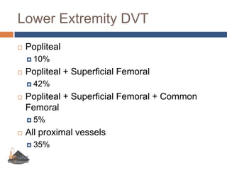
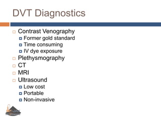
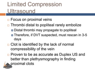
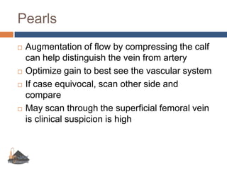
Deep Venous UltrasoundUniversity of Florida-JacksonvilleDepartment of Emergency MedicinePetra Duran-Gehring, M.D.
ObjectivesDescribe the indications and limitations of focused ultrasound for the detection of deep venous thrombosisUnderstand the standard ultrasound protocol when performing a focused examDefine the relevant local anatomyDevelop an understanding of doppler physics and instrumentationRecognize the relevant focused findings and pitfalls when evaluation for deep vein thrombosis
Deep Venous Thromboembolism Incidence in U.S.: 1 in 1000 people/year10% of proximal DVTs will lead to PE50% of untreated proximal DVTs will lead to PE within 3 months>80% of PEs due to DVTs
DVT Risk FactorsRecent TraumaRecent SurgeryImmobilityCancerEstrogen PregnancyOCPsPrior DVT/PEFamily history of hypercoagulabityProtein C or S deficiencyFactor V lieden or Antithrombin III deficiencyAntiphospholipin or anticardiolipin antibodyHomocysteineLupus anticoagulant
Physical ExamUnilateral leg swellingTenderness to palpationRednessWarmthPalpable cords- rareHomann’s sign- rarePratt’s signPoor sensitivity and Specificity
Lower Extremity DVTPopliteal10%Popliteal + Superficial Femoral42%Popliteal + Superficial Femoral + Common Femoral5%All proximal vessels35%
DVT DiagnosticsContrast VenographyFormer gold standardTime consumingIV dye exposurePlethysmographyCTMRIUltrasoundLow costPortableNon-invasive
Ultrasound ProtocolsDuplexComprehensiveColor flowDopplerTime consuming (about 45 mins)Limited CompressionFocused techniqueBedside examLook for clot only inCommon femoral veinPopliteal vein
Limited Compression UltrasoundFocus on proximal veinsThrombi distal to popliteal rarely embolizeDistal thrombi may propagate to poplitealTherefore, if DVT suspected, must rescan in 3-5 daysClot is identified by the lack of normal compressibility of the veinProven to be as accurate as Duplex US and better than plethysmography in finding proximal clots
Lower Extremity Venous AnatomyCommon FemoralCommon FemoralSuperficial (saphenous)DeepDeep Femoral (Profunda)Superficial FemoralPoplitealAnterior TibialPeronealPosterior TibialDeep Femoral Superficial FemoralPopliteal
Common Femoral AnatomyCommon Femoral VeinFemoral Artery
Femoral Junction AnatomyCommon Femoral VeinSaphenous VeinFemoral Artery
Femoral Bifurcation AnatomyCommon Femoral VeinFemoral ArteryProfundaFemoris
Superficial Femoral AnatomySuperficial Femoral VeinFemoral Artery
Popliteal AnatomyPopliteal VeinPopliteal Artery
Scanning TechniqueLinear array probe 6-10 mHzMedium footprintIf pt is obese, may need to use a lower frequency sector probePositioningReverse trendelenbergSemi-sitting with hips in 30 degrees flexion
Ultrasonic DVT FindingsNon-compressibilityEchogenic material with lumenDecreased blood flowDespite augmentation
CompressionCompress vein using transducerComplete apposition of the vein walls needed to rule out DVTIf compression is not achieved with pressure sufficient to deform adjacent artery, thrombus present
Common FemoralPt placed in supine positionLeg externally rotatedProbe indicator to pt’s right
Femoral VeinPlace probe in inguinal creaseUse color flow doppler to distinguish vesselsScan from CFV through the SFVCompress as you go
Femoral Vein DVT
PoplitealPositionProneDecubitusSeated on edge of gurneyKnee bent to increase venous fillingReverse trendelenburgProbe indicator to pt’s right
PoplitealPlace probe 10-12 cm above bend in kneeUse color flow doppler to distinguish vesselsScan through to the trifurcation of the poplitealCompress as you go
Popliteal Vein DVT
Scan ProtocolBegin by palpating femoral pulsePlace transducer over inguinal ligament with probe indicator to pt’s rightScan through the common femoral to the bifurcation (about 10 cm)Move to posterior knee bendScan through popliteal to the trifurcationTake clips to illustrate compressibilityMay need to image the contralateral side if results questionable
PearlsAugmentation of flow by compressing the calf can help distinguish the vein from arteryOptimize gain to best see the vascular systemIf case equivocal, scan other side and compareMay scan through the superficial femoral vein is clinical suspicion is high
Questions???

9 vascular us

  • 1.
    Deep Venous UltrasoundUniversityof Florida-JacksonvilleDepartment of Emergency MedicinePetra Duran-Gehring, M.D.
  • 2.
    ObjectivesDescribe the indicationsand limitations of focused ultrasound for the detection of deep venous thrombosisUnderstand the standard ultrasound protocol when performing a focused examDefine the relevant local anatomyDevelop an understanding of doppler physics and instrumentationRecognize the relevant focused findings and pitfalls when evaluation for deep vein thrombosis
  • 3.
    Deep Venous ThromboembolismIncidence in U.S.: 1 in 1000 people/year10% of proximal DVTs will lead to PE50% of untreated proximal DVTs will lead to PE within 3 months>80% of PEs due to DVTs
  • 4.
    DVT Risk FactorsRecentTraumaRecent SurgeryImmobilityCancerEstrogen PregnancyOCPsPrior DVT/PEFamily history of hypercoagulabityProtein C or S deficiencyFactor V lieden or Antithrombin III deficiencyAntiphospholipin or anticardiolipin antibodyHomocysteineLupus anticoagulant
  • 5.
    Physical ExamUnilateral legswellingTenderness to palpationRednessWarmthPalpable cords- rareHomann’s sign- rarePratt’s signPoor sensitivity and Specificity
  • 6.
    Lower Extremity DVTPopliteal10%Popliteal+ Superficial Femoral42%Popliteal + Superficial Femoral + Common Femoral5%All proximal vessels35%
  • 7.
    DVT DiagnosticsContrast VenographyFormergold standardTime consumingIV dye exposurePlethysmographyCTMRIUltrasoundLow costPortableNon-invasive
  • 8.
    Ultrasound ProtocolsDuplexComprehensiveColor flowDopplerTimeconsuming (about 45 mins)Limited CompressionFocused techniqueBedside examLook for clot only inCommon femoral veinPopliteal vein
  • 9.
    Limited Compression UltrasoundFocuson proximal veinsThrombi distal to popliteal rarely embolizeDistal thrombi may propagate to poplitealTherefore, if DVT suspected, must rescan in 3-5 daysClot is identified by the lack of normal compressibility of the veinProven to be as accurate as Duplex US and better than plethysmography in finding proximal clots
  • 10.
    Lower Extremity VenousAnatomyCommon FemoralCommon FemoralSuperficial (saphenous)DeepDeep Femoral (Profunda)Superficial FemoralPoplitealAnterior TibialPeronealPosterior TibialDeep Femoral Superficial FemoralPopliteal
  • 11.
    Common Femoral AnatomyCommonFemoral VeinFemoral Artery
  • 12.
    Femoral Junction AnatomyCommonFemoral VeinSaphenous VeinFemoral Artery
  • 13.
    Femoral Bifurcation AnatomyCommonFemoral VeinFemoral ArteryProfundaFemoris
  • 14.
    Superficial Femoral AnatomySuperficialFemoral VeinFemoral Artery
  • 15.
  • 16.
    Scanning TechniqueLinear arrayprobe 6-10 mHzMedium footprintIf pt is obese, may need to use a lower frequency sector probePositioningReverse trendelenbergSemi-sitting with hips in 30 degrees flexion
  • 17.
    Ultrasonic DVT FindingsNon-compressibilityEchogenicmaterial with lumenDecreased blood flowDespite augmentation
  • 18.
    CompressionCompress vein usingtransducerComplete apposition of the vein walls needed to rule out DVTIf compression is not achieved with pressure sufficient to deform adjacent artery, thrombus present
  • 19.
    Common FemoralPt placedin supine positionLeg externally rotatedProbe indicator to pt’s right
  • 20.
    Femoral VeinPlace probein inguinal creaseUse color flow doppler to distinguish vesselsScan from CFV through the SFVCompress as you go
  • 21.
  • 22.
    PoplitealPositionProneDecubitusSeated on edgeof gurneyKnee bent to increase venous fillingReverse trendelenburgProbe indicator to pt’s right
  • 23.
    PoplitealPlace probe 10-12cm above bend in kneeUse color flow doppler to distinguish vesselsScan through to the trifurcation of the poplitealCompress as you go
  • 24.
  • 25.
    Scan ProtocolBegin bypalpating femoral pulsePlace transducer over inguinal ligament with probe indicator to pt’s rightScan through the common femoral to the bifurcation (about 10 cm)Move to posterior knee bendScan through popliteal to the trifurcationTake clips to illustrate compressibilityMay need to image the contralateral side if results questionable
  • 26.
    PearlsAugmentation of flowby compressing the calf can help distinguish the vein from arteryOptimize gain to best see the vascular systemIf case equivocal, scan other side and compareMay scan through the superficial femoral vein is clinical suspicion is high
  • 27.

Editor's Notes

  • #12 You will notice again how large the vein is in comparison to the artery here in the femoral triangle. Note that the vein and artery lie side by side, but please note that this relationship may not occur in all your patients. It is common for the vein to reside under the artery, making your ability to distinguish artery from vein essential.
  • #13 You will notice again how large the vein is in comparison to the artery here in the femoral triangle. Note that the vein and artery lie side by side, but please note that this relationship may not occur in all your patients. It is common for the vein to reside under the artery, making your ability to distinguish artery from vein essential.
  • #14 You will notice again how large the vein is in comparison to the artery here in the femoral triangle. Note that the vein and artery lie side by side, but please note that this relationship may not occur in all your patients. It is common for the vein to reside under the artery, making your ability to distinguish artery from vein essential.
  • #15 You will notice again how large the vein is in comparison to the artery here in the femoral triangle. Note that the vein and artery lie side by side, but please note that this relationship may not occur in all your patients. It is common for the vein to reside under the artery, making your ability to distinguish artery from vein essential.