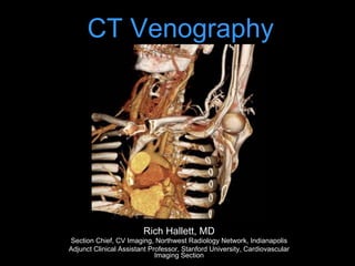
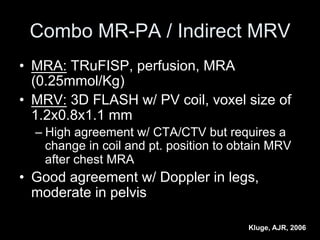
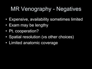
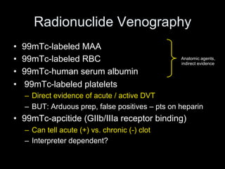
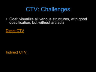
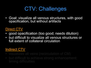
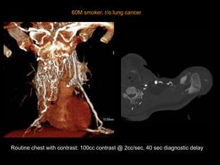
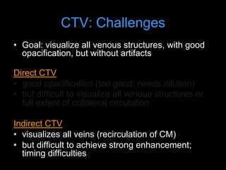
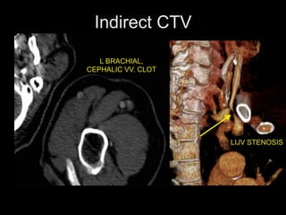
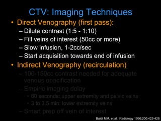
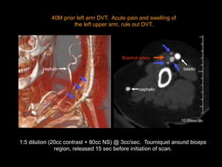
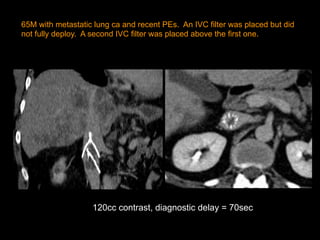
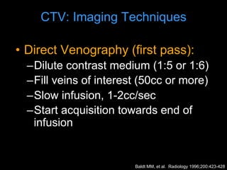
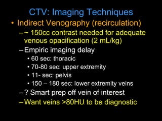
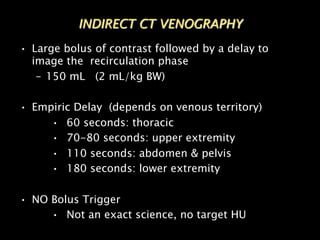
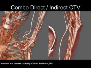
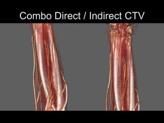
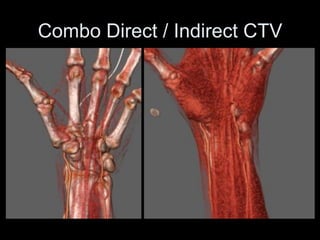
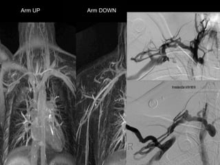
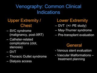
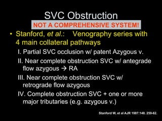
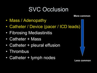
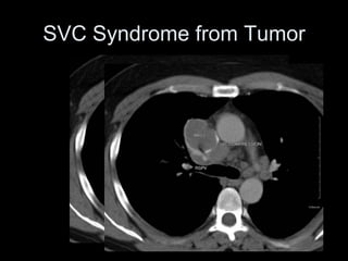
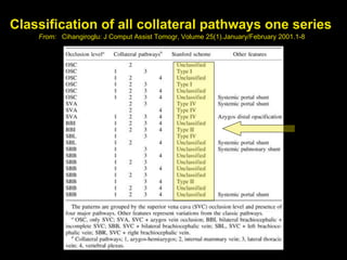
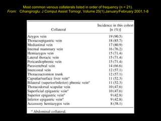
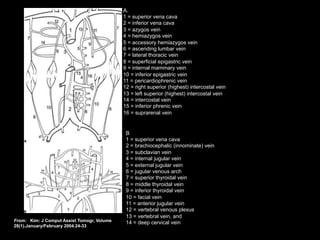
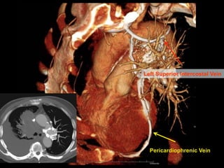
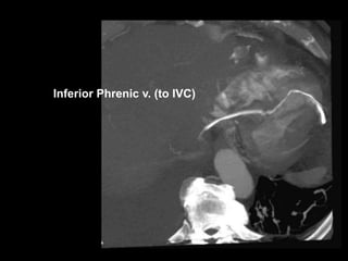
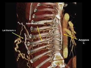
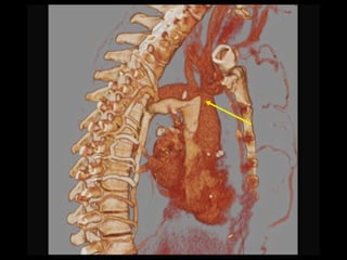
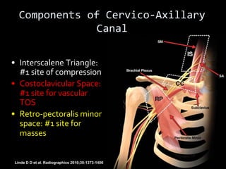
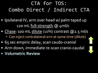
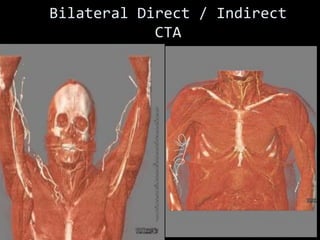
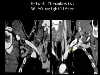
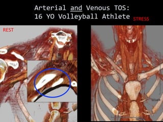
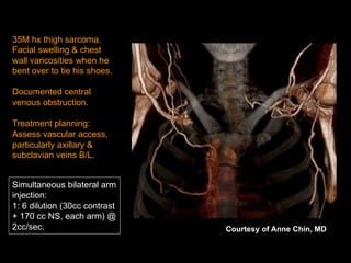
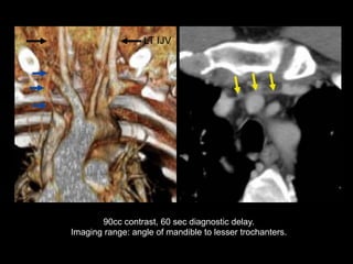
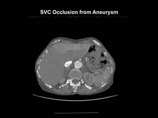
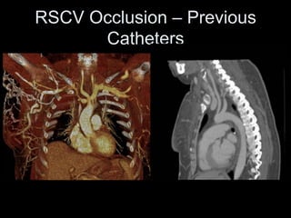
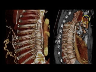
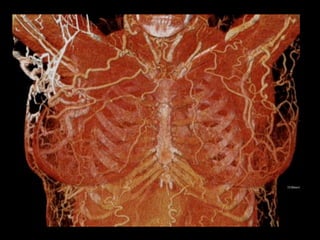
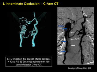
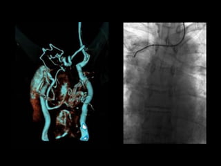
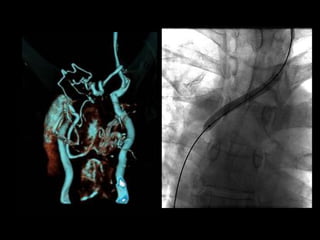
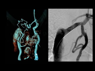
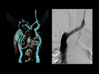
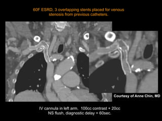
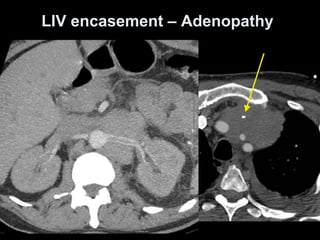
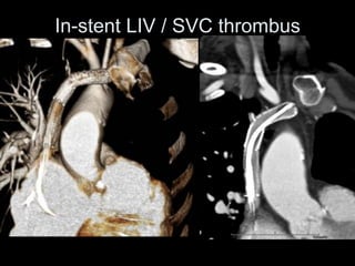
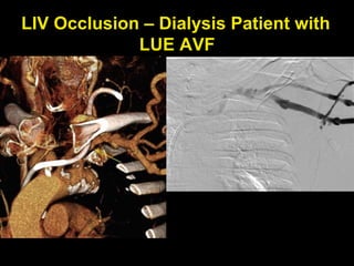
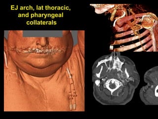
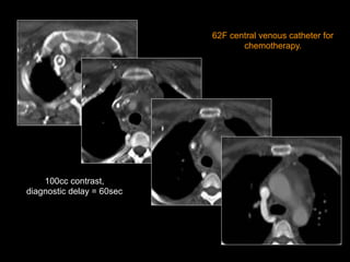
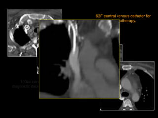
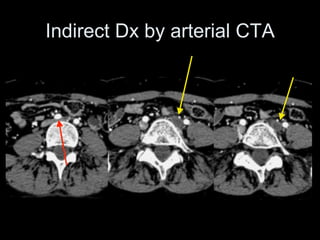
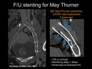
CT Venography
Rich Hallett, MD
Section Chief, CV Imaging, Northwest Radiology Network, Indianapolis
Adjunct Clinical Assistant Professor, Stanford University, Cardiovascular
Imaging Section
Introduction
• CT venography (CTV) is a technique targeted to
assess venous anatomy, determine venous
patency & delineate collateral circulation
• Non-invasive, simple protocols, wide anatomic
coverage, short acquisition time, and ability to be
combined with arterial-phase CTA
Lecture Outline
• Basic Clinical Options for Venous Imaging
– Venous Imaging Modalities
• CT Scan Protocols
– Indirect CTV
– Direct CTV
• Selected Regional Applications
– UE
– Chest
Venous Imaging Modalities – The
competition
• Doppler Ultrasound (US)
• MR Venography
• Catheter venography
• Nuclear venography
Doppler US
• Well established clinical utility
• No ionizing radiation
• Portable
• Inexpensive
• Flow direction information
• Operator / Patient dependent
• Some areas inaccessible (pelvis, SVC)
• Collateral pathways not well delineated
Doppler US
• Sens/ Spec ~ 95% for fem-
pop DVT in ideal situations
Performance of Doppler vs. CTV in ICU
patients – LE DVT
Sens Spec
Indiect CTV 70 96
Doppler US 70 100
Taffoni, AJR, 2004
MR Venography - Positives
• Excellent for pelvic venous system, CNS
• May not require contrast
• SI ratio thrombus:blood higher for MRV vs. CTV
– 3.7-8:1 vs 1.8-3.2 *
• For PE: Sens 80-95%, Spec 95%, depends on
technique (Perf imaging best)+
• For DVT: Sens ~92%, Spec ~95%
• 0.25 mmol/kg Gd better than 0.125 mmol/kg
+Sampson, Eur Radiol 2007; 17:175-181
* Kluge, AJR, 2006
Combo MR-PA / Indirect MRV
• MRA: TRuFISP, perfusion, MRA
(0.25mmol/Kg)
• MRV: 3D FLASH w/ PV coil, voxel size of
1.2x0.8x1.1 mm
– High agreement w/ CTA/CTV but requires a
change in coil and pt. position to obtain MRV
after chest MRA
• Good agreement w/ Doppler in legs,
moderate in pelvis
Kluge, AJR, 2006
MR Venography - Negatives
• Expensive, availability sometimes limited
• Exam may be lengthy
• Pt. cooperation?
• Spatial resolution (vs other choices)
• Limited anatomic coverage
Radionuclide Venography
• 99mTc-labeled MAA
• 99mTc-labeled RBC
• 99mTc-human serum albumin
• 99mTc-labeled platelets
– Direct evidence of acute / active DVT
– BUT: Arduous prep, false positives – pts on heparin
• 99mTc-apcitide (GIIb/IIIa receptor binding)
– Can tell acute (+) vs. chronic (-) clot
– Interpreter dependent?
Anatomic agents,
indirect evidence
Catheter Venography
• Considered the gold standard
• Invasive (but can treat lesions)
• You only see what you can fill
• Risks:
– Minor Complications: 18%
– Thrombosis: 2%
– Bronchospasm, Contrast reactions, etc
CTV: Challenges
• Goal: visualize all venous structures, with good
opacification, but without artifacts
Direct CTV
• good opacification (too good; needs dilution)
• but difficult to show all venous structures or full
extent of collateral circulation
Indirect CTV
• shows all veins; but difficult to achieve strong
enhancement; timing
CTV: Challenges
• Goal: visualize all venous structures, with good
opacification, but without artifacts
Direct CTV
• good opacification (too good; needs dilution)
• but difficult to visualize all venous structures or
full extent of collateral circulation
Indirect CTV
• visualizes all veins (recirculation of CM)
• but difficult to achieve strong enhancement;
timing difficulties
60M smoker, r/o lung cancer
Routine chest with contrast: 100cc contrast @ 2cc/sec, 40 sec diagnostic delay
CTV: Challenges
• Goal: visualize all venous structures, with good
opacification, but without artifacts
Direct CTV
• good opacification (too good; needs dilution)
• but difficult to visualize all venous structures or
full extent of collateral circulation
Indirect CTV
• visualizes all veins (recirculation of CM)
• but difficult to achieve strong enhancement;
timing difficulties
L BRACHIAL,
CEPHALIC VV. CLOT
LIJV STENOSIS
Indirect CTV
CTV: Imaging Techniques
• Direct Venography (first pass):
– Dilute contrast (1:5 - 1:10)
– Fill veins of interest (50cc or more)
– Slow infusion, 1-2cc/sec
– Start acquisition towards end of infusion
• Indirect Venography (recirculation)
– 100-150cc contrast needed for adequate
venous opacification
– Empiric imaging delay
• 60 seconds: upper extremity and pelvic veins
• 3 to 3.5 min: lower extremity veins
– Smart prep off vein of interest
Baldt MM, et al. Radiology 1996;200:423-428
40M prior left arm DVT. Acute pain and swelling of
the left upper arm, rule out DVT.
1:5 dilution (20cc contrast + 80cc NS) @ 3cc/sec. Tourniquet around biceps
region, released 15 sec before initiation of scan.
basilic
Brachial artery
basilic
cephalic
cephalic
CTV: Imaging Techniques
• Direct Venography (first pass):
– Dilute contrast (1:5 or 1:6)
– Fill veins of interest (50cc or more)
– Slow infusion, 1-2cc/sec
– Start acquisition towards end of infusion
• Indirect Venography (recirculation)
– 100-150cc contrast needed for adequate
venous opacification
– Empiric imaging delay
• 60 seconds: upper extremity and pelvic veins
• 3 to 3.5 min: lower extremity veins
– Smart prep off vein of interest
Baldt MM, et al. Radiology 1996;200:423-428
65M with metastatic lung ca and recent PEs. An IVC filter was placed but did
not fully deploy. A second IVC filter was placed above the first one.
120cc contrast, diagnostic delay = 70sec
CTV: Imaging Techniques
• Direct Venography (first pass):
–Dilute contrast medium (1:5 or 1:6)
–Fill veins of interest (50cc or more)
–Slow infusion, 1-2cc/sec
–Start acquisition towards end of
infusion
Baldt MM, et al. Radiology 1996;200:423-428
CTV: Imaging Techniques
• Indirect Venography (recirculation)
–~ 150cc contrast needed for adequate
venous opacification (2 mL/kg)
–Empiric imaging delay
• 60 sec: thoracic
• 70-80 sec: upper extremity
• 11- sec: pelvis
• 150 – 180 sec: lower extremity veins
–? Smart prep off vein of interest
–Want veins >80HU to be diagnostic
• Large bolus of contrast followed by a delay to
image the recirculation phase
– 150 mL (2 mL/kg BW)
• Empiric Delay (depends on venous territory)
• 60 seconds: thoracic
• 70-80 seconds: upper extremity
• 110 seconds: abdomen & pelvis
• 180 seconds: lower extremity
• NO Bolus Trigger
• Not an exact science, no target HU
INDIRECT CT VENOGRAPHY
Combo Direct / Indirect CTV
• R/O LUE venous
malformation; L hand
and arm swelling
• 120 mL @ 5 mL/s
followed by 100 mL
1:10 dilution at 2.5
mL/s via L hand IV
• Caudocranial
acquisition
Combo Direct / Indirect CTV
Protocol and dataset courtesy of Scott Alexander, MD
Combo Direct / Indirect CTV
CTA for TOS:
Combo Direct / Indirect CTA
• Ipsilateral IV, arm over head w/ palm taped up
• Bolus: 120 mL full-strength @ 4ml/s
• Chase: 100 mL dilute (10%) contrast @2.5 ml/s
• Can inject contralateral arm at same time (dilute)
• 65 sec empiric delay, scan caudo-cranial
• Arm down, immediate re-scan cranio-caudal
• Volumetric Review
MRA for TOS: Blood Pool MRA
• Anatomic imaging: Oblique sag and cor T1/T2
• Relaxed and Challenged imaging:
§ Gadofosveset (blood pool agent)
§ Breath-hold FSPGR, ECG-gated, high resolution (1.8
mm ST, 448 x 448 matrix) CORONAL acquisition
§ Challenged: Arm Abducted
§ Relaxed: Arm Down
Arm UP Arm DOWN
Venography: Common Clinical
Indications
Upper Extremity /
Chest
– SVC syndrome
(malignancy, post-XRT)
– Catheter-related
complications (clot,
stenosis)
– DVT
– Thoracic Outlet syndrome
– Dialysis access
Lower Extremity
– DVT (+/- PE study)
– May-Thurner syndrome
– Pre-transplant evaluation
General
- Venous stent evaluation
- Vascular Malformations –
treatment planning
SVC Obstruction
• Stanford, et al.: Venography series with
4 main collateral pathways
I. Partial SVC occlusion w/ patent Azygous v.
II. Near complete obstruction SVC w/ antegrade
flow azygous à RA
III. Near complete obstruction SVC w/
retrograde flow azygous
IV. Complete obstruction SVC + one or more
major tributaries (e.g. azygous v.)
Stanford W, et al AJR 1987:148. 259-62.
NOT A COMPREHENSIVE SYSTEM!
SVC Occlusion
• Mass / Adenopathy
• Catheter / Device (pacer / ICD leads)
• Fibrosing Mediastinitis
• Catheter + Mass
• Catheter + pleural effusion
• Thrombus
• Catheter + lymph nodes
More common
Less common
SVC Syndrome from Tumor
Classification of all collateral pathways one series
From: Cihangiroglu: J Comput Assist Tomogr, Volume 25(1).January/February 2001.1-8
Most common venous collaterals listed in order of frequency (n = 21).
From: Cihangiroglu: J Comput Assist Tomogr, Volume 25(1).January/February 2001.1-8
A.
1 = superior vena cava
2 = inferior vena cava
3 = azygos vein
4 = hemiazygos vein
5 = accessory hemiazygos vein
6 = ascending lumbar vein
7 = lateral thoracic vein
8 = superficial epigastric vein
9 = internal mammary vein
10 = inferior epigastric vein
11 = pericardiophrenic vein
12 = right superior (highest) intercostal vein
13 = left superior (highest) intercostal vein
14 = intercostal vein
15 = inferior phrenic vein
16 = suprarenal vein
B
1 = superior vena cava
2 = brachiocephalic (innominate) vein
3 = subclavian vein
4 = internal jugular vein
5 = external jugular vein
6 = jugular venous arch
7 = superior thyroidal vein
8 = middle thyroidal vein
9 = inferior thyroidal vein
10 = facial vein
11 = anterior jugular vein
12 = vertebral venous plexus
13 = vertebral vein, and
14 = deep cervical vein
From: Kim: J Comput Assist Tomogr, Volume
28(1).January/February 2004.24-33
Left Superior Intercostal Vein
Pericardiophrenic Vein
Inferior Phrenic v. (to IVC)
Lat thoracic v.
Intercostal veins
Azygous
v.
Internal Mammary Veins
Thoraco-acromio-
Clavicular vv.
Areolar Venous Plexus
Paravertebral vv.
Capsular / Liver surface vv.
Systemic – portal collaterals
Venous collaterals organized by plexus
systems – Easier, more complete to report
Cihangiroglu: J Comput Assist Tomogr, Volume 25(1).January/February 2001.1-8
The poster child for revised venous
plexus nomenclature……
Chest / Upper extremity cases
Thoracic Outlet Syndrome (TOS)
• Symptoma(c compression/entrapment
of neurovascular structures by bone
and/or so7 (ssue as they pass through
the cervicoaxillary canal
• 90% Neurogenic (PT, postural Tx, NSAIDs)
• 10% Vascular
• Venous > Arterial
Linda D D et al. Radiographics 2010;30:1373-1400
Components of Cervico-Axillary
Canal
• Interscalene Triangle:
#1 site of compression
• Costoclavicular Space:
#1 site for vascular
TOS
• Retro-pectoralis minor
space: #1 site for
masses
CTA for TOS:
Combo Direct / Indirect CTA
• Ipsilateral IV, arm over head w/ palm taped up
• Bolus: 120 mL full-strength @ 4ml/s
• Chase: 100 mL dilute (10%) contrast @2.5 ml/s
• Can inject contralateral arm at same time (dilute)
• 65 sec empiric delay, scan caudo-cranial
• Arm down, immediate re-scan cranio-caudal
• Volumetric Review
Bilateral Direct / Indirect
CTA
Venous TOS: Effort Thrombosis
• Paget-Schroetter syndrome (PSS)
• AKA axillo-subclavian venous
thrombosis
• Overhead athletes
• PE in up to 1/3!! *
• Post-thrombotic syndrome (later)
* Perlowski AA.Vasc Med (2010) vol. 15 (6) pp. 469-79
Effort Thrombosis:
36 YO weightlifter
Post-Op 1st rib resection
Arterial TOS
• Overhead athletes
• SX: Coolness, weakness, diffuse arm
pain (ischemic neuritis)
• Cause: Repetitive compression injury
– Anatomic predisposition (tight CCS)
– Post-traumatic, bony callus
– Scalene hypertrophy
Arterial and Venous TOS:
16 YO Volleyball Athlete
REST
STRESS
SVC and central veins
LUNG CA
with SVC syndrome
RSCV OCCLUSION – 47 F Dialysis Pt
35M hx thigh sarcoma.
Facial swelling & chest
wall varicosities when he
bent over to tie his shoes.
Documented central
venous obstruction.
Treatment planning:
Assess vascular access,
particularly axillary &
subclavian veins B/L.
Simultaneous bilateral arm
injection:
1: 6 dilution (30cc contrast
+ 170 cc NS, each arm) @
2cc/sec. Courtesy of Anne Chin, MD
90cc contrast, 60 sec diagnostic delay.
Imaging range: angle of mandible to lesser trochanters.
LT IJV
SVC Occlusion from Aneurysm
RSCV Occlusion – Previous
Catheters
LT IJ injection 1:2 dilution (12cc contrast
+ 12cc NS @ 2cc/sec) acquired on flat-
panel detector Dyna-CT.
SVC
RIMV
L innominate Occlusion - C-Arm CT
Courtesy of Anne Chin, MD
IV cannula in left arm. 100cc contrast + 20cc
NS flush, diagnostic delay = 60sec.
60F ESRD, 3 overlapping stents placed for venous
stenosis from previous catheters.
Courtesy of Anne Chin, MD
LIV encasement – Adenopathy
In-stent LIV / SVC thrombus
LIV Occlusion – Dialysis Patient with
LUE AVF
EJ arch, lat thoracic,
and pharyngeal
collaterals
100cc contrast,
diagnostic delay = 60sec
62F central venous catheter for
chemotherapy.
100cc contrast,
diagnostic delay = 60sec
62F central venous catheter for
chemotherapy.
MISC UE Cases
RUE Hemangiomatosis
UE AVMs
Pelvis / LE Cases
MAY-THURNER :
SUPERFICIAL VENOUS
VARICOSITIES
41 YO F, May - Thurner
Lack of Augmentation – suspect upstream obstruction
LCIV
External iliac vv.
S/P Mechnical Lysis, TPA, and PTA
Indirect Dx by arterial CTA
• 120 cc contrast
• Monitoring delay = 40sec
• Smart prep at infrarenal IVC
28F May-Thurner syndrome,
CIV/EIV stent placement
3 years ago
Courtesy of Anne Chin, MD
F/U stenting for May Thurner
Vascular Mapping
Extremity
Hemangiomatosis
Venous Mapping
Major drainage routes:
LEFT 12th IC VEIN
Left Gonadal V.
Greater Saphenous V.
Protocol:
CTA Runoff; + 40 sec interscan delay;
Caudocranial scan 16x0.75mm
IVC Aneurysm
IVC Aneurysm
• Rare
• Saccular > fusiform
• Cause unknown, may be related to anomalous
connections in embryologic venous systems
– Acquired (trauma, AV fistulae)
– May be associated with other congenital CV
anomalies
• Sx: Thrombosis (7/16), pain, rupture, leg
swelling
– Massive penile bleeding (1/16)
– PE if thrombus
• CTV is a robust, non-invasive technique to visualize venous
anatomy, and can be combined with arterial phase CTA
• Direct CTV: better opacification, less CM needed, but only the
injected and downstream veins will be visualized
• Indirect CTV: all venous anatomy is delineated, empiric delay
or smart-prep at ROI, opacification occasionally unpredictable
• Combo CTV : Perhaps the best choice for excellent and
consistent venous opacification
• Provides accurate 3D visualization of venous anatomy for
treatment planning
Conclusions
Thanks to:
Dominik Fleischmann, MD
Frandics Chan, MD PhD
Loud PA, et al. Radiology 2001; 219:498-502. (Sens / Spec of CTV good
compared to Doppler)
Begemann PG, et al. J Comput Assist Tomogr 2003; 27:399-409.
(Sensitivity=100%; Specificity=97% compared with ultrasonography)
Baldt MM, et al. Radiology 1996; 200:423-428. (Sensitivity=100%;
Specificiity=96% compared with conventional venography)
Sampson, FC, et al. Eur Radiol 2007; 17:175-181. (Pooled sensitivity=91.5%;
Pooled specificity=94.8% compared with conventional venography)
Key References
Kluge, A. et al. AJR 2006; 186:1686 – 1696 (Combo MRA/MRV for PE/DVT)
Kim, HC, et al. J Comput Assist Tomogr 2004; 28:24-33 (Collateral Pathways)
Cihangiroglu M, et al. J Comput Assis Tomogr 2001; 25: 1-8 (collaterals in
SVC Obstruction)
Lawler LP, et al. Radiographics 2002; 22:S45-S60 (normal and accessory
chest venous pathways)
Demos TC, et al. AJR 2004; 182:1139-1150 (Venous anomalies of chest)
Key References

Upper Extrem CT Venography_Hallett_2017_sm.pdf

  • 1.
    CT Venography Rich Hallett,MD Section Chief, CV Imaging, Northwest Radiology Network, Indianapolis Adjunct Clinical Assistant Professor, Stanford University, Cardiovascular Imaging Section
  • 2.
    Introduction • CT venography(CTV) is a technique targeted to assess venous anatomy, determine venous patency & delineate collateral circulation • Non-invasive, simple protocols, wide anatomic coverage, short acquisition time, and ability to be combined with arterial-phase CTA
  • 3.
    Lecture Outline • BasicClinical Options for Venous Imaging – Venous Imaging Modalities • CT Scan Protocols – Indirect CTV – Direct CTV • Selected Regional Applications – UE – Chest
  • 4.
    Venous Imaging Modalities– The competition • Doppler Ultrasound (US) • MR Venography • Catheter venography • Nuclear venography
  • 5.
    Doppler US • Wellestablished clinical utility • No ionizing radiation • Portable • Inexpensive • Flow direction information • Operator / Patient dependent • Some areas inaccessible (pelvis, SVC) • Collateral pathways not well delineated
  • 6.
    Doppler US • Sens/Spec ~ 95% for fem- pop DVT in ideal situations
  • 7.
    Performance of Dopplervs. CTV in ICU patients – LE DVT Sens Spec Indiect CTV 70 96 Doppler US 70 100 Taffoni, AJR, 2004
  • 8.
    MR Venography -Positives • Excellent for pelvic venous system, CNS • May not require contrast • SI ratio thrombus:blood higher for MRV vs. CTV – 3.7-8:1 vs 1.8-3.2 * • For PE: Sens 80-95%, Spec 95%, depends on technique (Perf imaging best)+ • For DVT: Sens ~92%, Spec ~95% • 0.25 mmol/kg Gd better than 0.125 mmol/kg +Sampson, Eur Radiol 2007; 17:175-181 * Kluge, AJR, 2006
  • 9.
    Combo MR-PA /Indirect MRV • MRA: TRuFISP, perfusion, MRA (0.25mmol/Kg) • MRV: 3D FLASH w/ PV coil, voxel size of 1.2x0.8x1.1 mm – High agreement w/ CTA/CTV but requires a change in coil and pt. position to obtain MRV after chest MRA • Good agreement w/ Doppler in legs, moderate in pelvis Kluge, AJR, 2006
  • 10.
    MR Venography -Negatives • Expensive, availability sometimes limited • Exam may be lengthy • Pt. cooperation? • Spatial resolution (vs other choices) • Limited anatomic coverage
  • 11.
    Radionuclide Venography • 99mTc-labeledMAA • 99mTc-labeled RBC • 99mTc-human serum albumin • 99mTc-labeled platelets – Direct evidence of acute / active DVT – BUT: Arduous prep, false positives – pts on heparin • 99mTc-apcitide (GIIb/IIIa receptor binding) – Can tell acute (+) vs. chronic (-) clot – Interpreter dependent? Anatomic agents, indirect evidence
  • 12.
    Catheter Venography • Consideredthe gold standard • Invasive (but can treat lesions) • You only see what you can fill • Risks: – Minor Complications: 18% – Thrombosis: 2% – Bronchospasm, Contrast reactions, etc
  • 13.
    CTV: Challenges • Goal:visualize all venous structures, with good opacification, but without artifacts Direct CTV • good opacification (too good; needs dilution) • but difficult to show all venous structures or full extent of collateral circulation Indirect CTV • shows all veins; but difficult to achieve strong enhancement; timing
  • 14.
    CTV: Challenges • Goal:visualize all venous structures, with good opacification, but without artifacts Direct CTV • good opacification (too good; needs dilution) • but difficult to visualize all venous structures or full extent of collateral circulation Indirect CTV • visualizes all veins (recirculation of CM) • but difficult to achieve strong enhancement; timing difficulties
  • 15.
    60M smoker, r/olung cancer Routine chest with contrast: 100cc contrast @ 2cc/sec, 40 sec diagnostic delay
  • 16.
    CTV: Challenges • Goal:visualize all venous structures, with good opacification, but without artifacts Direct CTV • good opacification (too good; needs dilution) • but difficult to visualize all venous structures or full extent of collateral circulation Indirect CTV • visualizes all veins (recirculation of CM) • but difficult to achieve strong enhancement; timing difficulties
  • 17.
    L BRACHIAL, CEPHALIC VV.CLOT LIJV STENOSIS Indirect CTV
  • 18.
    CTV: Imaging Techniques •Direct Venography (first pass): – Dilute contrast (1:5 - 1:10) – Fill veins of interest (50cc or more) – Slow infusion, 1-2cc/sec – Start acquisition towards end of infusion • Indirect Venography (recirculation) – 100-150cc contrast needed for adequate venous opacification – Empiric imaging delay • 60 seconds: upper extremity and pelvic veins • 3 to 3.5 min: lower extremity veins – Smart prep off vein of interest Baldt MM, et al. Radiology 1996;200:423-428
  • 19.
    40M prior leftarm DVT. Acute pain and swelling of the left upper arm, rule out DVT. 1:5 dilution (20cc contrast + 80cc NS) @ 3cc/sec. Tourniquet around biceps region, released 15 sec before initiation of scan. basilic Brachial artery basilic cephalic cephalic
  • 20.
    CTV: Imaging Techniques •Direct Venography (first pass): – Dilute contrast (1:5 or 1:6) – Fill veins of interest (50cc or more) – Slow infusion, 1-2cc/sec – Start acquisition towards end of infusion • Indirect Venography (recirculation) – 100-150cc contrast needed for adequate venous opacification – Empiric imaging delay • 60 seconds: upper extremity and pelvic veins • 3 to 3.5 min: lower extremity veins – Smart prep off vein of interest Baldt MM, et al. Radiology 1996;200:423-428
  • 21.
    65M with metastaticlung ca and recent PEs. An IVC filter was placed but did not fully deploy. A second IVC filter was placed above the first one. 120cc contrast, diagnostic delay = 70sec
  • 22.
    CTV: Imaging Techniques •Direct Venography (first pass): –Dilute contrast medium (1:5 or 1:6) –Fill veins of interest (50cc or more) –Slow infusion, 1-2cc/sec –Start acquisition towards end of infusion Baldt MM, et al. Radiology 1996;200:423-428
  • 23.
    CTV: Imaging Techniques •Indirect Venography (recirculation) –~ 150cc contrast needed for adequate venous opacification (2 mL/kg) –Empiric imaging delay • 60 sec: thoracic • 70-80 sec: upper extremity • 11- sec: pelvis • 150 – 180 sec: lower extremity veins –? Smart prep off vein of interest –Want veins >80HU to be diagnostic
  • 24.
    • Large bolusof contrast followed by a delay to image the recirculation phase – 150 mL (2 mL/kg BW) • Empiric Delay (depends on venous territory) • 60 seconds: thoracic • 70-80 seconds: upper extremity • 110 seconds: abdomen & pelvis • 180 seconds: lower extremity • NO Bolus Trigger • Not an exact science, no target HU INDIRECT CT VENOGRAPHY
  • 25.
    Combo Direct /Indirect CTV • R/O LUE venous malformation; L hand and arm swelling • 120 mL @ 5 mL/s followed by 100 mL 1:10 dilution at 2.5 mL/s via L hand IV • Caudocranial acquisition
  • 26.
    Combo Direct /Indirect CTV Protocol and dataset courtesy of Scott Alexander, MD
  • 27.
    Combo Direct /Indirect CTV
  • 28.
    Combo Direct /Indirect CTV
  • 29.
    CTA for TOS: ComboDirect / Indirect CTA • Ipsilateral IV, arm over head w/ palm taped up • Bolus: 120 mL full-strength @ 4ml/s • Chase: 100 mL dilute (10%) contrast @2.5 ml/s • Can inject contralateral arm at same time (dilute) • 65 sec empiric delay, scan caudo-cranial • Arm down, immediate re-scan cranio-caudal • Volumetric Review
  • 30.
    MRA for TOS:Blood Pool MRA • Anatomic imaging: Oblique sag and cor T1/T2 • Relaxed and Challenged imaging: § Gadofosveset (blood pool agent) § Breath-hold FSPGR, ECG-gated, high resolution (1.8 mm ST, 448 x 448 matrix) CORONAL acquisition § Challenged: Arm Abducted § Relaxed: Arm Down
  • 31.
  • 32.
    Venography: Common Clinical Indications UpperExtremity / Chest – SVC syndrome (malignancy, post-XRT) – Catheter-related complications (clot, stenosis) – DVT – Thoracic Outlet syndrome – Dialysis access Lower Extremity – DVT (+/- PE study) – May-Thurner syndrome – Pre-transplant evaluation General - Venous stent evaluation - Vascular Malformations – treatment planning
  • 33.
    SVC Obstruction • Stanford,et al.: Venography series with 4 main collateral pathways I. Partial SVC occlusion w/ patent Azygous v. II. Near complete obstruction SVC w/ antegrade flow azygous à RA III. Near complete obstruction SVC w/ retrograde flow azygous IV. Complete obstruction SVC + one or more major tributaries (e.g. azygous v.) Stanford W, et al AJR 1987:148. 259-62. NOT A COMPREHENSIVE SYSTEM!
  • 34.
    SVC Occlusion • Mass/ Adenopathy • Catheter / Device (pacer / ICD leads) • Fibrosing Mediastinitis • Catheter + Mass • Catheter + pleural effusion • Thrombus • Catheter + lymph nodes More common Less common
  • 35.
  • 36.
    Classification of allcollateral pathways one series From: Cihangiroglu: J Comput Assist Tomogr, Volume 25(1).January/February 2001.1-8
  • 37.
    Most common venouscollaterals listed in order of frequency (n = 21). From: Cihangiroglu: J Comput Assist Tomogr, Volume 25(1).January/February 2001.1-8
  • 38.
    A. 1 = superiorvena cava 2 = inferior vena cava 3 = azygos vein 4 = hemiazygos vein 5 = accessory hemiazygos vein 6 = ascending lumbar vein 7 = lateral thoracic vein 8 = superficial epigastric vein 9 = internal mammary vein 10 = inferior epigastric vein 11 = pericardiophrenic vein 12 = right superior (highest) intercostal vein 13 = left superior (highest) intercostal vein 14 = intercostal vein 15 = inferior phrenic vein 16 = suprarenal vein B 1 = superior vena cava 2 = brachiocephalic (innominate) vein 3 = subclavian vein 4 = internal jugular vein 5 = external jugular vein 6 = jugular venous arch 7 = superior thyroidal vein 8 = middle thyroidal vein 9 = inferior thyroidal vein 10 = facial vein 11 = anterior jugular vein 12 = vertebral venous plexus 13 = vertebral vein, and 14 = deep cervical vein From: Kim: J Comput Assist Tomogr, Volume 28(1).January/February 2004.24-33
  • 39.
    Left Superior IntercostalVein Pericardiophrenic Vein
  • 40.
  • 41.
  • 43.
  • 44.
    Paravertebral vv. Capsular /Liver surface vv. Systemic – portal collaterals
  • 45.
    Venous collaterals organizedby plexus systems – Easier, more complete to report Cihangiroglu: J Comput Assist Tomogr, Volume 25(1).January/February 2001.1-8
  • 46.
    The poster childfor revised venous plexus nomenclature……
  • 47.
    Chest / Upperextremity cases
  • 48.
    Thoracic Outlet Syndrome(TOS) • Symptoma(c compression/entrapment of neurovascular structures by bone and/or so7 (ssue as they pass through the cervicoaxillary canal • 90% Neurogenic (PT, postural Tx, NSAIDs) • 10% Vascular • Venous > Arterial
  • 49.
    Linda D Det al. Radiographics 2010;30:1373-1400 Components of Cervico-Axillary Canal • Interscalene Triangle: #1 site of compression • Costoclavicular Space: #1 site for vascular TOS • Retro-pectoralis minor space: #1 site for masses
  • 50.
    CTA for TOS: ComboDirect / Indirect CTA • Ipsilateral IV, arm over head w/ palm taped up • Bolus: 120 mL full-strength @ 4ml/s • Chase: 100 mL dilute (10%) contrast @2.5 ml/s • Can inject contralateral arm at same time (dilute) • 65 sec empiric delay, scan caudo-cranial • Arm down, immediate re-scan cranio-caudal • Volumetric Review
  • 51.
    Bilateral Direct /Indirect CTA
  • 52.
    Venous TOS: EffortThrombosis • Paget-Schroetter syndrome (PSS) • AKA axillo-subclavian venous thrombosis • Overhead athletes • PE in up to 1/3!! * • Post-thrombotic syndrome (later) * Perlowski AA.Vasc Med (2010) vol. 15 (6) pp. 469-79
  • 53.
  • 54.
    Post-Op 1st ribresection
  • 55.
    Arterial TOS • Overheadathletes • SX: Coolness, weakness, diffuse arm pain (ischemic neuritis) • Cause: Repetitive compression injury – Anatomic predisposition (tight CCS) – Post-traumatic, bony callus – Scalene hypertrophy
  • 56.
    Arterial and VenousTOS: 16 YO Volleyball Athlete REST STRESS
  • 57.
  • 58.
  • 59.
    RSCV OCCLUSION –47 F Dialysis Pt
  • 61.
    35M hx thighsarcoma. Facial swelling & chest wall varicosities when he bent over to tie his shoes. Documented central venous obstruction. Treatment planning: Assess vascular access, particularly axillary & subclavian veins B/L. Simultaneous bilateral arm injection: 1: 6 dilution (30cc contrast + 170 cc NS, each arm) @ 2cc/sec. Courtesy of Anne Chin, MD
  • 62.
    90cc contrast, 60sec diagnostic delay. Imaging range: angle of mandible to lesser trochanters. LT IJV
  • 63.
  • 64.
    RSCV Occlusion –Previous Catheters
  • 68.
    LT IJ injection1:2 dilution (12cc contrast + 12cc NS @ 2cc/sec) acquired on flat- panel detector Dyna-CT. SVC RIMV L innominate Occlusion - C-Arm CT Courtesy of Anne Chin, MD
  • 73.
    IV cannula inleft arm. 100cc contrast + 20cc NS flush, diagnostic delay = 60sec. 60F ESRD, 3 overlapping stents placed for venous stenosis from previous catheters. Courtesy of Anne Chin, MD
  • 74.
  • 75.
    In-stent LIV /SVC thrombus
  • 76.
    LIV Occlusion –Dialysis Patient with LUE AVF
  • 77.
    EJ arch, latthoracic, and pharyngeal collaterals
  • 78.
    100cc contrast, diagnostic delay= 60sec 62F central venous catheter for chemotherapy.
  • 79.
    100cc contrast, diagnostic delay= 60sec 62F central venous catheter for chemotherapy.
  • 80.
  • 81.
  • 82.
  • 83.
  • 84.
  • 86.
    41 YO F,May - Thurner Lack of Augmentation – suspect upstream obstruction
  • 87.
  • 89.
  • 90.
    Indirect Dx byarterial CTA
  • 91.
    • 120 cccontrast • Monitoring delay = 40sec • Smart prep at infrarenal IVC 28F May-Thurner syndrome, CIV/EIV stent placement 3 years ago Courtesy of Anne Chin, MD F/U stenting for May Thurner
  • 92.
  • 93.
    Extremity Hemangiomatosis Venous Mapping Major drainageroutes: LEFT 12th IC VEIN Left Gonadal V. Greater Saphenous V. Protocol: CTA Runoff; + 40 sec interscan delay; Caudocranial scan 16x0.75mm
  • 94.
  • 95.
    IVC Aneurysm • Rare •Saccular > fusiform • Cause unknown, may be related to anomalous connections in embryologic venous systems – Acquired (trauma, AV fistulae) – May be associated with other congenital CV anomalies • Sx: Thrombosis (7/16), pain, rupture, leg swelling – Massive penile bleeding (1/16) – PE if thrombus
  • 96.
    • CTV isa robust, non-invasive technique to visualize venous anatomy, and can be combined with arterial phase CTA • Direct CTV: better opacification, less CM needed, but only the injected and downstream veins will be visualized • Indirect CTV: all venous anatomy is delineated, empiric delay or smart-prep at ROI, opacification occasionally unpredictable • Combo CTV : Perhaps the best choice for excellent and consistent venous opacification • Provides accurate 3D visualization of venous anatomy for treatment planning Conclusions
  • 97.
    Thanks to: Dominik Fleischmann,MD Frandics Chan, MD PhD
  • 98.
    Loud PA, etal. Radiology 2001; 219:498-502. (Sens / Spec of CTV good compared to Doppler) Begemann PG, et al. J Comput Assist Tomogr 2003; 27:399-409. (Sensitivity=100%; Specificity=97% compared with ultrasonography) Baldt MM, et al. Radiology 1996; 200:423-428. (Sensitivity=100%; Specificiity=96% compared with conventional venography) Sampson, FC, et al. Eur Radiol 2007; 17:175-181. (Pooled sensitivity=91.5%; Pooled specificity=94.8% compared with conventional venography) Key References Kluge, A. et al. AJR 2006; 186:1686 – 1696 (Combo MRA/MRV for PE/DVT) Kim, HC, et al. J Comput Assist Tomogr 2004; 28:24-33 (Collateral Pathways) Cihangiroglu M, et al. J Comput Assis Tomogr 2001; 25: 1-8 (collaterals in SVC Obstruction) Lawler LP, et al. Radiographics 2002; 22:S45-S60 (normal and accessory chest venous pathways)
  • 99.
    Kluge, A. etal. AJR 2006; 186:1686 – 1696 (Combo MRA/MRV for PE/DVT) Kim, HC, et al. J Comput Assist Tomogr 2004; 28:24-33 (Collateral Pathways) Cihangiroglu M, et al. J Comput Assis Tomogr 2001; 25: 1-8 (collaterals in SVC Obstruction) Lawler LP, et al. Radiographics 2002; 22:S45-S60 (normal and accessory chest venous pathways) Demos TC, et al. AJR 2004; 182:1139-1150 (Venous anomalies of chest) Key References