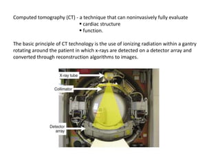
CT provides noninvasive evaluation of cardiac structure and function. It uses ionizing radiation and reconstruction algorithms to form images. Advancements include increasing detector rows for wider coverage and shorter scan times. CT can assess coronary calcium scoring, coronary angiography, bypass graft/stent patency, cardiac morphology/function, and pericardial diseases. It is useful for diagnosing chest pain, coronary anomalies, and evaluating surgical candidates. Risks include radiation exposure and contrast nephropathy, so appropriate patient selection is important.
A detailed description of ct coronary angiography and calcium scoring with various aspects regarding the preparation, procedure, limitations and a short review regarding post CABG imaging.
Basic physics of multidetector computed tomography ( CT Scan) - how ct scan works, different generations of ct, how image is generated and displayed and image artifacts related to CT Scan.
Image Quality, Artifacts and it's Remedies in CT-Avinesh ShresthaAvinesh Shrestha
CT is one of the frequently used diagnostic imaging modalities in Radiology. Knowledge about image quality and artifacts is essential when diagnosing a patient with the help of CT images. Moreover, Radiology Technologist's should be very well aware about the ways to identify and eliminate or minimize the artifacts in CT for better image quality.
CT TRIPLE PHASE SHOWING LIVER ANATOMY THREE PHASES OF EXAMINATION LIKE ARTERIAL PHASE,VENOUS PHASE DELAYED PHASE AND PORTAL PHASE CLEARLY AND LOBS OF LIVER EXPLAINED WELL.SOME TRIPLEPHASE EXPLAINING TECHNIQUES PROTOCOLS AND QUESIONEERS
Triphasic CT (TPCT) Scan of the liver is essential in view of the dual blood supply of the liver. TPCT allows characterisaiton of all liver lesions and close to pathological correlaiton by non invasive imaging alone. Additionally providing segmental vascular analysis as a surgicical guide.
Computed tomography angiography (CTA) of the coronary arteries is a useful noninvasive tool to rule out significant coronary artery disease (CAD) in many clinical situations. Recent guidelines of stable CAD and non-ST segment elevation myocardial infarction endorse the use of CTA in symptomatic patients with low to intermediate likelihood of the disease, given the particularly high negative predictive value of the technique. However, in patients with high pre-test likelihood of CAD, the technique is not recommended, and one of the reasons is the high probability of coronary calcification in these patients, which interferes with the analysis of the images and reduces the specificity and negative predictive value of CTA.
(TOSHIBA CTEU140095) - Article from Toshiba's VISIONS Magazine#25, March 2015
Usefulness of Non-Enhanced 3-Dementional CT with Partial Maximum Intensity Pr...science journals
Computed Tomography (CT) with contrast material is often used for preoperative assessment and planning of embolotherapy in the treatment of Pulmonary Arteriovenous Malformations (PAVMs).
Youssef Abdelwahed: Preprocedural CT – which patient needs itEuro CTO Club
14th Experts "Live" CTO
September 2nd- 3rd, 2022 - Mainz, Germany
Main Session - Session 2:
Selecting the patient and planning the procedure B
Preprocedural CT – which patient needs it
Youssef Abdelwahed, Berlin, Germany
Room:
Guteberg Hall (Auditorium) - Friday 11:10
Chairmen:
Alexander Bufe, Krefeld, Germany;
Leszek Bryniarski, Krakow, Poland;
Hans Bonnier, Nuenen, Belgium
What is New in Cardiac CT? In Search of the Comprehensive and Conclusive Hear...Apollo Hospitals
Coronary CT Angiography (CT) with its noninvasive cross sectional information has seen remarkable growth in recent years. With the introduction of the new generation scanners, like the 320-slice CT, it has risen to a whole new level. Percent diameter stenosis determined with the use of 320-slice CT shows good correlation with Invasive catheter angiogram (ICA) without significant underestimation or overestimation. Plaque composition on CT regardless of lesion severity has emerged as a strong predictor of major cardiac events. The percentage stenosis mismatch between CT and ICA can be explained by the 2 dimensional nature of ICA and its interpretive inconsistencies. In the upcoming years, we need to evolve from focusing on lumen stenosis to a comprehensive assessment of CAD and its impact on patient outcome.
Computed tomography (CT scan) is a medical imaging procedure that uses computer-processed X-rays to produce tomographic images or 'slices' of specific areas of the body. These cross-sectional images are used for diagnostic and therapeutic purposes in various medical disciplines.
Report Back from SGO 2024: What’s the Latest in Cervical Cancer?bkling
Are you curious about what’s new in cervical cancer research or unsure what the findings mean? Join Dr. Emily Ko, a gynecologic oncologist at Penn Medicine, to learn about the latest updates from the Society of Gynecologic Oncology (SGO) 2024 Annual Meeting on Women’s Cancer. Dr. Ko will discuss what the research presented at the conference means for you and answer your questions about the new developments.
These lecture slides, by Dr Sidra Arshad, offer a quick overview of physiological basis of a normal electrocardiogram.
Learning objectives:
1. Define an electrocardiogram (ECG) and electrocardiography
2. Describe how dipoles generated by the heart produce the waveforms of the ECG
3. Describe the components of a normal electrocardiogram of a typical bipolar leads (limb II)
4. Differentiate between intervals and segments
5. Enlist some common indications for obtaining an ECG
Study Resources:
1. Chapter 11, Guyton and Hall Textbook of Medical Physiology, 14th edition
2. Chapter 9, Human Physiology - From Cells to Systems, Lauralee Sherwood, 9th edition
3. Chapter 29, Ganong’s Review of Medical Physiology, 26th edition
4. Electrocardiogram, StatPearls - https://www.ncbi.nlm.nih.gov/books/NBK549803/
5. ECG in Medical Practice by ABM Abdullah, 4th edition
6. ECG Basics, http://www.nataliescasebook.com/tag/e-c-g-basics
Flu Vaccine Alert in Bangalore Karnatakaaddon Scans
As flu season approaches, health officials in Bangalore, Karnataka, are urging residents to get their flu vaccinations. The seasonal flu, while common, can lead to severe health complications, particularly for vulnerable populations such as young children, the elderly, and those with underlying health conditions.
Dr. Vidisha Kumari, a leading epidemiologist in Bangalore, emphasizes the importance of getting vaccinated. "The flu vaccine is our best defense against the influenza virus. It not only protects individuals but also helps prevent the spread of the virus in our communities," he says.
This year, the flu season is expected to coincide with a potential increase in other respiratory illnesses. The Karnataka Health Department has launched an awareness campaign highlighting the significance of flu vaccinations. They have set up multiple vaccination centers across Bangalore, making it convenient for residents to receive their shots.
To encourage widespread vaccination, the government is also collaborating with local schools, workplaces, and community centers to facilitate vaccination drives. Special attention is being given to ensuring that the vaccine is accessible to all, including marginalized communities who may have limited access to healthcare.
Residents are reminded that the flu vaccine is safe and effective. Common side effects are mild and may include soreness at the injection site, mild fever, or muscle aches. These side effects are generally short-lived and far less severe than the flu itself.
Healthcare providers are also stressing the importance of continuing COVID-19 precautions. Wearing masks, practicing good hand hygiene, and maintaining social distancing are still crucial, especially in crowded places.
Protect yourself and your loved ones by getting vaccinated. Together, we can help keep Bangalore healthy and safe this flu season. For more information on vaccination centers and schedules, residents can visit the Karnataka Health Department’s official website or follow their social media pages.
Stay informed, stay safe, and get your flu shot today!
TEST BANK for Operations Management, 14th Edition by William J. Stevenson, Ve...kevinkariuki227
TEST BANK for Operations Management, 14th Edition by William J. Stevenson, Verified Chapters 1 - 19, Complete Newest Version.pdf
HOT NEW PRODUCT! BIG SALES FAST SHIPPING NOW FROM CHINA!! EU KU DB BK substit...GL Anaacs
Contact us if you are interested:
Email / Skype : kefaya1771@gmail.com
Threema: PXHY5PDH
New BATCH Ku !!! MUCH IN DEMAND FAST SALE EVERY BATCH HAPPY GOOD EFFECT BIG BATCH !
Contact me on Threema or skype to start big business!!
Hot-sale products:
NEW HOT EUTYLONE WHITE CRYSTAL!!
5cl-adba precursor (semi finished )
5cl-adba raw materials
ADBB precursor (semi finished )
ADBB raw materials
APVP powder
5fadb/4f-adb
Jwh018 / Jwh210
Eutylone crystal
Protonitazene (hydrochloride) CAS: 119276-01-6
Flubrotizolam CAS: 57801-95-3
Metonitazene CAS: 14680-51-4
Payment terms: Western Union,MoneyGram,Bitcoin or USDT.
Deliver Time: Usually 7-15days
Shipping method: FedEx, TNT, DHL,UPS etc.Our deliveries are 100% safe, fast, reliable and discreet.
Samples will be sent for your evaluation!If you are interested in, please contact me, let's talk details.
We specializes in exporting high quality Research chemical, medical intermediate, Pharmaceutical chemicals and so on. Products are exported to USA, Canada, France, Korea, Japan,Russia, Southeast Asia and other countries.
Explore natural remedies for syphilis treatment in Singapore. Discover alternative therapies, herbal remedies, and lifestyle changes that may complement conventional treatments. Learn about holistic approaches to managing syphilis symptoms and supporting overall health.
Tom Selleck Health: A Comprehensive Look at the Iconic Actor’s Wellness Journeygreendigital
Tom Selleck, an enduring figure in Hollywood. has captivated audiences for decades with his rugged charm, iconic moustache. and memorable roles in television and film. From his breakout role as Thomas Magnum in Magnum P.I. to his current portrayal of Frank Reagan in Blue Bloods. Selleck's career has spanned over 50 years. But beyond his professional achievements. fans have often been curious about Tom Selleck Health. especially as he has aged in the public eye.
Follow us on: Pinterest
Introduction
Many have been interested in Tom Selleck health. not only because of his enduring presence on screen but also because of the challenges. and lifestyle choices he has faced and made over the years. This article delves into the various aspects of Tom Selleck health. exploring his fitness regimen, diet, mental health. and the challenges he has encountered as he ages. We'll look at how he maintains his well-being. the health issues he has faced, and his approach to ageing .
Early Life and Career
Childhood and Athletic Beginnings
Tom Selleck was born on January 29, 1945, in Detroit, Michigan, and grew up in Sherman Oaks, California. From an early age, he was involved in sports, particularly basketball. which played a significant role in his physical development. His athletic pursuits continued into college. where he attended the University of Southern California (USC) on a basketball scholarship. This early involvement in sports laid a strong foundation for his physical health and disciplined lifestyle.
Transition to Acting
Selleck's transition from an athlete to an actor came with its physical demands. His first significant role in "Magnum P.I." required him to perform various stunts and maintain a fit appearance. This role, which he played from 1980 to 1988. necessitated a rigorous fitness routine to meet the show's demands. setting the stage for his long-term commitment to health and wellness.
Fitness Regimen
Workout Routine
Tom Selleck health and fitness regimen has evolved. adapting to his changing roles and age. During his "Magnum, P.I." days. Selleck's workouts were intense and focused on building and maintaining muscle mass. His routine included weightlifting, cardiovascular exercises. and specific training for the stunts he performed on the show.
Selleck adjusted his fitness routine as he aged to suit his body's needs. Today, his workouts focus on maintaining flexibility, strength, and cardiovascular health. He incorporates low-impact exercises such as swimming, walking, and light weightlifting. This balanced approach helps him stay fit without putting undue strain on his joints and muscles.
Importance of Flexibility and Mobility
In recent years, Selleck has emphasized the importance of flexibility and mobility in his fitness regimen. Understanding the natural decline in muscle mass and joint flexibility with age. he includes stretching and yoga in his routine. These practices help prevent injuries, improve posture, and maintain mobilit
Prix Galien International 2024 Forum ProgramLevi Shapiro
June 20, 2024, Prix Galien International and Jerusalem Ethics Forum in ROME. Detailed agenda including panels:
- ADVANCES IN CARDIOLOGY: A NEW PARADIGM IS COMING
- WOMEN’S HEALTH: FERTILITY PRESERVATION
- WHAT’S NEW IN THE TREATMENT OF INFECTIOUS,
ONCOLOGICAL AND INFLAMMATORY SKIN DISEASES?
- ARTIFICIAL INTELLIGENCE AND ETHICS
- GENE THERAPY
- BEYOND BORDERS: GLOBAL INITIATIVES FOR DEMOCRATIZING LIFE SCIENCE TECHNOLOGIES AND PROMOTING ACCESS TO HEALTHCARE
- ETHICAL CHALLENGES IN LIFE SCIENCES
- Prix Galien International Awards Ceremony
Pulmonary Thromboembolism - etilogy, types, medical- Surgical and nursing man...VarunMahajani
Disruption of blood supply to lung alveoli due to blockage of one or more pulmonary blood vessels is called as Pulmonary thromboembolism. In this presentation we will discuss its causes, types and its management in depth.
The prostate is an exocrine gland of the male mammalian reproductive system
It is a walnut-sized gland that forms part of the male reproductive system and is located in front of the rectum and just below the urinary bladder
Function is to store and secrete a clear, slightly alkaline fluid that constitutes 10-30% of the volume of the seminal fluid that along with the spermatozoa, constitutes semen
A healthy human prostate measures (4cm-vertical, by 3cm-horizontal, 2cm ant-post ).
It surrounds the urethra just below the urinary bladder. It has anterior, median, posterior and two lateral lobes
It’s work is regulated by androgens which are responsible for male sex characteristics
Generalised disease of the prostate due to hormonal derangement which leads to non malignant enlargement of the gland (increase in the number of epithelial cells and stromal tissue)to cause compression of the urethra leading to symptoms (LUTS
Ethanol (CH3CH2OH), or beverage alcohol, is a two-carbon alcohol
that is rapidly distributed in the body and brain. Ethanol alters many
neurochemical systems and has rewarding and addictive properties. It
is the oldest recreational drug and likely contributes to more morbidity,
mortality, and public health costs than all illicit drugs combined. The
5th edition of the Diagnostic and Statistical Manual of Mental Disorders
(DSM-5) integrates alcohol abuse and alcohol dependence into a single
disorder called alcohol use disorder (AUD), with mild, moderate,
and severe subclassifications (American Psychiatric Association, 2013).
In the DSM-5, all types of substance abuse and dependence have been
combined into a single substance use disorder (SUD) on a continuum
from mild to severe. A diagnosis of AUD requires that at least two of
the 11 DSM-5 behaviors be present within a 12-month period (mild
AUD: 2–3 criteria; moderate AUD: 4–5 criteria; severe AUD: 6–11 criteria).
The four main behavioral effects of AUD are impaired control over
drinking, negative social consequences, risky use, and altered physiological
effects (tolerance, withdrawal). This chapter presents an overview
of the prevalence and harmful consequences of AUD in the U.S.,
the systemic nature of the disease, neurocircuitry and stages of AUD,
comorbidities, fetal alcohol spectrum disorders, genetic risk factors, and
pharmacotherapies for AUD.
The POPPY STUDY (Preconception to post-partum cardiovascular function in prim...
Ct angio ppt
1. Computed tomography (CT) - a technique that can noninvasively fully evaluate
cardiac structure
function.
The basic principle of CT technology is the use of ionizing radiation within a gantry
rotating around the patient in which x-rays are detected on a detector array and
converted through reconstruction algorithms to images.
2. Types of CT
1. EBCT (Electron beam CT)
2. MDCT ( Multidetector CT)
The most notable technical advance is progressive increase in the number of detector
rows (or slices). Each row is a narrow channel, approximately 0.625 mm in width,
through which x-rays are detected on scintillation crystals. The number of detector
rows aligned in an array has increased from a single detector to 4, 16, and 64
(present standard technology) and now on to “wide” detectors of 256 to 320 rows.
3. The increase in the number of rows leads to wider coverage (more of the heart
viewed simultaneously, e.g., 64 rows of 0.625-mm width produces
approximately 4 cm of coverage, leading to shorter scan acquisition times and
consequently reduced radiation exposure and contrast requirements.
4. EBCT :
EBCT uses an electron beam (current 630 mA, voltage 130 kV) that is deflected
via a magnetic coil and focused to strike a series of four tungsten targets
located beneath the patient. The electron beam is magnetically swept along
the tungsten targets at a 210-degree arc. EBCT has no moving parts.
5. MDCT :
Multidetector CT (MDCT) scanners produce images by rotating an x-ray tube
around a circular gantry through which the patient advances on a moving table.
6. Scan Modes
There are two basic scan modes in cardiac CT, helical (spiral) and axial (sequential, step
& shoot) scanning.
Helical (spiral) scanning : Most current MDCT scanners use spiral, retrospectively gated
acquisition techniques. Helical scanning involves continuous radiation exposure and
table movement (the patient is moved through the rotating x-ray beam), during which
the detector arrays receive projection data from multiple contiguous slices of the
patient.
Axial (sequential, step & shoot) scanning : axial imaging involves sequential scanner
“snapshots,” in between which the x-ray tube is turned off and the table is moved to a
different position for the next image to be acquired.
7.
8. ECG gating
a. Prospective triggering: The trigger signal is derived from the patient’s ECG based
on a prospective estimation of the RR interval. The scan is usually triggered to
begin at a defined point after the R wave, usually allowing image acquisition to
occur during diastole.
Advantage: ● dose efficient (80% reduction in x-ray exposure)
Disadvantage : ● limited portion of cardiac cycle data set obtained
● greatly depends on the regularity of patient’s heart rate
9. b. Retrospective gating : Collects data during the entire cardiac cycle. Once scan is
complete , data from specific periods of the cardiac cycle are used for image
reconstruction by retrospective referencing to the ECG signal.
Advantage : allows assessment of cardiac function via four-dimensional
reconstruction.
Disadvantage : higher radiation dose exposure.
10. Contrast enhanced imaging :
Administration of iodinated contrast media increases the attenuation of the blood
pool, improving vessel delineation and tissue characterization. When using contrast,
image acquisition must be timed such that images are acquired when the blood pool
enhancement in the target structure is maximal.
Various techniques exist to time the arrival of the contrast bolus in the arterial
tree and initiate imaging.
11. Indications:
A. Evaluation of chest pain in patients at low to intermediate pretest probability of
disease and persistent chest pain after an equivocal stress test.
B. Suspicion of coronary artery anomalies. MDCT has very high sensitivity and
specificity for coronary anomalies.
C. Pulmonary vein evaluation can be performed, often before or after pulmonary vein
isolation for atrial fibrillation.
D. Evaluation of cardiac masses when other modalities such as TTE, TEE, or MRI are
unrevealing.
E. Evaluation of pericardial disease when other modalities such as TTE, TEE, or MRI
are unrevealing.
F. Assessment of anatomy in complex congenital heart disease.
12. G. Presurgical evaluation, particularly before redo open heart surgery. MDCT can aid in
describing prior bypass graft location, identifying safe sites for surgical approach.
H. Assessing graft patency after prior bypass surgery is feasible in many cases, though
sometimes limited by artifacts related to calcium and surgical clips.
I. Evaluation of aortic disease. MDCT is the test of choice for evaluating aortic
aneurysm and suspected aortic dissection.
J. Evaluation of suspected pulmonary embolism
13. CONTRAINDICATIONS:
Unlike with cardiac MRI, few absolute contra indications exist for cardiac CT. However,
there are important risks associated with radiation and/or contrast exposure that must be
weighed against the benefits of the scan.
Absolute contraindications :
A. Renal insufficiency. Given the potential for contrast nephropathy, patients with
significant renal insufficiency (i.e., Cr > 1.6 mg/dL) should not undergo contrast-enhanced
CT unless the information from the scan is critical and the risks/benefits
are thoroughly discussed with the patient.
B. Known history of anaphylactic contrast reactions A prior anaphylactic response to
contrast is generally felt to be an absolute contraindication to intravenous iodinated
contrast administration at many institutions.
C. Pregnancy
D. Clinical instability
14. Relative contraindications
A. Contrast (iodine) allergy. Patients with allergic reactions to contrast should be
pretreated with diphenhydramine and steroids before contrast administration.
B. Recent intravenous iodinated contrast administration. Patients who have
received an intravenous dose of iodinated contrast should avoid contrast-enhanced
CT scanning for 24 hours to reduce the risk of contrast nephropathy.
C. Hyperthyroidism. Iodinated contrast is contraindicated in the setting of
uncontrolled hyperthyroidism due to possible precipitation of thyrotoxicosis.
D. Atrial fibrillation or any irregular heart rhythm, is a contraindication to coronary
CT angiography due to image degradation from suboptimal ECG gating.
E. Inability to breath hold for at least 10 seconds. Image quality will be significantly
reduced due to respiratory motion artifact if the patient cannot comply with breath
hold instructions.
F. Morbid obesity
G. Severe coronary calcium
15. SAFETY
A. Radiation exposure : Radiation doses of cardiac CT scans vary greatly depending
on the scan parameter settings, scan range (cranial-caudal length of the scan),
gender (women receive more radiation due to breast tissue), and patient body
habitus (obesity increases exposure).
● chest x-ray is 0.04 to 0.10 mSv,
● average annual background radiation 3 to 3.6 mSv.
● Invasive diagnostic coronary angiography 2.1 to 4 mSv.
● coronary CT angiography 4 to 11 mSv. With use of prospective-ECG triggering,
axial imaging modes, and software adaptations, recent studies have reported the
feasibility of CT coronary angiography with comparable image quality and
substantially reduced radiation doses (i.e., 1.1 to 3.0 mSv). This remains an area of
active investigation.
16. B. Contrast nephropathy : Iodinated contrast media can cause renal ischemia by
reducing renal blood flow or increasing oxygen demand and may also have a direct
toxic effect on tubular epithelial cells. If a contrast-enhanced CT study is necessary in
patients with significant renal insufficiency, prophylactic measures should be taken
● saline hydration
● n-acetylcysteine
● use of low osmolar agents
● sodium bicarbonate infusion
17.
18. CLINICAL APPLICATIONS
A. Coronary calcium scoring
Coronary calcium is a surrogate marker for coronary atherosclerotic plaque. Coronary
artery calcium score is directly proportional to the overall extent of atherosclerosis,
although typically only a minority (approximately 20%) of plaque is calcified.
Complete absence of coronary artery calcium makes the presence of significant
coronary luminal obstruction highly unlikely and indicates a very low risk of future
coronary events.
Men, CKD, diabetics tend to have higher coronary calcium scores.
Contrast is not necessary because calcium is readily identified secondary to its very
high x-ray attenuation coefficient (high Hounsfield unit score).
19.
20. The Agatston coronary artery calcium (CAC) score is the most frequently
used scoring system. It is derived by measuring the area of each calcified coronary
lesion and multiplying it by a coefficient of 1 to 4, depending on the maximum
CT attenuation within that lesion.
Volume score
Mass score
A coronary calcium coverage score : multivessel coronary calcium, the number of
calcified lesions and diffuse spotty pattern (small foci <3 mm) are associated with a
higher clinical risk.
The CAC score can be classified into five groups:
1) zero, no coronary calcification;
2) 100, mild coronary calcification;
3) > 100 to 399, moderate calcification;
4) >400 to 999, severe calcification;
5) > 1000, extensive calcification.
21. In comparison with a CAC score of zero, the presence of any CAC is associated with a
fourfold risk of coronary events over 3 to 5 years.
In patients at intermediate clinical risk for coronary events (e.g., by Framingham
score), the CAC score can help to reclassify patients to a higher or lower risk group. For
instance, a CAC score of zero confirms low risk of events. Conversely, a CAC score of
greater than 400 is observed with a significant cardiac event rate (greater than 2 %/year)
in patients who appear to be intermediate risk by Framingham score.
Because statins have no documented effect on CAC progression, there is no value in
repeating CAC in persons with a score of greater than 100 or the 75th percentile.
However, not every atherosclerotic plaque is calcified, and even the detection of
a large amount of calcium does not imply the presence of significant stenoses.
Therefore, it adds only incrementally to traditional risk assessment and should
not be used in isolation. The test is most useful in intermediate risk populations,
in which a high or low score may reclassify individuals to a higher or lower risk
group. Unselected screening is not recommended.
22. CAC and Stenosis Severity : Significant coronary artery stenosis (>50%) by angiography is
frequently associated with the presence of coronary artery calcium. However, the
severity of angiographic coronary artery stenosis is not directly related to the total CAC.
CAC and Myocardial Ischemia : Good correlation between CT and myocardial perfusion
SPECT for identifying both subclinical CAD and silent myocardial ischemia in a generally
asymptomatic population who had risk factors for CAD development. Few patients with
CACS <400 had a perfusion defect, whereas nearly half of the patients with CACS >400
had an abnormal SPECT.
23. Coronary CT angiography : The primary clinical application of cardiac CT is the
performance of noninvasive coronary CT angiography among patients with symptoms
suggestive of myocardial ischemia. The overall accuracy of 64-row CT angiography
included a sensitivity of 87% to 99% and specificity of 93% to 96%.
Coronary CT angiography for evaluating CAD is most useful in low- to intermediate-risk
patients with angina or anginal equivalent. The negative predictive value of coronary CT
angiography is uniformly high in studies, approaching 93% to 100%; in other words,
coronary CT angiography is an excellent modality for ruling out coronary disease.
24.
25. Patients who are generally poor candidates for coronary CT angiography include
1. those likely to have heavily calcified coronary arteries (older than 75, endstage
renal disease, Paget's disease), atrial fibrillation /flutter, frequent ventricular
ectopic beats, or uncontrolled tachycardia.
2. Known severe CAD is generally a contraindication to coronary CT angiography.
However, cardiac CT has been shown to have high sensitivity and specificity
for assessment of bypass graft patency in patients with previous coronary artery
bypass grafting (CABG)
3. Patients with prior coronary artery stents are generally poor candidates for CAD,
although selected patients with proximal LAD or left main stents may be
successfully imaged. Current CT technology does not allow for the accurate
quantification of in-stent stenosis severity.
26. Noncalcified plaque appears as a low to intermediate attenuation irregularity in the
vessel wall. Calcified plaques are bright, high-attenuation lesions in the vessel wall and
may be associated with positive remodeling of the vessel. Densely calcified plaques are
often associated with calcium blooming artifact, which can lead to overestimation of
luminal stenosis severity.
The accuracy of coronary CT angiography is highest in the larger proximal to
medium vessels, which are more likely to benefit from an invasive management
strategy. Coronary stenoses are generally categorized as mild (less than 50%
diameter stenosis), moderate (50% t070% stenosis), or severe (greater than 70%
stenosis). Similar to results with invasive coronary angiography, the determination of an
anatomic stenosis is only modestly predictive of inducible ischemia. A 50% or greater
stenosis on cardiac CT has a 30% to 50% likelihood of demonstrable ischemia on
myocardial perfusion imaging .
27. Detection of Noncalcified Plaque
Defined as any coronary arterial wall lesion with an x-ray attenuation detectably below
the iodine contrast medium but higher than surrounding tissue.
Such plaque is difficult to quantify, with limited accuracy and reproducibility. Detection
requires maximal spatial and temporal resolution and minimized image noise with
higher radiation exposures. Compared with intravascular ultrasound, the sensitivity of
coronary MDCT is approximately 80%.
Detection of vulnerable plaques
Plaque features proposed to be associated with greater risk for plaque rupture or acute
coronary syndromes include ●low-attenuation plaque (plaque <30 HU), ●outward
arterial remodeling (artery diameter ratio of the involved segment to a proximal
reference of 1.1 or greater), and ●a spotty pattern (<3 mm in size) of calcification.
In particular, both low-attenuation plaque and outward arterial remodeling have been
associated with increased risk of acute coronary events.
A threefold worse cardiovascular prognosis has been found in the setting of a greater
number of coronary vessels and of coronary artery segments involved with plaque.
28. Bypass graft imaging :
1. Graft location : MDCT can accurately characterize the origin, course, and touchdown
of prior bypass grafts
2. Graft patency : Using a protocol similar to that used for coronary artery assessment
(less than 1 mm slice thickness), patency of both arterial and venous bypass
grafts can be assessed. Recent studies have suggested that the sensitivity and
specificity of MDCT for detecting stenosis or occlusion of bypass grafts, when
compared with invasive angiography, is 97% and 97%, respectively. Occasionally,
artifacts related to metallic clips can interfere with assessment of the distal
anastomosis of an arterial graft (internal mammary or radial artery graft).
29.
30. Stent patency:
Image artifact from metallic stents limits the application in patients with prior
coronary stent procedures. Small stents are difficult to evaluate and prone to
noninterpretability. However, 90% accuracy can be obtained in stents 3 mm or greater
in diameter with the use of sharp kernel and wide display window. Quantitative
assessment of within-stent contrast density may assist in the diagnosis.
31. Coronary artery anomalies :
MDCT is an excellent modality for assessing patients with known or suspected coronary
artery anomalies. MDCT can accurately assess the origin and course of anomalous
coronaries, and can describe the relationship of the coronary artery to neighboring
structures. Although MRI can also be used to assess anomalous coronaries without
the need for radiation exposure, the spatial resolution, ease of data acquisition, and
reliable image quality of MDCT make it a reasonable first choice. Intramyocardial
bridging can also be detected with high sensitivity.
32. Cardiac morphology/function :
Contrast-enhanced MDCT can provide high resolution morphologic images of the
cardiac chambers as well as accurate assessment of right and left ventricular systolic
function. However, other imaging modalities such as echocardiography or MRI, which
do not require radiation exposure, are generally preferred initially for assessing cardiac
morphology.
1. Patients with prior myocardial infarction can have fibrous replacement of
myocardium with or without calcification, ventricular wall thinning, aneurysm
formation and cavitary thrombus.
2. Ventricular dysplasia is characterized by: fibrous and/or fatty replacement of
myocardium, ventricular wall thinning and/or focal aneurysm formation, and
ventricular cavity dilation with regional or global wall motion abnormalities.
3. Mass : CT provides somewhat less information about tissue type than cardiac
MRI.
33.
34. Pericardial diseases :
The pericardium appears as a thin line (1 to 2 mm) surrounding the heart, usually
visible with a small amount of adjacent pericardiaI fat. The pericardium normally
enhances with contrast administration; hyperenhancement of the pericardium in the
appropriate clinical setting is characteristic of pericarditis.
1. By CT, congenital absence of the pericardium is easily diagnosed.
2. Findings of pericardial constriction on CT include irregular pericardiaI thickening
and calcification, conical or tubular compression of one or both ventricles,
enlargement of one or both atria, dilation of the IVC, and a characteristic diastolic
bounce of the interventricular septum.
3. Pericardial effusions can be reliably detected by CT. Pericardial tamponade is
better evaluated by echocardiography, however, due to its ability to provide
hemodynamic information.
4. A pericardiaI cyst will appear as a well circumscribed paracardiac mass with
characteristic water attenuation (H.U. = 0), usually in the right costophrenic
angle.
5. Both primary neoplasms and, more commonly, metastatic neoplasms can be
visualized in the pericardium.
35.
36. Congenital heart disease :
MDCT may be used in selected patients in whom echocardiography is non-diagnostic
or inadequate and MRI is not available.
The ability to evaluate cardiovascular anatomy in multiple planes is often helpful for
delineating cardiac morphology in congenital heart disease, particularly with regard
to the relationship of the great vessels, pulmonary veins, and coronary arteries.
Specific situations in which MDCT is helpful include
1. "hard-to-find" adult shunt detection (sinus venosus atrial septal defect, patent
ductus arteriosus);
2. visualization of pulmonary arteries in cyanotic congenital heart disease;
3. precise definition of aortic anatomy in Marfan's syndrome or coarctation;
4. definition of partial or total anomalous pulmonary venous drainage.
37. Diseases of the aorta constitute a common and important indication for CT
examinations.
Contrast-enhanced MDCT is nearly 100% sensitive and specific for
evaluating acute aortic syndromes.
1. Acute aortic dissection is characterized on CT by visualization of a dissection flap
(i.e., separation of the intima from the media) that forms true and false lumens. The
CT study can characterize the origin and extent of the dissection, classify it as Type A
or B, assess for concomitant aneurysmal aortic dilatation, and identify branch vessels
involvement.
2. Aortic intramural hematomas are believed to be caused by spontaneous
hemorrhage of the vaso vasorum into the medial layer. They appear as crescent-
shaped areas of increased attenuation with eccentric aortic wall thickening. Unlike
dissections, hematomas do not spiral around the aorta.
3. Aortic aneurysm is a permanent dilation of 150% of the normal aortic caliber
(usually greater than 5 cm in the thoracic aorta and greater than 3 cm in the
abdominal aorta).
4. Penetrating atherosclerotic ulcer. These tend to be focal lesions of the descending
thoracic aorta that appear as contrast-filled irregular outpouchings of the aortic wall.
38.
39.
40. Evaluation of pulmonary veins :
In the context of electrophysiology interventions such as pulmonary vein isolation (PVI),
preprocedural MDCT can be used to define pulmonary venous anatomy and identify
supernumerary veins, and postprocedural MDCT can be used to evaluate for pulmonary
vein stenosis. Additionally, in the setting of congenital heart disease, CT can be used to
identify anomalous pulmonary venous return.
41. Evaluation of pulmonary embolism :
MDCT is highly accurate in detecting PE, which appear as a filling defect in the
pulmonary arteries. This modality is most sensitive for proximal (main segmental
branches) thrombi, and small, distal emboli may be missed.
42. Valvular heart disease :
Visualization of the valve leaflets, particularly the aortic valve, is feasible with newer-
generation scanners due to their improved temporal resolution. Nonenhanced
MDCT is also useful for assessing prosthetic mechanical valve leaflet motion.
43. Surgical planning :
The utility of MDCT in surgical planning before cardiothoracic surgery, particularly for
reoperations, is increasingly recognized. Preoperative scans can evaluate the
proximity of mediastinal structures to the sternum (i.e., aorta, right ventricle, bypass
grafts); the degree of aortic calcification (i.e., to guide cannulation sites); and
concomitantly provide information about cardiac morphology (e.g., presence of a
ventricular aneurysm). Ongoing studies are evaluating whether this added
information might reduce intraoperative and perioperative complications.
44. Peripheral arteries :
MDCT can also be used to evaluate peripheral arteries, including the carotid, renal,
visceral, and lower-extremity vessels. Indeed, imaging these vessels is generally more
straightforward than coronary imaging, due to their large caliber and minimal motion.
CT can be used for planning and follow-up of vascular disease in these peripheral
vascular beds.
45. Emerging Applications
Detection of myocardial scar and viability
Features suggestive of prior myocardial infarction and nonviable myocardium
On first-pass imaging : ●Myocardial hypoattenuation (<50% of the surrounding
myocardium) and, ●Myocardial thickness <5 mm.
Late myocardial enhancement imaging with infusion of additional contrast medium and a
delay of approximately 10 minutes. The kinetics of iodinated contrast material is similar to
that of gadolinium, with accumulation within the interstitial space of myocardial fibrosis.
Under delayed imaging, contrast preferentially accumulates within areas of scarring and
can be detected on delayed imaging. Delayed enhancement on cardiac CT indicates
regions of myocardium with reduced likelihood of functional recovery and patients whose
ejection fraction will remain lower after myocardial infarction, particularly when a
transmural pattern of delayed enhancement is present.
46.
47. 2010 ACCF/AHA Guidelines for assessment of cardiovascular risk in asymptomatic
adults
Recommendations for Calcium Scoring Methods
Class IIa
Measurement of CAC is reasonable for cardiovascular risk assessment in asymptomatic
adults at intermediate risk (10% to 20% 10-year risk) (Level of Evidence: B)
Class IIb
Measurement of CAC may be reasonable for cardiovascular risk assessment in persons
at low to intermediate risk (6% to 10% 10-year risk) (Level of Evidence: B)
Class III: No Benefit
Persons at low risk (<6% 10-year risk) should not undergo CAC measurement for
cardiovascular risk assessment (Level of Evidence: B)
Recommendations for Coronary CT Angiography
Class III: No Benefit
Coronary computed tomography angiography is not recommended for cardiovascular
risk assessment in asymptomatic adults (Level of Evidence: C)