This document discusses various aspects of visual field defects and ocular reflexes. It describes hemianopia, which is the loss of vision in half of the visual field, and its two types: homonymous and heteronymous. It also explains the pupillary light reflex, including the direct and consensual responses when light enters the eye. The document outlines the six extraocular muscles that control eye movement and their functions. It discusses accommodation, convergence, and pupillary constriction as the three components of near response. Various methods for testing visual acuity and mapping the visual field are also summarized.
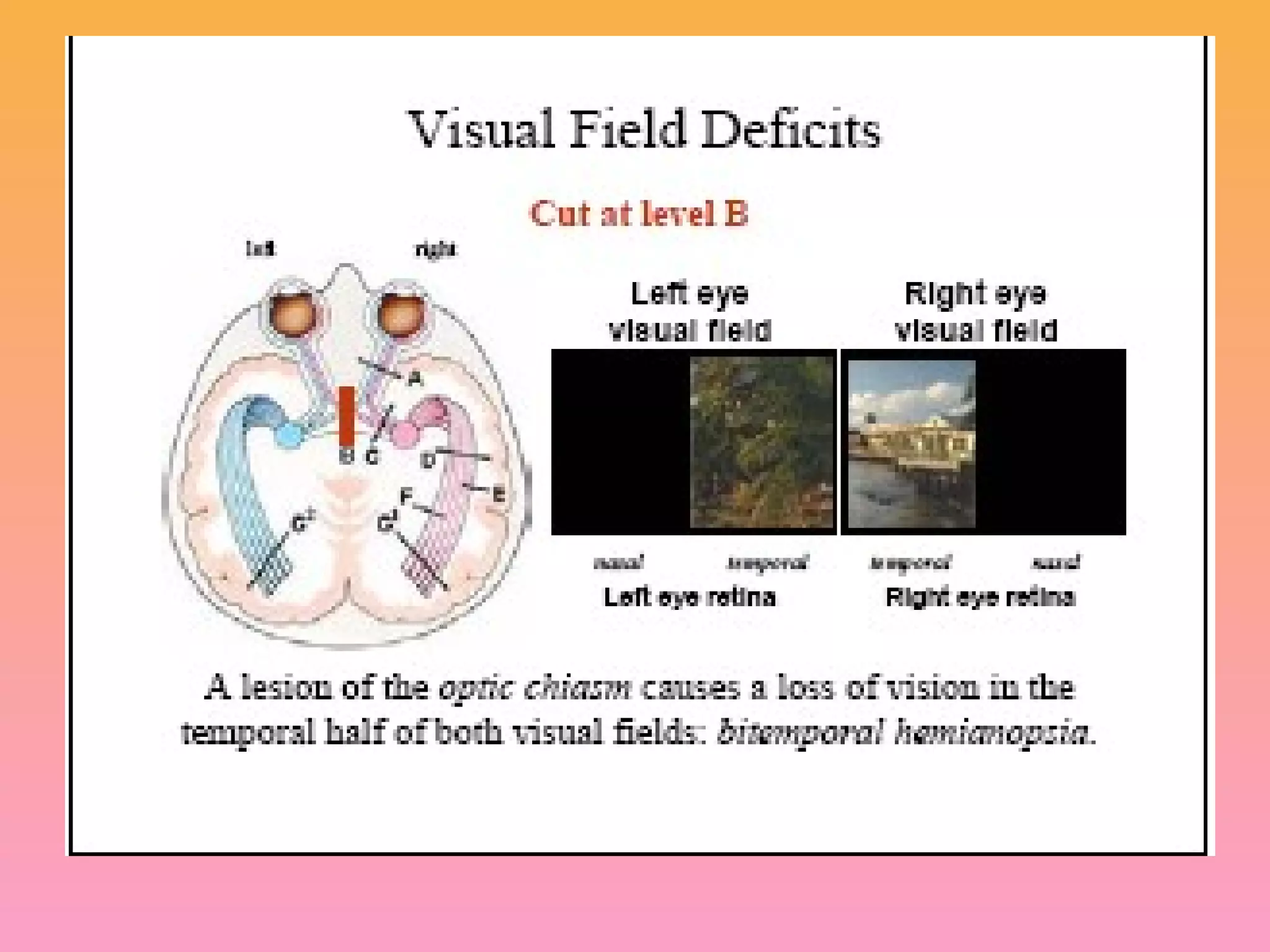
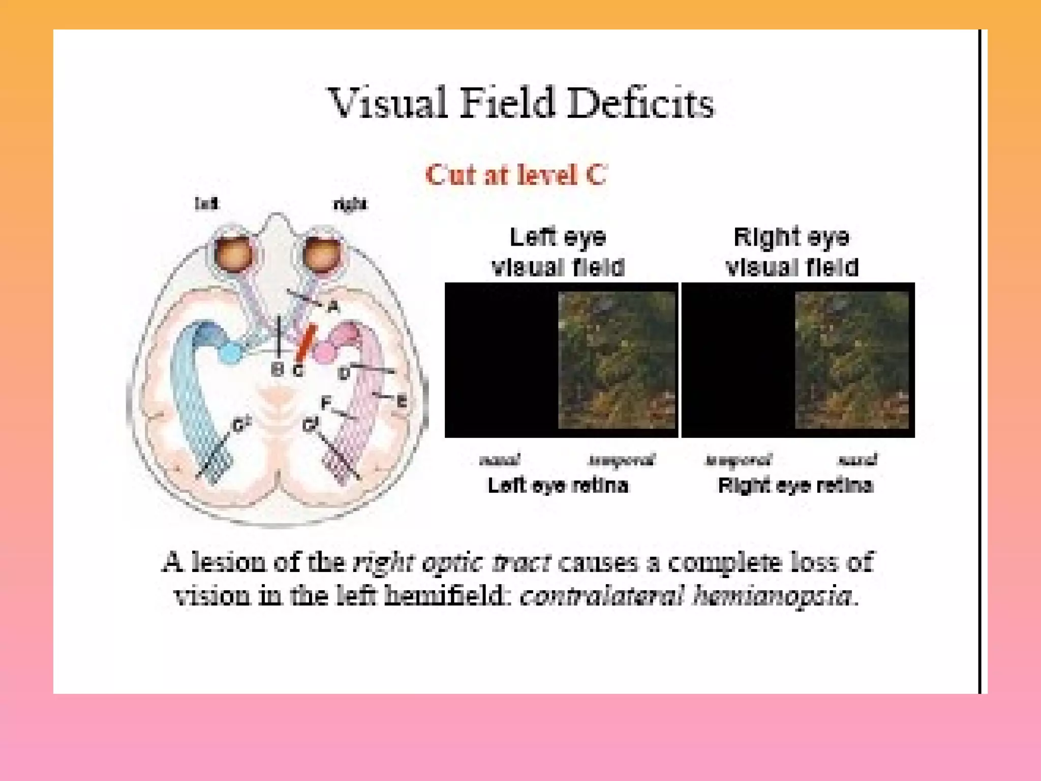
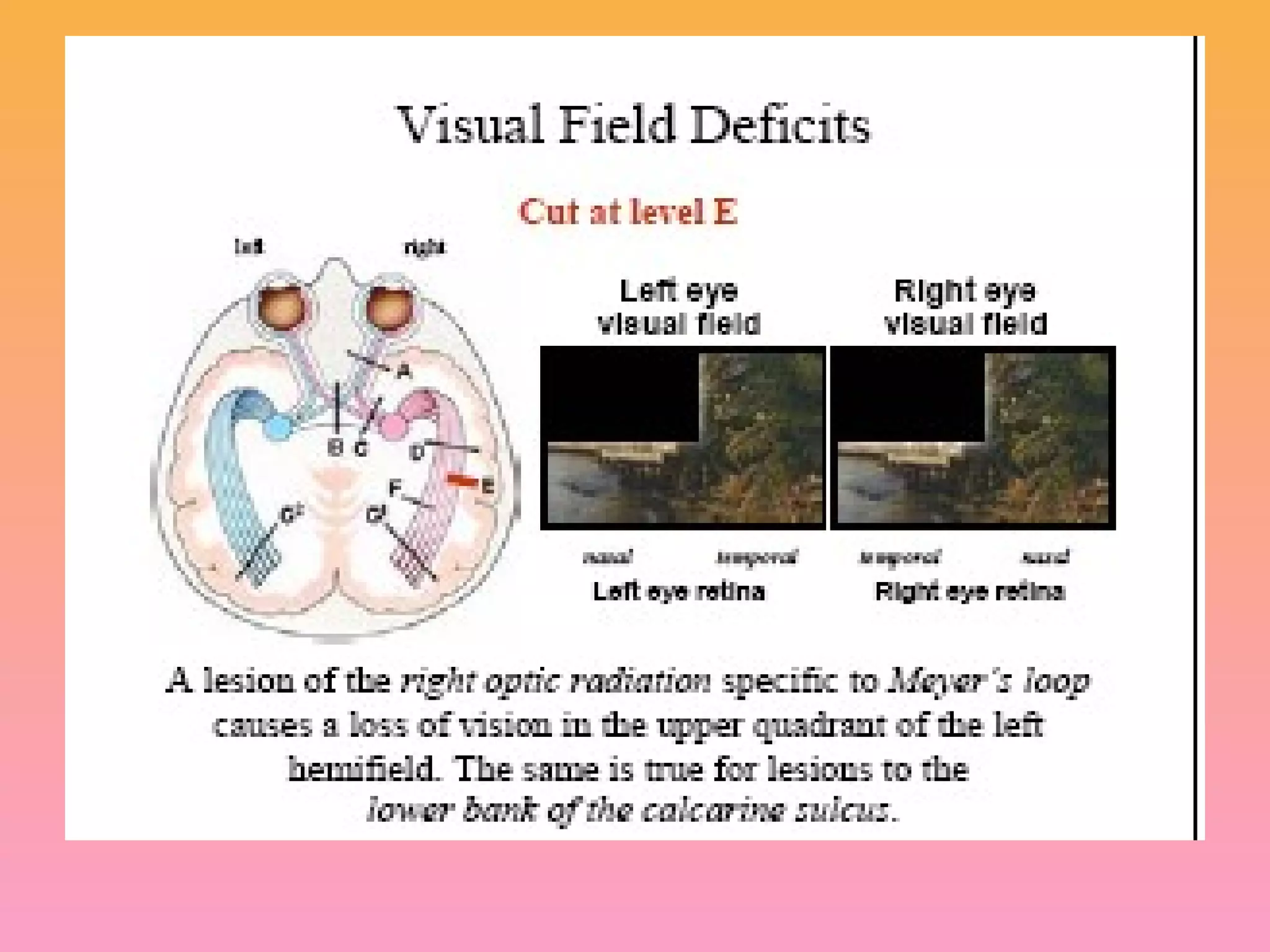
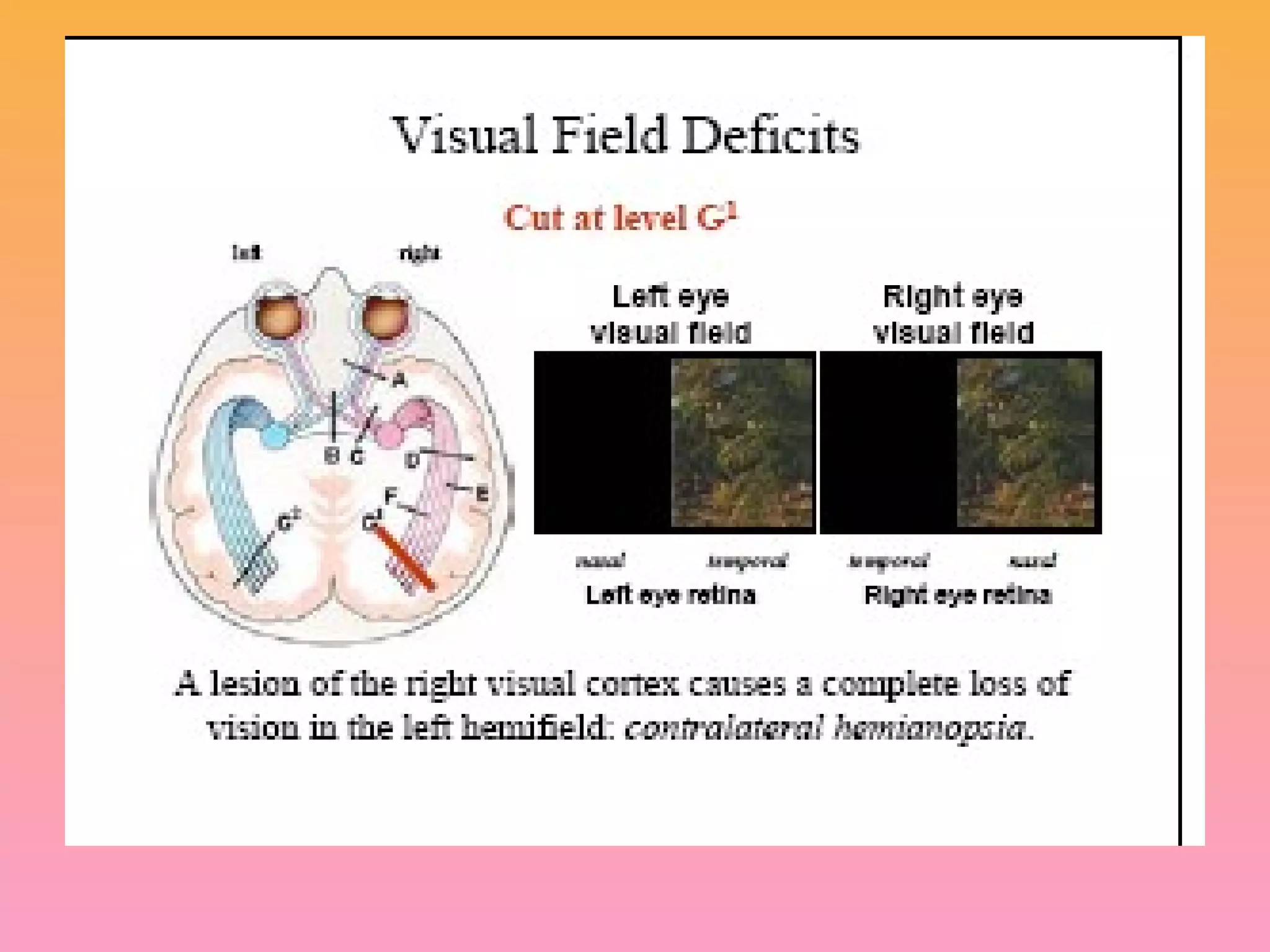
Introduction to visual field defects and types: hemianopia (homonymous & heteronymous).
Explanation of the pupillary light reflex: direct and consensual responses to light changes.
Explanation of the pupillary light reflex: direct and consensual responses to light changes.
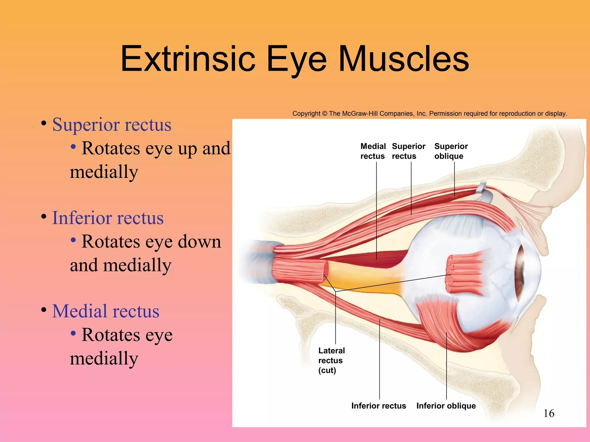
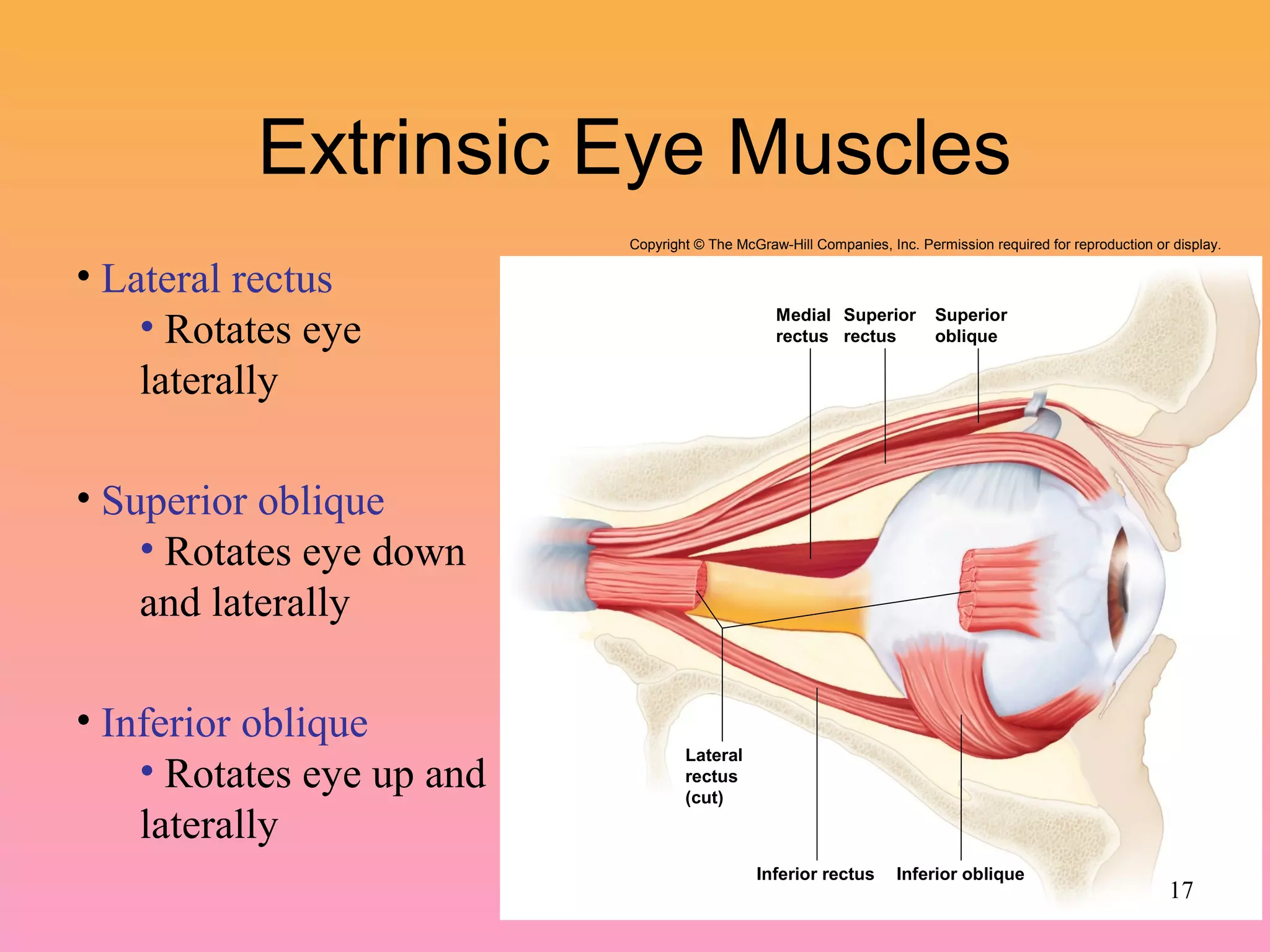
Details of extrinsic eye muscles: functions of rectus (superior, inferior, medial, lateral) and oblique muscles.Accommodation: ability to focus on varying distances, involving constriction, lens thickening, and convergence.
Definition and testing of visual acuity using Snellen charts; 20/20 or 6/6 vision is ideal.
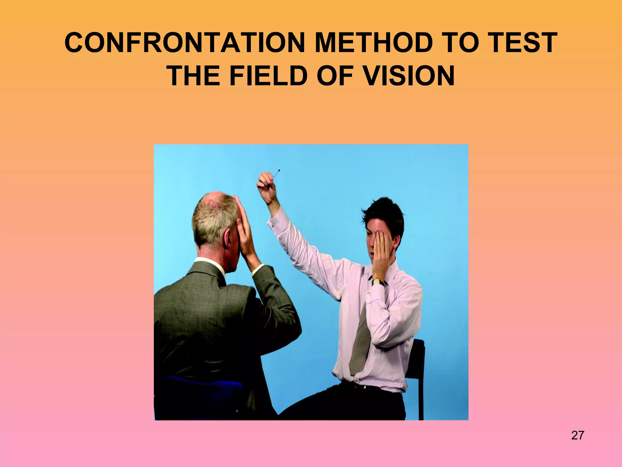
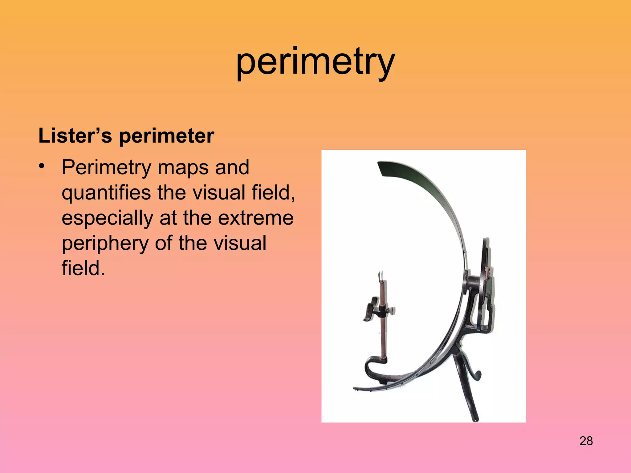
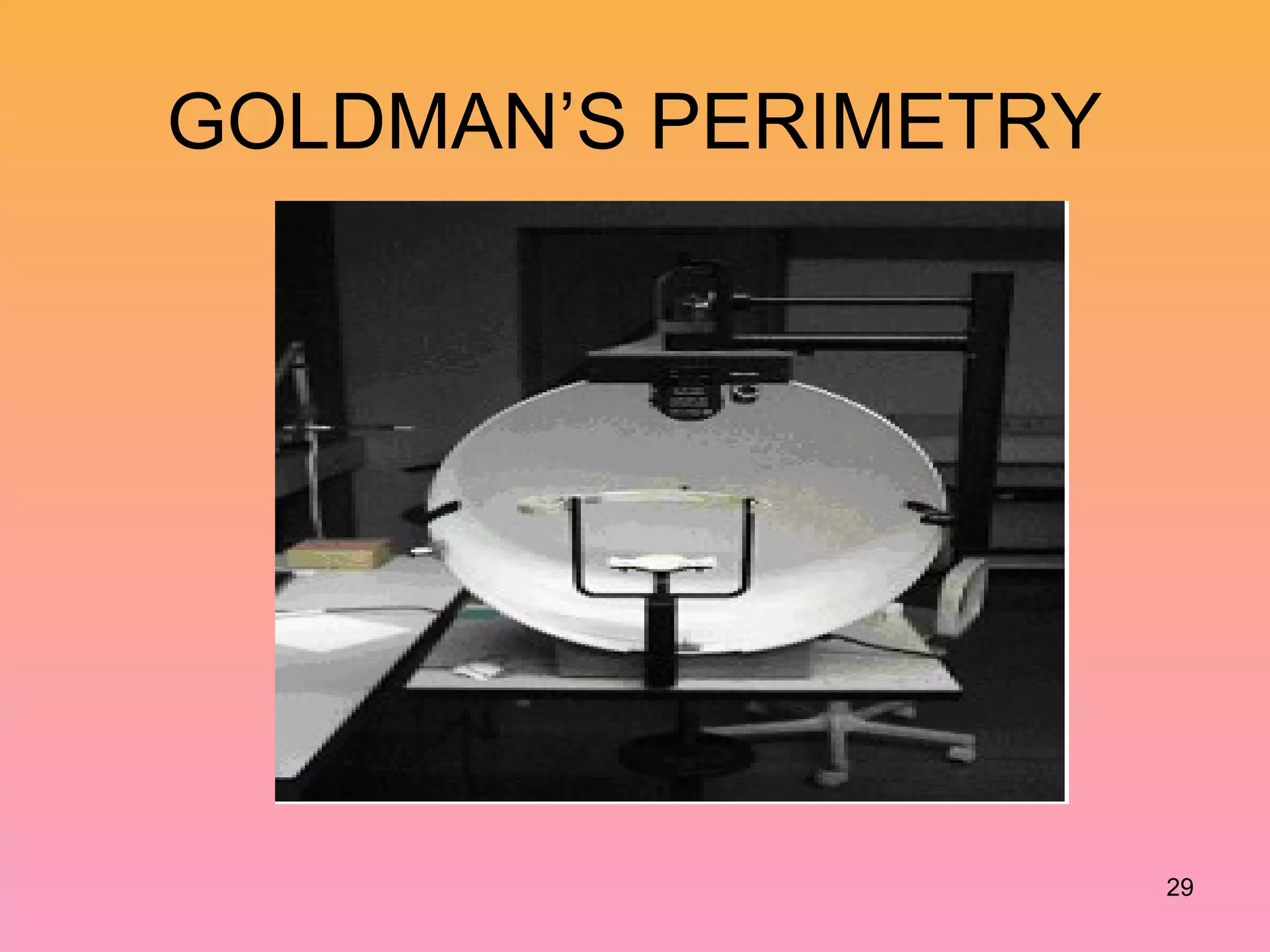
Methods for testing visual fields, including perimetry, and discussion of the physiologic blind spot.
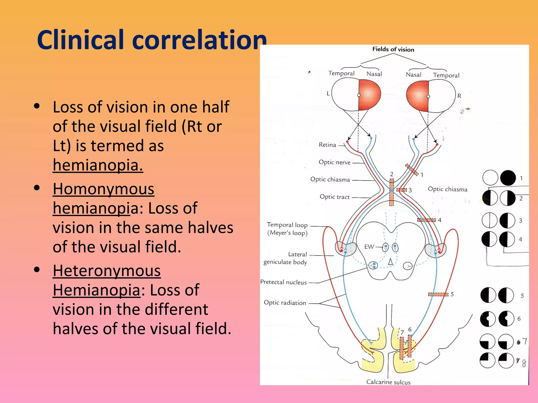
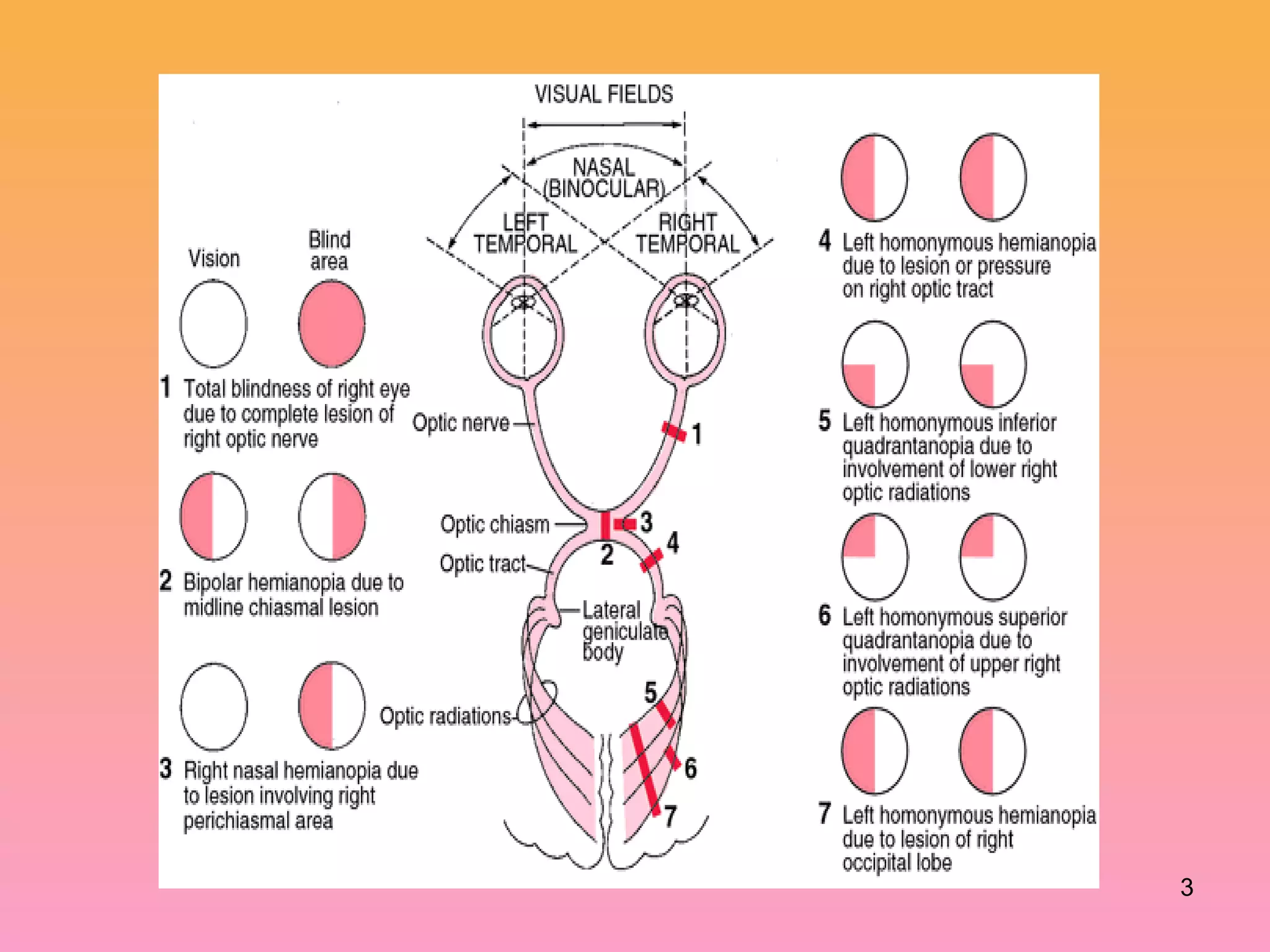
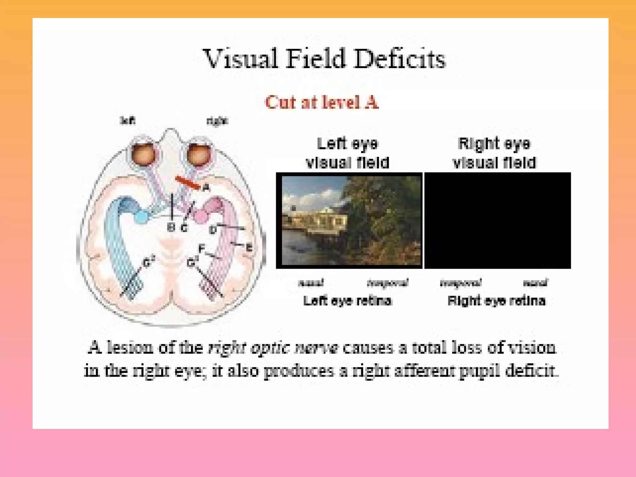
Clinical correlation
• Lossof vision in one half
of the visual field (Rt or
Lt) is termed as
hemianopia.
• Homonymous
hemianopia: Loss of
vision in the same halves
of the visual field.
• Heteronymous
Hemianopia: Loss of
vision in the different
halves of the visual field.
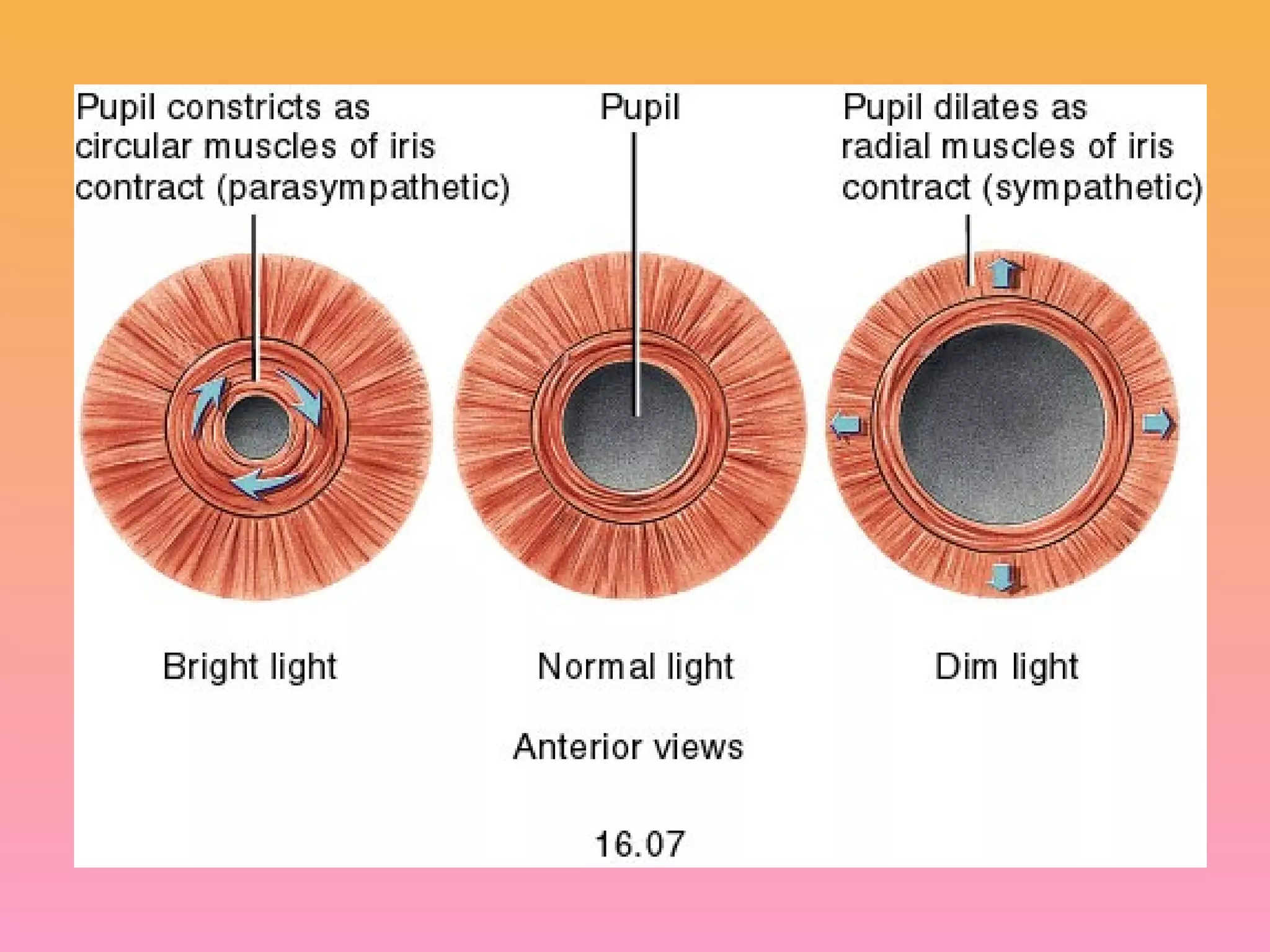
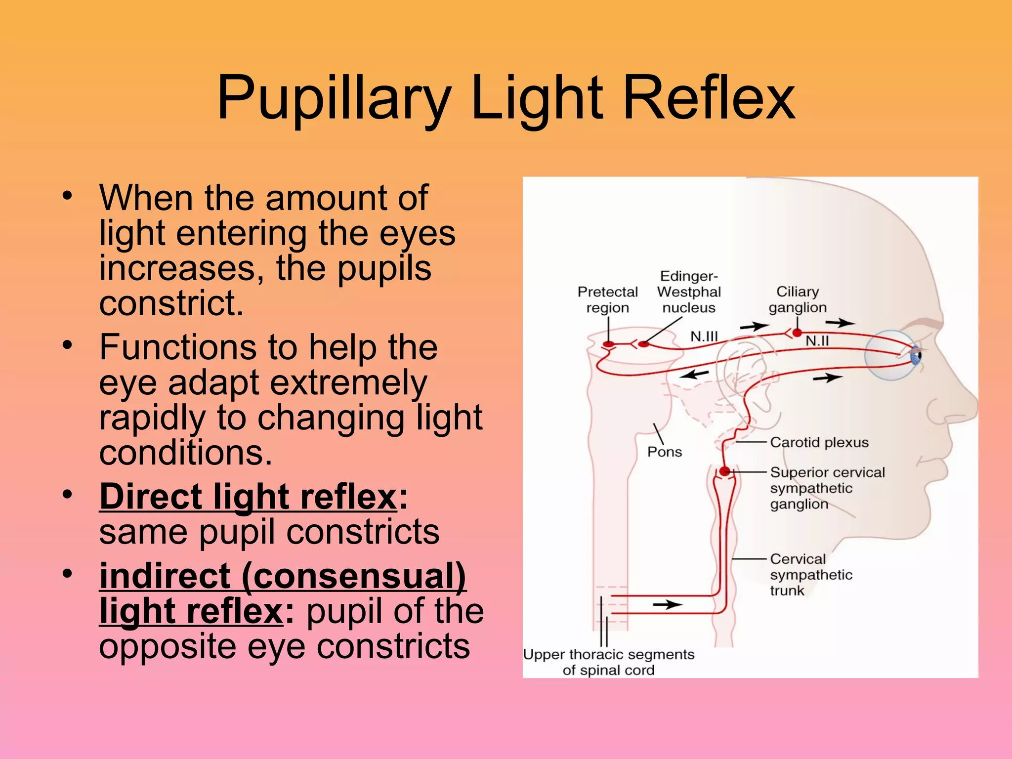
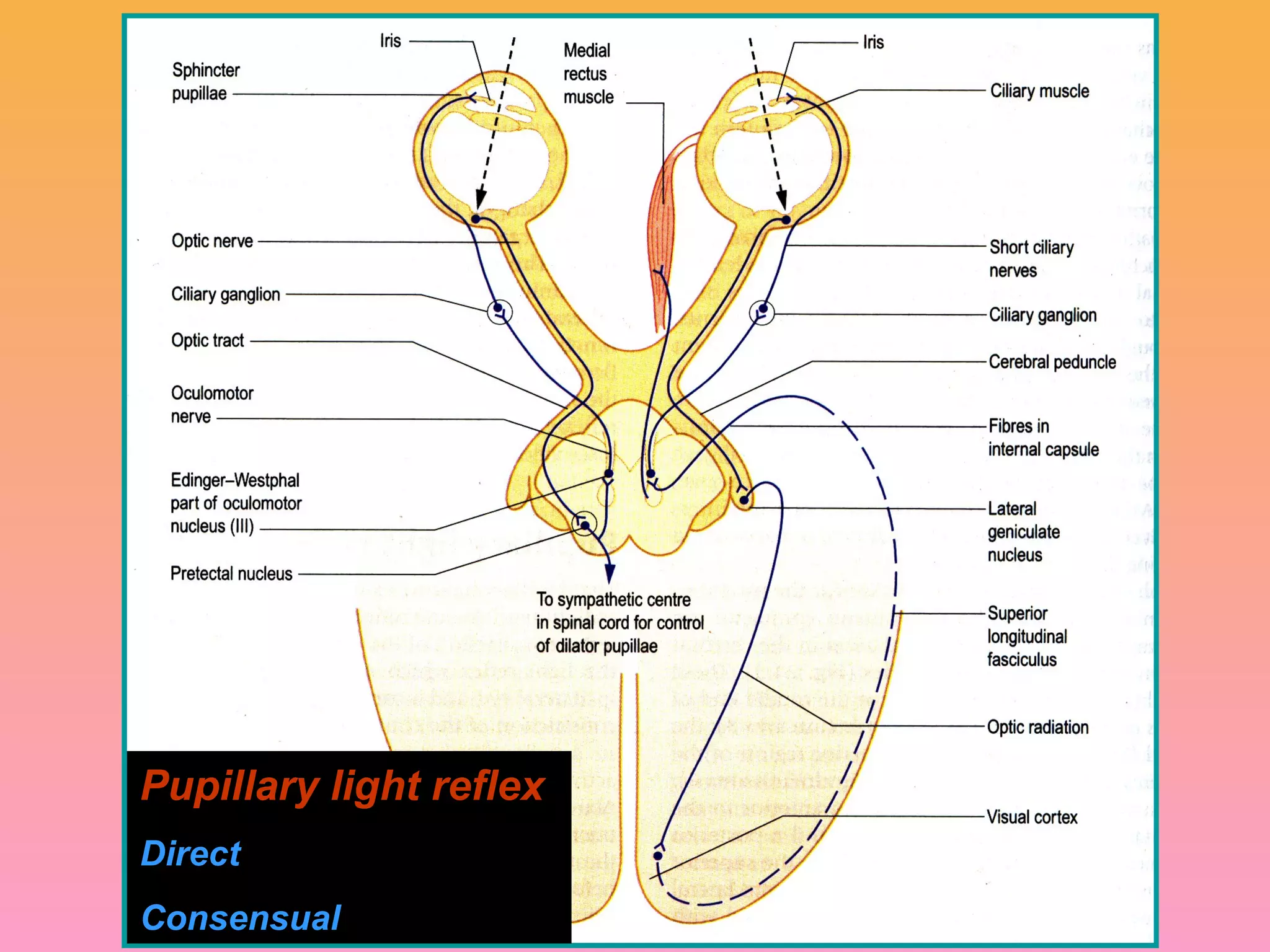
Pupillary Light Reflex
•When the amount of
light entering the eyes
increases, the pupils
constrict.
• Functions to help the
eye adapt extremely
rapidly to changing light
conditions.
• Direct light reflex:
same pupil constricts
• indirect (consensual)
light reflex: pupil of the
opposite eye constricts
12.
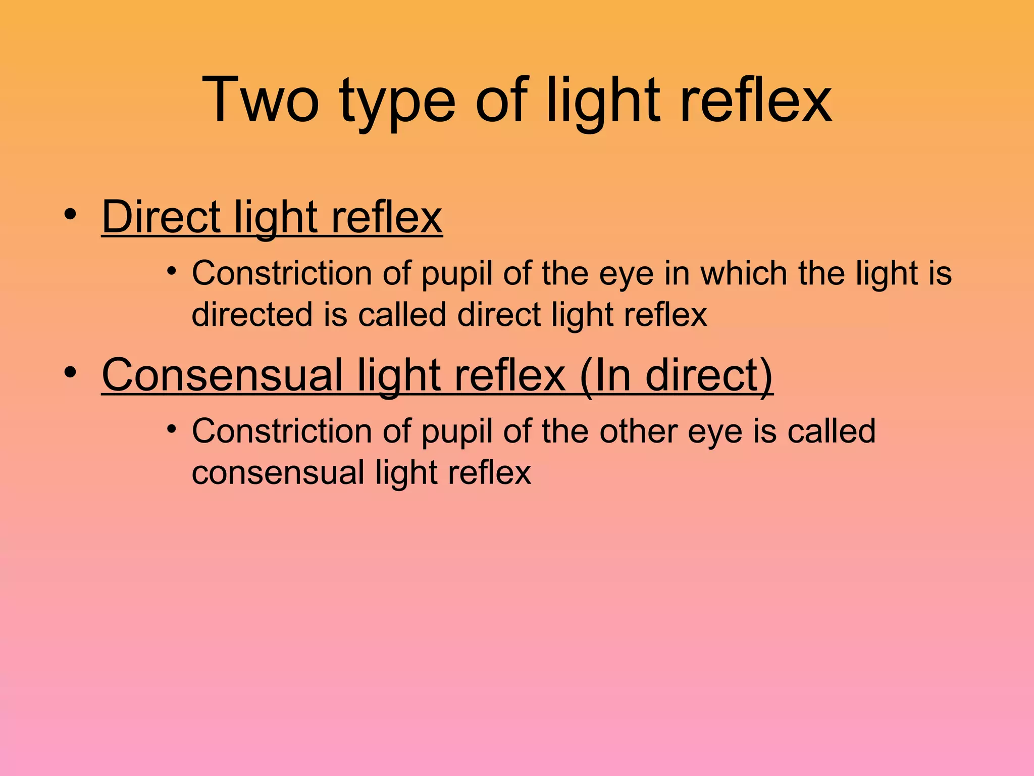
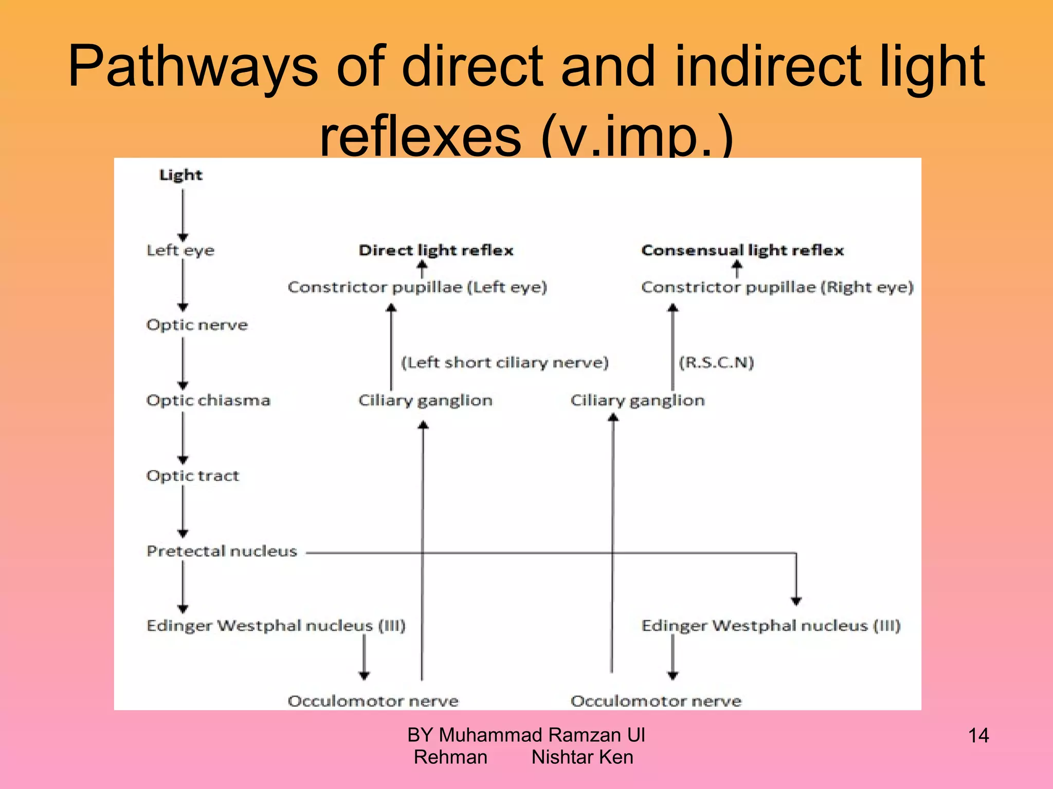
Two type oflight reflex
• Direct light reflex
• Constriction of pupil of the eye in which the light is
directed is called direct light reflex
• Consensual light reflex (In direct)
• Constriction of pupil of the other eye is called
consensual light reflex
14.
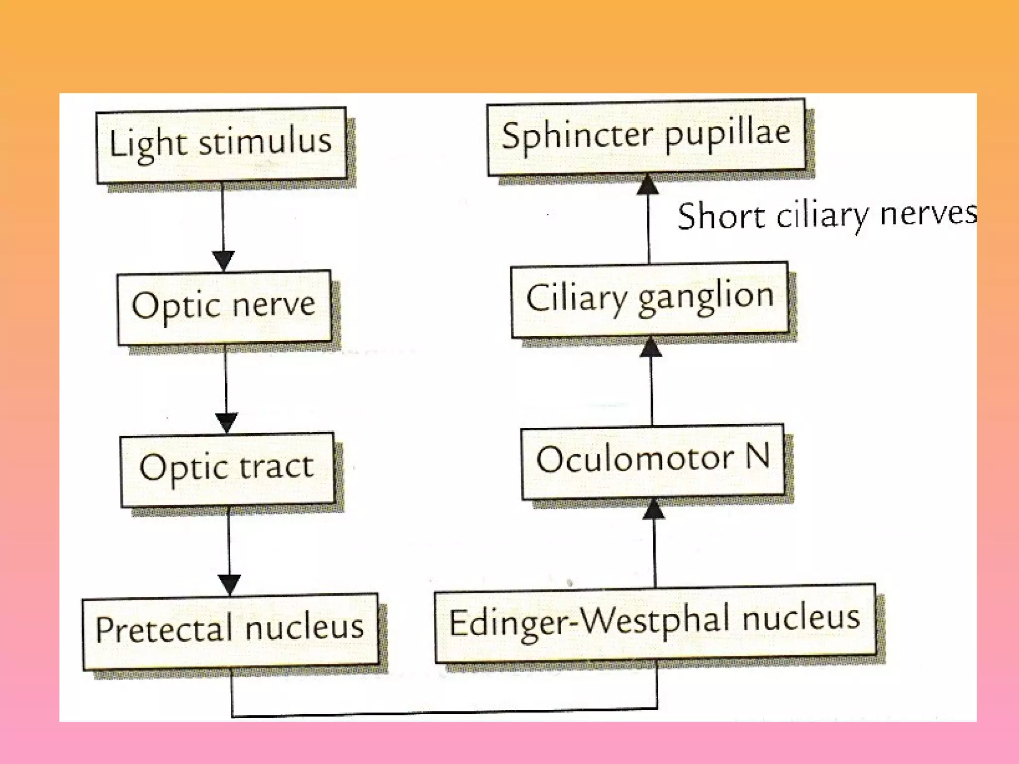
Pathways of directand indirect light
reflexes (v.imp.)
BY Muhammad Ramzan Ul
Rehman Nishtar Ken
14
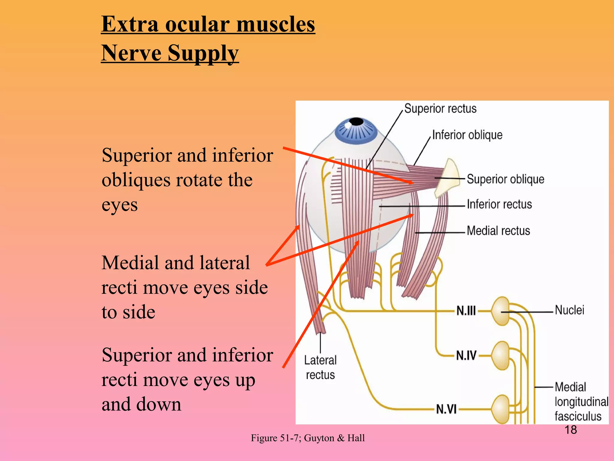
Medial and lateral
rectimove eyes side
to side
Superior and inferior
recti move eyes up
and down
Superior and inferior
obliques rotate the
eyes
Extra ocular muscles
Nerve Supply
Figure 51-7; Guyton & Hall
18
19.
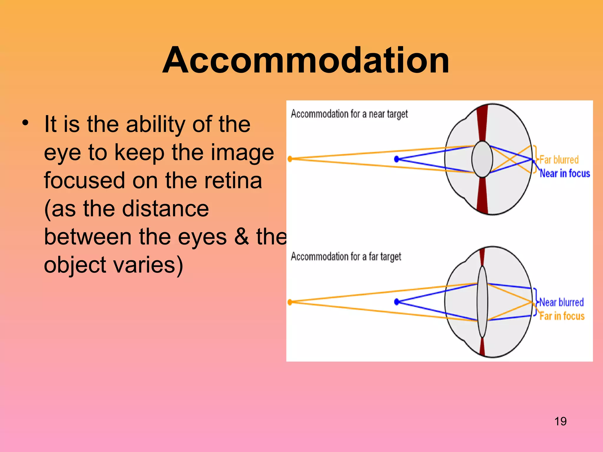
Accommodation
• It isthe ability of the
eye to keep the image
focused on the retina
(as the distance
between the eyes & the
object varies)
19
NEAR RESPONSE oraccomodation
for near vision
• The three components of near response
are:
1. accommodation,
2. convergence of the eyeballs &
3. Pupillary constriction
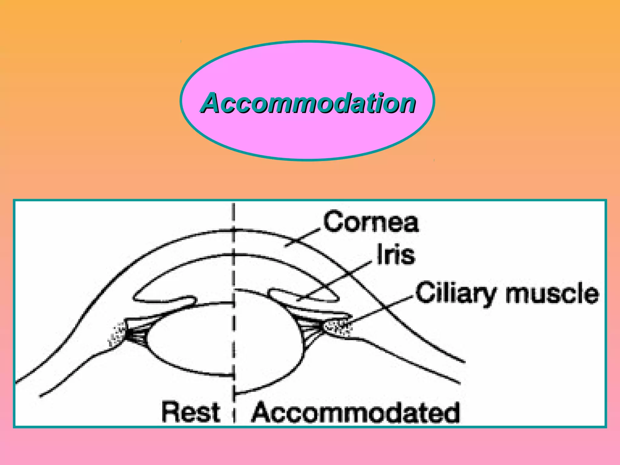
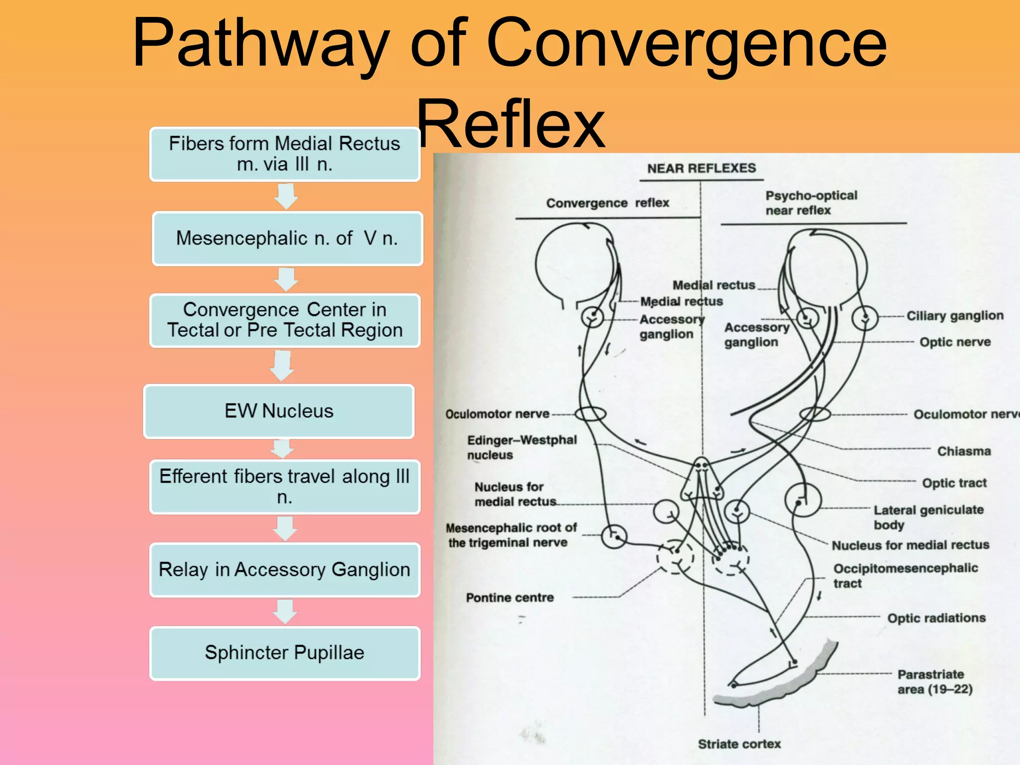
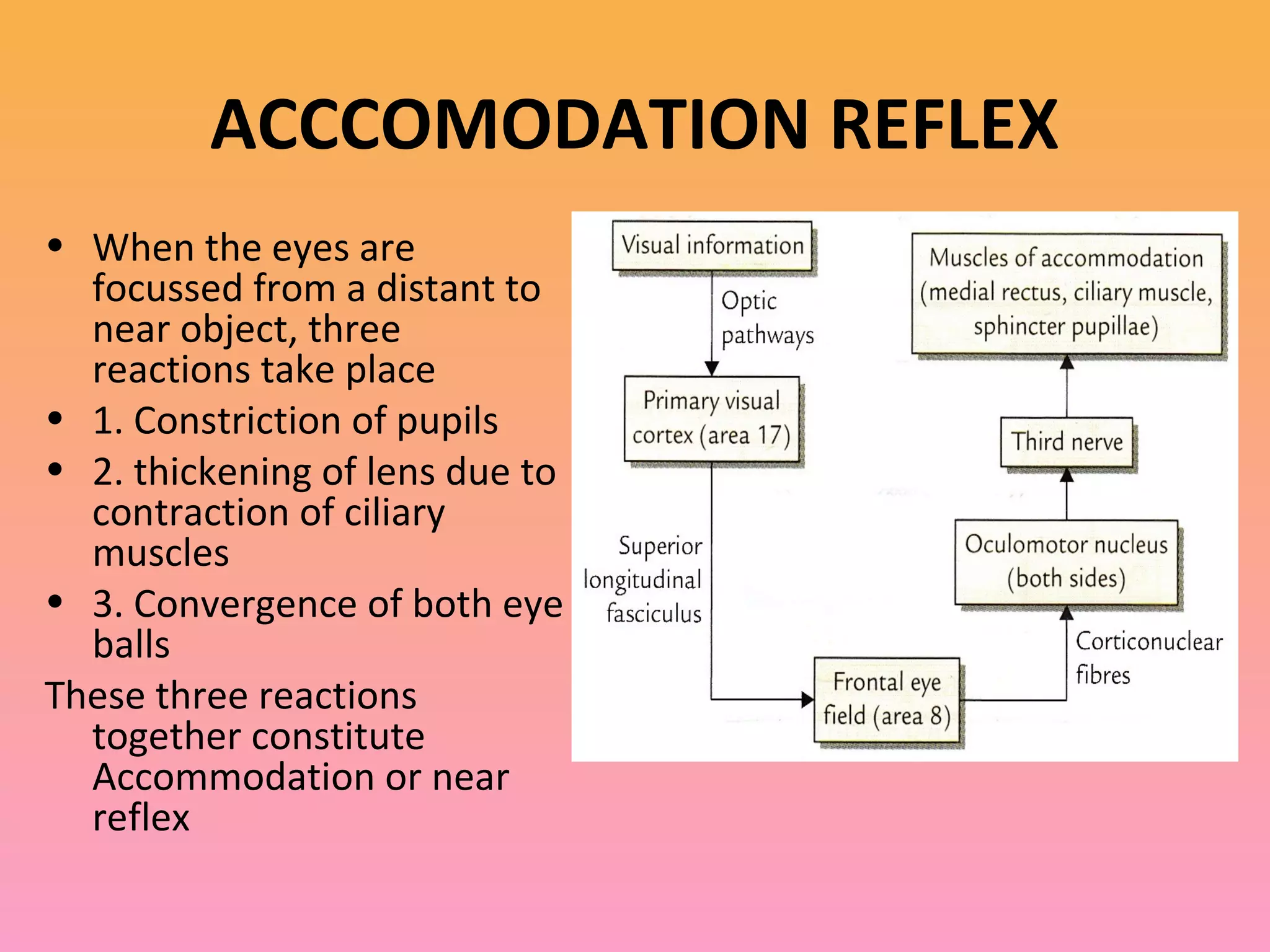
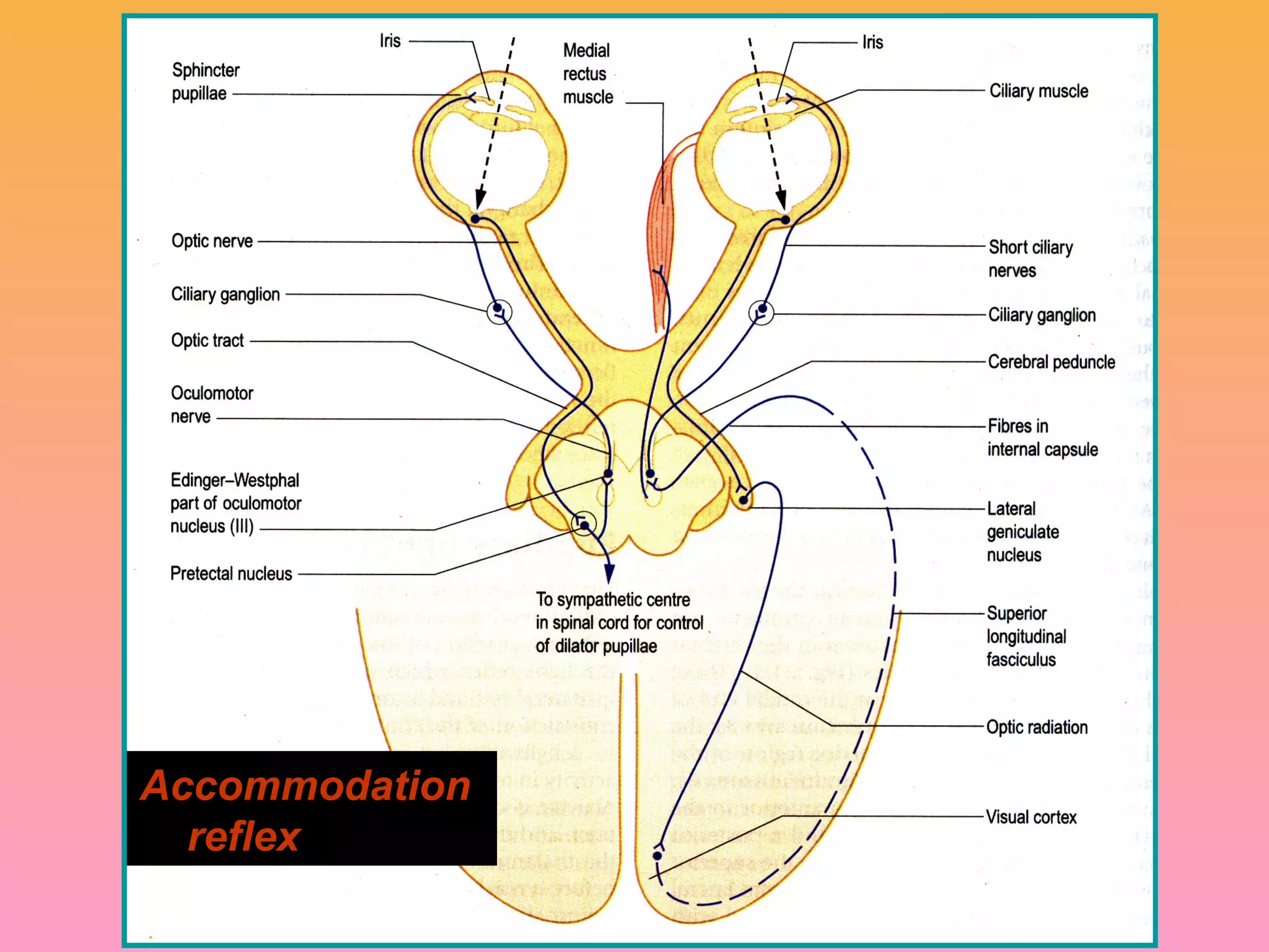
ACCCOMODATION REFLEX
• Whenthe eyes are
focussed from a distant to
near object, three
reactions take place
• 1. Constriction of pupils
• 2. thickening of lens due to
contraction of ciliary
muscles
• 3. Convergence of both eye
balls
These three reactions
together constitute
Accommodation or near
reflex
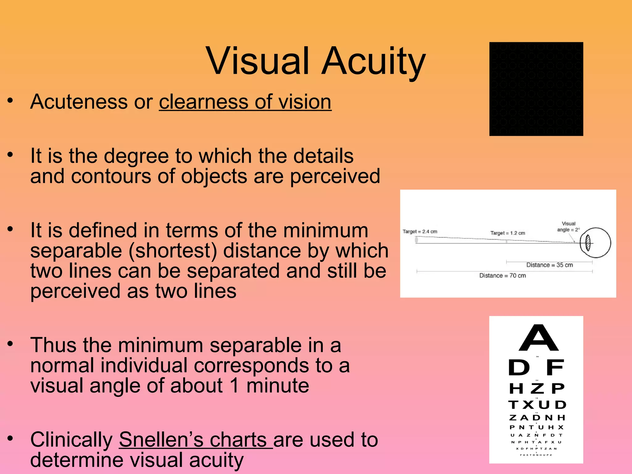
Visual Acuity
• Acutenessor clearness of vision
• It is the degree to which the details
and contours of objects are perceived
• It is defined in terms of the minimum
separable (shortest) distance by which
two lines can be separated and still be
perceived as two lines
• Thus the minimum separable in a
normal individual corresponds to a
visual angle of about 1 minute
• Clinically Snellen’s charts are used to
determine visual acuity
26.
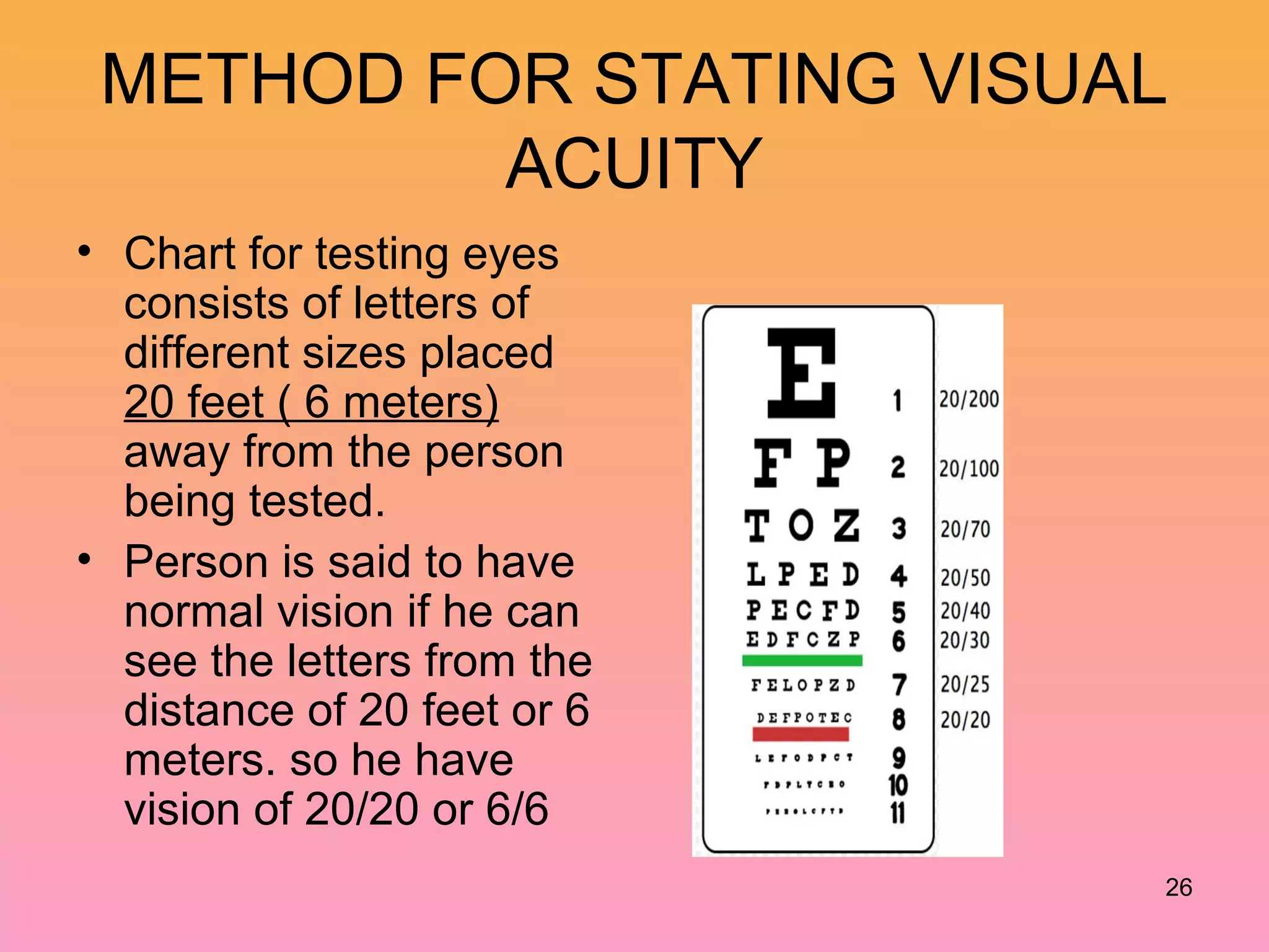
METHOD FOR STATINGVISUAL
ACUITY
• Chart for testing eyes
consists of letters of
different sizes placed
20 feet ( 6 meters)
away from the person
being tested.
• Person is said to have
normal vision if he can
see the letters from the
distance of 20 feet or 6
meters. so he have
vision of 20/20 or 6/6
26
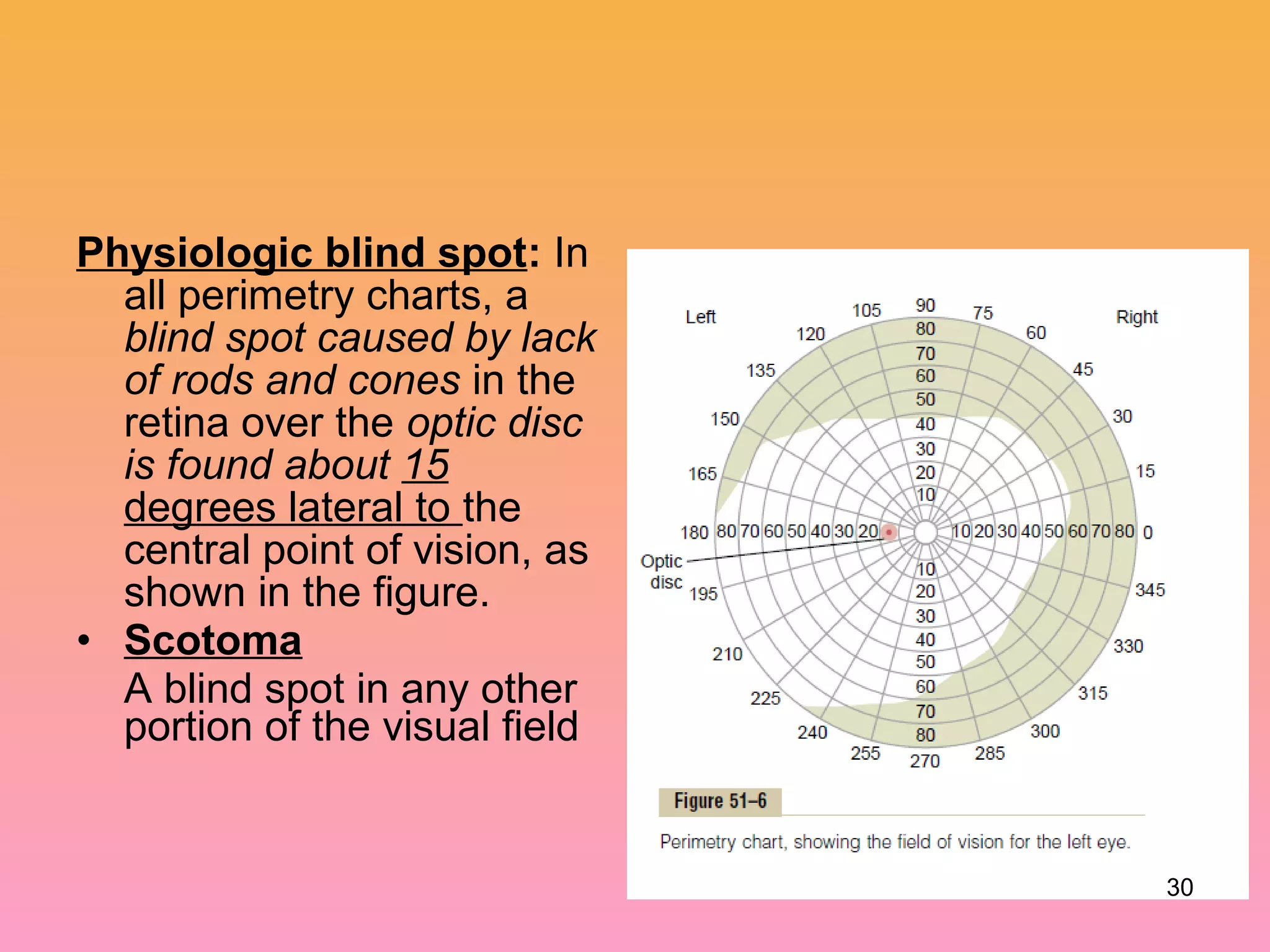
Physiologic blind spot:In
all perimetry charts, a
blind spot caused by lack
of rods and cones in the
retina over the optic disc
is found about 15
degrees lateral to the
central point of vision, as
shown in the figure.
• Scotoma
A blind spot in any other
portion of the visual field
30
Editor's Notes
#23 Affeerent fibers from MR via III n.
To Mesencephalic nuclei of 5th n
To convergence center in Tectal or Pre Tectal region
From convergence center to EW nucleus
Efferent fibers travel along the III n.
Relay in accessory ganglion
Reaches the sphincter pupillae