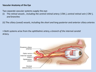
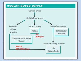
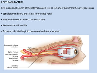
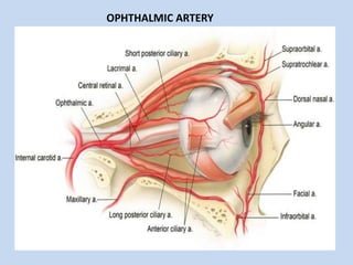
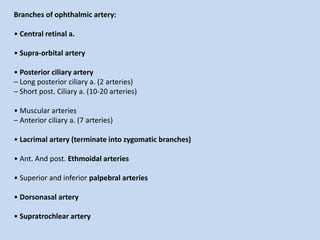
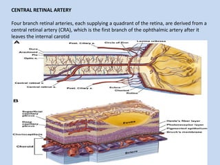
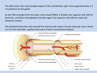
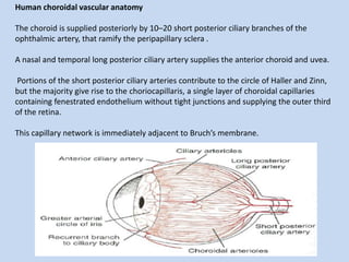
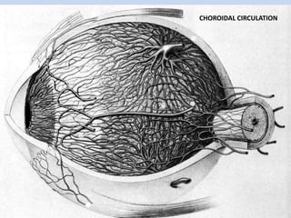
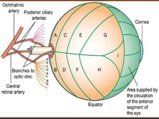
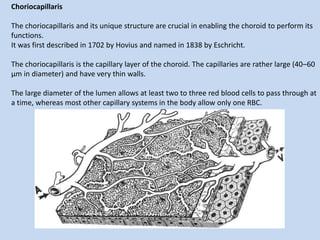
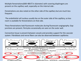
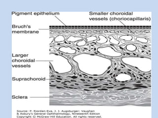
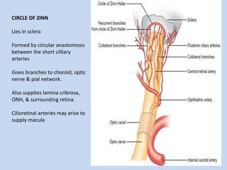
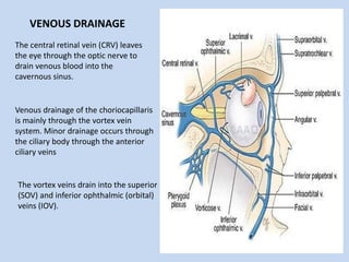
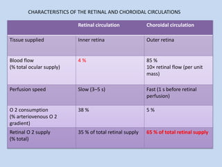
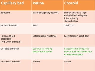
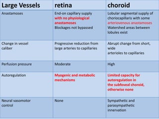
OCULAR CIRCULATION
PRESENTED BY
MAJ ANJANI KUMAR
RESIDENT (OPHTH)
MODERATOR
LT COL M JHA
CL SPL (OPHTH)
References
Skalicky, Simon
Vascular Anatomy of the Eye
Two separate vascular systems supply the eye:
(i) The retinal vessels , including the central retinal artery ( CRA ), central retinal vein ( CRV ),
and branches
(ii) The ciliary (uveal) vessels, including the short and long posterior and anterior ciliary arteries
• Both systems arise from the ophthalmic artery, a branch of the internal carotid
Artery.
65-85%
800-1000ml /sec
5%
80ml/sec
OPHTHALMIC ARTERY
First intracranial branch of the internal carotid just as the artery exits from the cavernous sinus
• optic foramen below and lateral to the optic nerve
• Pass over the optic nerve to its medial side
• Between the MR and SO
• Terminates by dividing into dorsonasal and supratrochlear
OPHTHALMIC ARTERY
Branches of ophthalmic artery:
• Central retinal a.
• Supra-orbital artery
• Posterior ciliary artery
– Long posterior ciliary a. (2 arteries)
– Short post. Ciliary a. (10-20 arteries)
• Muscular arteries
– Anterior ciliary a. (7 arteries)
• Lacrimal artery (terminate into zygomatic branches)
• Ant. And post. Ethmoidal arteries
• Superior and inferior palpebral arteries
• Dorsonasal artery
• Supratrochlear artery
CENTRAL RETINAL ARTERY
Four branch retinal arteries, each supplying a quadrant of the retina, are derived from a
central retinal artery (CRA), which is the first branch of the ophthalmic artery after it
leaves the internal carotid
The CRA enters the ventromedial aspect of the retrobulbar optic nerve approximately 1.2
cm posterior to the globe.
As the CRA emerges from the optic nerve head (ONH), it divides into superior and inferior
branches, and then immediately branches again into superior and inferior nasal and
temporal vessels.
The temporal branches arch around the macula and create a foveal avascular zone, which
is a 0.4 mm diameter, capillary-free zone of pure cone photoreceptors.
Arteries and veins remain in the nerve fiber layer, while arterioles and venules extend into
the deeper layers of the retina,forming two major microvascular networks;
(1) superficial capillaries -in the ganglion cell and nerve fiber layers
(2) deep, more dense, capillaries in the inner nuclear layer.
In the perifoveal and peripheral regions of the retina, these capillary networks thin to a
single layer
Retinal vessels
(i) Central retinal artery
• Retinal arteries have a well-developed smooth muscle layer and lack an internal elastic
lamina
• The CRA supplies the inner retina ; the outer retina is avascular, nourished from the
choroid
• 10–20 % of individuals have a cilioretinal artery , arising from the choroidal circulation;
this typically enters the inner retina at the temporal optic disc margin and supplies some
of the macula
(ii) Retinal capillaries and veins
• Capillaries are arranged in lamellae within the inner retina
• Astrocytes surround retinal vessels and maintain their integrity
Ciliary vessels
The ciliary vessels include the vascular beds of the uveal tract .
(i) The anterior ciliary vessels
• Seven anterior ciliary arteries provide the major blood supply to the anterior uvea .
• Two travel with each rectus muscle (the lateral rectus has only one) and pierce the
sclera anteriorly.
• They anastomose with the long posterior ciliary arteries to form the major iridial circle
• This forms a ring around the iris peripheral margin supplying the iris and ciliary body .
The posterior ciliary vessels
• 10–20 short posterior ciliary arteries enter the sclera to form an anastomotic
ring ( circle of Zinn - Haller ) around the optic nerve. This supplies the anterior optic nerve
and posterior choroid .
• Two long posterior ciliary arteries supply the iris , ciliary body , and anterior choroid
• Venous blood from the choroid and anterior uvea drains through four vortex vein.
The choroid
The choroid is a highly vascular uveal layer between the retina and sclera.
• It provides oxygen and nutrients to the outer retina and is a heat sink absorbing
excessive light energy focused onto the retina .
• The anterior surface, the choriocapillaris , is a dense, lobular, single-layered capillary
network .
• Feeding arteries and draining venules located deep to the choriocapillaris supply the
choroid in a segmented fashion .
Human choroidal vascular anatomy
The choroid is supplied posteriorly by 10–20 short posterior ciliary branches of the
ophthalmic artery, that ramify the peripapillary sclera .
A nasal and temporal long posterior ciliary artery supplies the anterior choroid and uvea.
Portions of the short posterior ciliary arteries contribute to the circle of Haller and Zinn,
but the majority give rise to the choriocapillaris, a single layer of choroidal capillaries
containing fenestrated endothelium without tight junctions and supplying the outer third
of the retina.
This capillary network is immediately adjacent to Bruch’s membrane.
CHOROIDAL CIRCULATION
Choriocapillaris
The choriocapillaris and its unique structure are crucial in enabling the choroid to perform its
functions.
It was first described in 1702 by Hovius and named in 1838 by Eschricht.
The choriocapillaris is the capillary layer of the choroid. The capillaries are rather large (40–60
μm in diameter) and have very thin walls.
The large diameter of the lumen allows at least two to three red blood cells to pass through at
a time, whereas most other capillary systems in the body allow only one RBC.
Multiple fenestrations(600–800 Å in diameter) with covering diaphragms are
present on the capillary wall, especially on the internal side .
Fenestrations are also noted on the other side of the capillary but are much less
frequent.
The endothelial cell nucleus usually lies on the outer side of the capillary, so less
room is available for fenestrations on that side.
These fenestrations leak fluorescein molecules during fluorescein angiography. Gap
junctions are present. Pericytes occasionally are seen on the outer wall.
Connective tissue is present between vessels and provides support for the vascular
system. Fibroblasts and nerve fibers can also be observed between capillaries
Choroidal circulation: Facts
Choroidal circulation constitutes 85% of the blood circulation of the eye.
Choroidal blood flow is higher than that in tissues like retina and brain.
Choroidal blood-flow ranges from 800 to 2000 mL/min/100 g of tissue.
Choroid provides the metabolic requirements of the full retinal thickness only in the
macular region.
In embryonic life, choroid serves as an additional site for the erythropoiesis.
The optic nerve head
• Most of the anterior optic nerve is supplied by the circle of Zinn-Haller and pial vessels
• There is a small physiological break in the blood - neural barrier at the lateral optic
nerve head, adjacent to the choroid (border tissue of Elschnig).
Choroidal extravascular solutes may diffuse into the nerve tissue there
.
• Branches of the central retinal artery supply the superficial optic nerve head .
CIRCLE OF ZINN
Lies in sclera
Formed by circular anastomosis
between the short cilliary
arteries
Gives branches to choroid, optic
nerve & pial network.
Also supplies lamina cribrosa,
ONH, & surrounding retina.
Cilioretinal arteries may arise to
supply macula
The central retinal vein (CRV) leaves
the eye through the optic nerve to
drain venous blood into the
cavernous sinus.
Venous drainage of the choriocapillaris
is mainly through the vortex vein
system. Minor drainage occurs through
the ciliary body through the anterior
ciliary veins
The vortex veins drain into the superior
(SOV) and inferior ophthalmic (orbital)
veins (IOV).
VENOUS DRAINAGE
Control of Circulation
With high metabolic requirements and relatively low flow, retinal and optic nerve
perfusion must remain constant despite changes in perfusion pressure.
1. Ocular perfusion pressure and intraocular pressure
BF =(Pa - IOP) / R
• A rise in IOP or reduction in mean arterial pressure reduces the ocular perfusion pressure
.
• This would cause reduced retinal or optic nerve perfusion if vascular resistance was
unchanged; however, autoregulation results in vascular dilation, reduced resistance, and
unchanged perfusion.
• In contrast the choroid has limited autoregulation, and perfusion reduces
when Pa drops or IOP rises.
This does not cause significant ischemia except in extreme changes in Pa or IOP.
AUTOREGULATION
• Retinal and optic nerve head vessels have the ability to autoregulate
.
• They maintain constant blood flow despite changes in oxygenation or perfusion pressure
• The endothelium regulates vascular tone in response to myogenic , metabolic , and light
stimuli:
(i) Myogenic stimuli (changes in vessel wall pressure)
• Decreased perfusion pressure results in vascular dilatation.
• Increased perfusion pressure results in reduced vascular dilatation.
(ii) Metabolic stimuli (changes in lactic acid, O 2 , and CO 2 levels).
• Low O 2 and high CO 2 tensions result in vascular dilatation.
• Low CO 2 and high O 2 tensions result in reduced vascular dilatation.
(iii) Light stimuli
• Flickering light increases retinal metabolism resulting in retinal capillary dilatation.
(iv) Mechanisms of autoregulation
• The vascular endothelium orchestrates vasodilation by release of prostacyclin and
nitric oxide .
Both cause endothelial cell relaxation in response to myogenic and metabolic stimuli.
• Endothelins released by the endothelium are also involved in control of vascular tone.
(v) Limited choroidal autoregulation
• The subfoveal choroid has a limited capacity for autoregulation.
• In general autoregulatory mechanisms are not found in the choroidal circulation.
• The choroid with high blood flow and O 2 supply can tolerate some perfusion
decrease without tissue compromise
CHARACTERISTICS OF THE RETINAL AND CHOROIDAL CIRCULATIONS
Retinal circulation Choroidal circulation
Tissue supplied Inner retina Outer retina
Blood flow
(% total ocular supply)
4 % 85 %
10× retinal flow (per unit
mass)
Perfusion speed Slow (3–5 s) Fast (1 s before retinal
perfusion)
O 2 consumption
(% arteriovenous O 2
gradient)
38 % 5 %
Retinal O 2 supply
(% total)
35 % of total retinal supply 65 % of total retinal supply
Capillary bed Retina Choroid
Structure Stratified capillary network choriocapillaris: a large
endothelial-lined space
interrupted by
stromal pillars
luminal diameter 5 um 10–20 um
Passage of red
blood cells
(7–8 um in diameter)
Deform under resistance Move freely in sheet flow
Endothelial barrier Continuous, forming
blood-retinal barrier
Fenestrated allowing free
flow of fluid and solutes into
extravascular space
Intramural pericytes Present Absent
Large Vessels retina choroid
Anastamoses End-on capillary supply
with no physiological
anastamoses
Blockages not bypassed
Lobular segmental supply of
choriocapillaris with some
arteriovenous anastamoses
Watershed areas between
lobules exist
Change in vessel
caliber
Progressive reduction from
large arteries to capillaries
Abrupt change from short,
wide
arterioles to capillaries
Perfusion pressure Moderate High
Autoregulation Myogenic and metabolic
mechanisms
Limited capacity for
autoregulation in
the subfoveal choroid,
otherwise none
Neural vasomotor
control
None Sympathetic and
parasympathetic
innervation
THANK YOU

Ocular circulation

  • 1.
    OCULAR CIRCULATION PRESENTED BY MAJANJANI KUMAR RESIDENT (OPHTH) MODERATOR LT COL M JHA CL SPL (OPHTH)
  • 2.
  • 3.
    Vascular Anatomy ofthe Eye Two separate vascular systems supply the eye: (i) The retinal vessels , including the central retinal artery ( CRA ), central retinal vein ( CRV ), and branches (ii) The ciliary (uveal) vessels, including the short and long posterior and anterior ciliary arteries • Both systems arise from the ophthalmic artery, a branch of the internal carotid Artery.
  • 4.
  • 5.
    OPHTHALMIC ARTERY First intracranialbranch of the internal carotid just as the artery exits from the cavernous sinus • optic foramen below and lateral to the optic nerve • Pass over the optic nerve to its medial side • Between the MR and SO • Terminates by dividing into dorsonasal and supratrochlear
  • 6.
  • 7.
    Branches of ophthalmicartery: • Central retinal a. • Supra-orbital artery • Posterior ciliary artery – Long posterior ciliary a. (2 arteries) – Short post. Ciliary a. (10-20 arteries) • Muscular arteries – Anterior ciliary a. (7 arteries) • Lacrimal artery (terminate into zygomatic branches) • Ant. And post. Ethmoidal arteries • Superior and inferior palpebral arteries • Dorsonasal artery • Supratrochlear artery
  • 8.
    CENTRAL RETINAL ARTERY Fourbranch retinal arteries, each supplying a quadrant of the retina, are derived from a central retinal artery (CRA), which is the first branch of the ophthalmic artery after it leaves the internal carotid
  • 9.
    The CRA entersthe ventromedial aspect of the retrobulbar optic nerve approximately 1.2 cm posterior to the globe. As the CRA emerges from the optic nerve head (ONH), it divides into superior and inferior branches, and then immediately branches again into superior and inferior nasal and temporal vessels. The temporal branches arch around the macula and create a foveal avascular zone, which is a 0.4 mm diameter, capillary-free zone of pure cone photoreceptors.
  • 10.
    Arteries and veinsremain in the nerve fiber layer, while arterioles and venules extend into the deeper layers of the retina,forming two major microvascular networks; (1) superficial capillaries -in the ganglion cell and nerve fiber layers (2) deep, more dense, capillaries in the inner nuclear layer. In the perifoveal and peripheral regions of the retina, these capillary networks thin to a single layer
  • 12.
    Retinal vessels (i) Centralretinal artery • Retinal arteries have a well-developed smooth muscle layer and lack an internal elastic lamina • The CRA supplies the inner retina ; the outer retina is avascular, nourished from the choroid • 10–20 % of individuals have a cilioretinal artery , arising from the choroidal circulation; this typically enters the inner retina at the temporal optic disc margin and supplies some of the macula (ii) Retinal capillaries and veins • Capillaries are arranged in lamellae within the inner retina • Astrocytes surround retinal vessels and maintain their integrity
  • 13.
    Ciliary vessels The ciliaryvessels include the vascular beds of the uveal tract . (i) The anterior ciliary vessels • Seven anterior ciliary arteries provide the major blood supply to the anterior uvea . • Two travel with each rectus muscle (the lateral rectus has only one) and pierce the sclera anteriorly. • They anastomose with the long posterior ciliary arteries to form the major iridial circle • This forms a ring around the iris peripheral margin supplying the iris and ciliary body .
  • 14.
    The posterior ciliaryvessels • 10–20 short posterior ciliary arteries enter the sclera to form an anastomotic ring ( circle of Zinn - Haller ) around the optic nerve. This supplies the anterior optic nerve and posterior choroid . • Two long posterior ciliary arteries supply the iris , ciliary body , and anterior choroid • Venous blood from the choroid and anterior uvea drains through four vortex vein.
  • 16.
    The choroid The choroidis a highly vascular uveal layer between the retina and sclera. • It provides oxygen and nutrients to the outer retina and is a heat sink absorbing excessive light energy focused onto the retina . • The anterior surface, the choriocapillaris , is a dense, lobular, single-layered capillary network . • Feeding arteries and draining venules located deep to the choriocapillaris supply the choroid in a segmented fashion .
  • 17.
    Human choroidal vascularanatomy The choroid is supplied posteriorly by 10–20 short posterior ciliary branches of the ophthalmic artery, that ramify the peripapillary sclera . A nasal and temporal long posterior ciliary artery supplies the anterior choroid and uvea. Portions of the short posterior ciliary arteries contribute to the circle of Haller and Zinn, but the majority give rise to the choriocapillaris, a single layer of choroidal capillaries containing fenestrated endothelium without tight junctions and supplying the outer third of the retina. This capillary network is immediately adjacent to Bruch’s membrane.
  • 18.
  • 20.
    Choriocapillaris The choriocapillaris andits unique structure are crucial in enabling the choroid to perform its functions. It was first described in 1702 by Hovius and named in 1838 by Eschricht. The choriocapillaris is the capillary layer of the choroid. The capillaries are rather large (40–60 μm in diameter) and have very thin walls. The large diameter of the lumen allows at least two to three red blood cells to pass through at a time, whereas most other capillary systems in the body allow only one RBC.
  • 21.
    Multiple fenestrations(600–800 Åin diameter) with covering diaphragms are present on the capillary wall, especially on the internal side . Fenestrations are also noted on the other side of the capillary but are much less frequent. The endothelial cell nucleus usually lies on the outer side of the capillary, so less room is available for fenestrations on that side. These fenestrations leak fluorescein molecules during fluorescein angiography. Gap junctions are present. Pericytes occasionally are seen on the outer wall. Connective tissue is present between vessels and provides support for the vascular system. Fibroblasts and nerve fibers can also be observed between capillaries
  • 23.
    Choroidal circulation: Facts Choroidalcirculation constitutes 85% of the blood circulation of the eye. Choroidal blood flow is higher than that in tissues like retina and brain. Choroidal blood-flow ranges from 800 to 2000 mL/min/100 g of tissue. Choroid provides the metabolic requirements of the full retinal thickness only in the macular region. In embryonic life, choroid serves as an additional site for the erythropoiesis.
  • 24.
    The optic nervehead • Most of the anterior optic nerve is supplied by the circle of Zinn-Haller and pial vessels • There is a small physiological break in the blood - neural barrier at the lateral optic nerve head, adjacent to the choroid (border tissue of Elschnig). Choroidal extravascular solutes may diffuse into the nerve tissue there . • Branches of the central retinal artery supply the superficial optic nerve head .
  • 25.
    CIRCLE OF ZINN Liesin sclera Formed by circular anastomosis between the short cilliary arteries Gives branches to choroid, optic nerve & pial network. Also supplies lamina cribrosa, ONH, & surrounding retina. Cilioretinal arteries may arise to supply macula
  • 26.
    The central retinalvein (CRV) leaves the eye through the optic nerve to drain venous blood into the cavernous sinus. Venous drainage of the choriocapillaris is mainly through the vortex vein system. Minor drainage occurs through the ciliary body through the anterior ciliary veins The vortex veins drain into the superior (SOV) and inferior ophthalmic (orbital) veins (IOV). VENOUS DRAINAGE
  • 27.
    Control of Circulation Withhigh metabolic requirements and relatively low flow, retinal and optic nerve perfusion must remain constant despite changes in perfusion pressure. 1. Ocular perfusion pressure and intraocular pressure BF =(Pa - IOP) / R • A rise in IOP or reduction in mean arterial pressure reduces the ocular perfusion pressure . • This would cause reduced retinal or optic nerve perfusion if vascular resistance was unchanged; however, autoregulation results in vascular dilation, reduced resistance, and unchanged perfusion. • In contrast the choroid has limited autoregulation, and perfusion reduces when Pa drops or IOP rises. This does not cause significant ischemia except in extreme changes in Pa or IOP.
  • 28.
    AUTOREGULATION • Retinal andoptic nerve head vessels have the ability to autoregulate . • They maintain constant blood flow despite changes in oxygenation or perfusion pressure • The endothelium regulates vascular tone in response to myogenic , metabolic , and light stimuli: (i) Myogenic stimuli (changes in vessel wall pressure) • Decreased perfusion pressure results in vascular dilatation. • Increased perfusion pressure results in reduced vascular dilatation. (ii) Metabolic stimuli (changes in lactic acid, O 2 , and CO 2 levels). • Low O 2 and high CO 2 tensions result in vascular dilatation. • Low CO 2 and high O 2 tensions result in reduced vascular dilatation. (iii) Light stimuli • Flickering light increases retinal metabolism resulting in retinal capillary dilatation.
  • 29.
    (iv) Mechanisms ofautoregulation • The vascular endothelium orchestrates vasodilation by release of prostacyclin and nitric oxide . Both cause endothelial cell relaxation in response to myogenic and metabolic stimuli. • Endothelins released by the endothelium are also involved in control of vascular tone. (v) Limited choroidal autoregulation • The subfoveal choroid has a limited capacity for autoregulation. • In general autoregulatory mechanisms are not found in the choroidal circulation. • The choroid with high blood flow and O 2 supply can tolerate some perfusion decrease without tissue compromise
  • 30.
    CHARACTERISTICS OF THERETINAL AND CHOROIDAL CIRCULATIONS Retinal circulation Choroidal circulation Tissue supplied Inner retina Outer retina Blood flow (% total ocular supply) 4 % 85 % 10× retinal flow (per unit mass) Perfusion speed Slow (3–5 s) Fast (1 s before retinal perfusion) O 2 consumption (% arteriovenous O 2 gradient) 38 % 5 % Retinal O 2 supply (% total) 35 % of total retinal supply 65 % of total retinal supply
  • 31.
    Capillary bed RetinaChoroid Structure Stratified capillary network choriocapillaris: a large endothelial-lined space interrupted by stromal pillars luminal diameter 5 um 10–20 um Passage of red blood cells (7–8 um in diameter) Deform under resistance Move freely in sheet flow Endothelial barrier Continuous, forming blood-retinal barrier Fenestrated allowing free flow of fluid and solutes into extravascular space Intramural pericytes Present Absent
  • 32.
    Large Vessels retinachoroid Anastamoses End-on capillary supply with no physiological anastamoses Blockages not bypassed Lobular segmental supply of choriocapillaris with some arteriovenous anastamoses Watershed areas between lobules exist Change in vessel caliber Progressive reduction from large arteries to capillaries Abrupt change from short, wide arterioles to capillaries Perfusion pressure Moderate High Autoregulation Myogenic and metabolic mechanisms Limited capacity for autoregulation in the subfoveal choroid, otherwise none Neural vasomotor control None Sympathetic and parasympathetic innervation
  • 33.