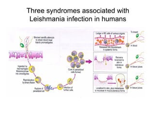
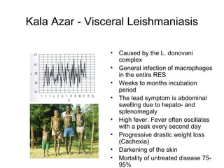
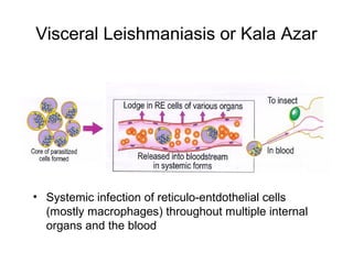
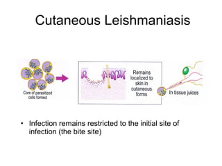
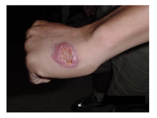
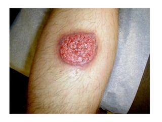
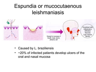
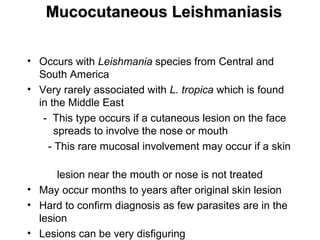
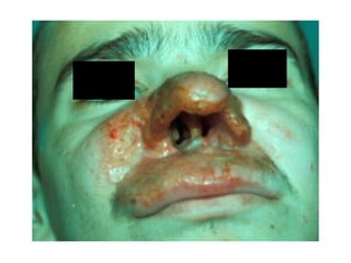
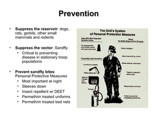
Leishmania
Leishmaniasias
Is a disease caused the protozoan
parasites of the genus Leishmania
Three syndromes associated with
Leishmania infection in humans
Kala Azar - Visceral Leishmaniasis
• Caused by the L. donovani
complex
• General infection of macrophages
in the entire RES
• Weeks to months incubation
period
• The lead symptom is abdominal
swelling due to hepato- and
splenomegaly
• High fever. Fever often oscillates
with a peak every second day
• Progressive drastic weight loss
(Cachexia)
• Darkening of the skin
• Mortality of untreated disease 75-
95%
Visceral Leishmaniasis or Kala Azar
• Systemic infection of reticulo-entdothelial cells
(mostly macrophages) throughout multiple internal
organs and the blood
Visceral LeishmaniasisVisceral Leishmaniasis
• Most severe form of the disease, may be fatal if left
untreated
• Usually associated with fever, weight loss, and an
enlarged spleen and liver
• Anemia (low RBC), leukopenia (low WBC), and
thrombocytopenia (low platelets) are common
• Lymphadenopathy may be present
• Visceral disease from the Middle East is usually milder
with less specific findings than visceral leishmaniasis
from other areas of the world
• Profile view of a
teenage boy suffering
from visceral
leishmaniasis. The boy
exhibits splenomegaly,
distended abdomen
and severe muscle
wasting.
Cutaneous Leishmaniasis
• Infection remains restricted to the initial site of
infection (the bite site)
Espundia or mucocutaenous
leishmaniasis
• Caused by L. braziliensis
• ~20% of infected patients develop ulcers of the
oral and nasal mucosa
Mucocutaneous LeishmaniasisMucocutaneous Leishmaniasis
• Occurs with Leishmania species from Central and
South America
• Very rarely associated with L. tropica which is found
in the Middle East
- This type occurs if a cutaneous lesion on the face
spreads to involve the nose or mouth
- This rare mucosal involvement may occur if a skin
lesion near the mouth or nose is not treated
• May occur months to years after original skin lesion
• Hard to confirm diagnosis as few parasites are in the
lesion
• Lesions can be very disfiguring
Espundia or mucocutaenous
leishmaniasis
• Caused by L. braziliensis
• ~20% of infected patients
develop ulcers of the oral and
nasal mucosa
• Progression of the ulceration is
slow but steady, ultimately
destroying all soft parts of the
nose, the lips, and the soft
palate
• Death can occur through
secondary bacterial infection
Adapted from Paulo Pimenta
Life cycle of Leishmania
Promastigote
Amastigote
Sandfly vector
Human host
Differentiation
Differentiation
Growth
Macrophage
Differentiation
Dwyer, D.M.
Dwyer, D.M.
Debrabant,A.
• Leishmania
(Leishman-
Donovan or LD
bodies). Lying in
macrophage cells
from liver. Giemsa.
×12000. Enlarged
by 9.6.
• A macrophage
filled with
Leishmania
amastigotes.
PreventionPrevention
• Suppress the reservoir: dogs,
rats, gerbils, other small
mammals and rodents
• Suppress the vector: Sandfly
• Critical to preventing
disease in stationary troop
populations
• Prevent sandfly bites:
Personal Protective Measures
• Most important at night
• Sleeves down
• Insect repellent w/ DEET
• Permethrin treated uniforms
• Permethrin treated bed nets

Leishmania

  • 1.
  • 2.
    Leishmaniasias Is a diseasecaused the protozoan parasites of the genus Leishmania
  • 3.
    Three syndromes associatedwith Leishmania infection in humans
  • 4.
    Kala Azar -Visceral Leishmaniasis • Caused by the L. donovani complex • General infection of macrophages in the entire RES • Weeks to months incubation period • The lead symptom is abdominal swelling due to hepato- and splenomegaly • High fever. Fever often oscillates with a peak every second day • Progressive drastic weight loss (Cachexia) • Darkening of the skin • Mortality of untreated disease 75- 95%
  • 5.
    Visceral Leishmaniasis orKala Azar • Systemic infection of reticulo-entdothelial cells (mostly macrophages) throughout multiple internal organs and the blood
  • 6.
    Visceral LeishmaniasisVisceral Leishmaniasis •Most severe form of the disease, may be fatal if left untreated • Usually associated with fever, weight loss, and an enlarged spleen and liver • Anemia (low RBC), leukopenia (low WBC), and thrombocytopenia (low platelets) are common • Lymphadenopathy may be present • Visceral disease from the Middle East is usually milder with less specific findings than visceral leishmaniasis from other areas of the world
  • 8.
    • Profile viewof a teenage boy suffering from visceral leishmaniasis. The boy exhibits splenomegaly, distended abdomen and severe muscle wasting.
  • 9.
    Cutaneous Leishmaniasis • Infectionremains restricted to the initial site of infection (the bite site)
  • 12.
    Espundia or mucocutaenous leishmaniasis •Caused by L. braziliensis • ~20% of infected patients develop ulcers of the oral and nasal mucosa
  • 13.
    Mucocutaneous LeishmaniasisMucocutaneous Leishmaniasis •Occurs with Leishmania species from Central and South America • Very rarely associated with L. tropica which is found in the Middle East - This type occurs if a cutaneous lesion on the face spreads to involve the nose or mouth - This rare mucosal involvement may occur if a skin lesion near the mouth or nose is not treated • May occur months to years after original skin lesion • Hard to confirm diagnosis as few parasites are in the lesion • Lesions can be very disfiguring
  • 14.
    Espundia or mucocutaenous leishmaniasis •Caused by L. braziliensis • ~20% of infected patients develop ulcers of the oral and nasal mucosa • Progression of the ulceration is slow but steady, ultimately destroying all soft parts of the nose, the lips, and the soft palate • Death can occur through secondary bacterial infection
  • 16.
    Adapted from PauloPimenta Life cycle of Leishmania Promastigote Amastigote Sandfly vector Human host Differentiation Differentiation Growth Macrophage Differentiation Dwyer, D.M. Dwyer, D.M. Debrabant,A.
  • 17.
    • Leishmania (Leishman- Donovan orLD bodies). Lying in macrophage cells from liver. Giemsa. ×12000. Enlarged by 9.6.
  • 18.
    • A macrophage filledwith Leishmania amastigotes.
  • 19.
    PreventionPrevention • Suppress thereservoir: dogs, rats, gerbils, other small mammals and rodents • Suppress the vector: Sandfly • Critical to preventing disease in stationary troop populations • Prevent sandfly bites: Personal Protective Measures • Most important at night • Sleeves down • Insect repellent w/ DEET • Permethrin treated uniforms • Permethrin treated bed nets

Editor's Notes

  • #7 Based on our experience with Desert Storm, well nourished American soldiers generally have a less symptomatic, relatively oligoparasitic infection that was not life threatening but posed some diagnostic challenges
  • #8 Two children with visceral leishmaniasis. The size of the spleen is marked on the abdomen. Normally the spleen does not protrude below the bottom rib. Photograph provided by COL Charles Oster
  • #11 Photograph provided by COL Naomi Aronson
  • #12 Photograph provided by COL Naomi Aronson
  • #14 Much of the destruction is from a hyperimmune reaction to the Leishmania infection. There are not many parasites in the affected tissue so confirming the diagnosis can be difficult. This form of leishmaniasis would be unusual in soldiers infected in South West Asia.
  • #16 Photograph provided courtesy of COL Donald Skillman
  • #20 Sandflies bite dusk to dawn so personal protection is most important during these times. Even light clothing covering skin is sufficient to prevent the bite of the sandfly. DEET is the most effective insect repellent. Sandflies are small enough to fly through bed netting unless it is treated with permethrin.