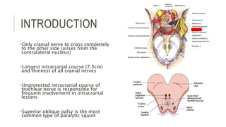
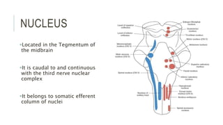
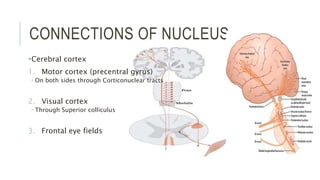
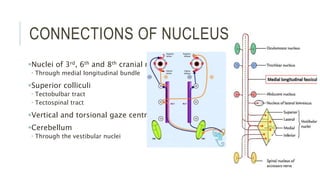
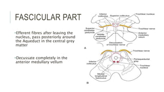
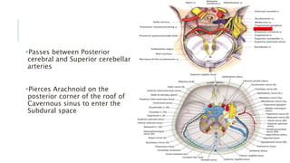
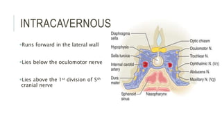
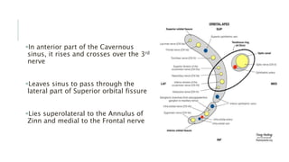
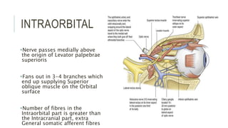
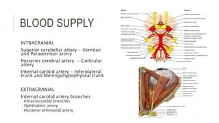
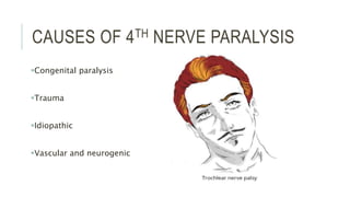
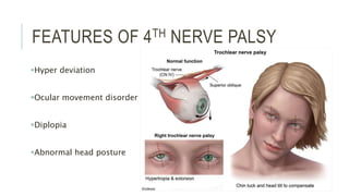
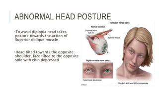
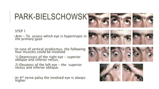
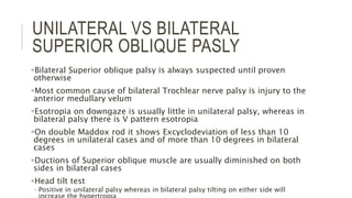
2. INTRODUCTION
Purely motor nerve, supplies only
superior oblique muscle of the eye
The nerve is named after trochlea, the
fibrous pulley through which the tendon
of the superior oblique muscle passes
Only cranial nerve to arise from the
dorsal aspect of the brain
Trochlear nerve contains fewer number
of axons compared to other cranial
nerves
3. INTRODUCTION
Only cranial nerve to cross completely
to the other side (arises from the
contralateral nucleus)
Longest intracranial course (7.5cm)
and thinnest of all cranial nerves
Unprotected intracranial course of
trochlear nerve is responsible for
frequent involvement in intracranial
lesions
Superior oblique palsy is the most
common type of paralytic squint
4. FUNCTIONAL COMPONENTS
Somatic efferent – movement of eyeball
through Superior oblique
General somatic afferent
Proprioceptive signals from Superior oblique
These impulses are relayed to mesencephalic
nucleus of Trigeminal nerve
5. NUCLEUS
Located in the Tegmentum of
the midbrain
It is caudal to and continuous
with the third nerve nuclear
complex
It belongs to somatic efferent
column of nuclei
6. RELATIONS OF THE NUCLEUS
Ventrolateral to the Cerebral
Aqueduct
At the level of superior border of
inferior colliculus
Dorsal to Medial longitudinal
bundle
7. CONNECTIONS OF NUCLEUS
Cerebral cortex
1. Motor cortex (precentral gyrus)
On both sides through Corticonuclear tracts
2. Visual cortex
Through Superior colliculus
3. Frontal eye fields
8. CONNECTIONS OF NUCLEUS
Nuclei of 3rd, 6th and 8th cranial nerve
Through medial longitudinal bundle
Superior colliculi
Tectobulbar tract
Tectospinal tract
Vertical and torsional gaze centres
Cerebellum
Through the vestibular nuclei
9. FASCICULAR PART
Efferent fibres after leaving the
nucleus, pass posteriorly around
the Aqueduct in the central grey
matter
Decussate completely in the
anterior medullary vellum
10. PRECAVERNOUS PART
Emerges from the anterior medullary
vellum just below inferior colliculus,
on the dorsal aspect of midbrain
It winds around Superior cerebellar
peduncle and Cerebral peduncle just
above the Pons
It runs beneath the free edge of
Tentorium
11. Passes between Posterior
cerebral and Superior cerebellar
arteries
Pierces Arachnoid on the
posterior corner of the roof of
Cavernous sinus to enter the
Subdural space
12. INTRACAVERNOUS PART
Runs forward in the lateral wall
Lies below the oculomotor nerve
Lies above the 1st division of 5th
cranial nerve
13. In anterior part of the Cavernous
sinus, it rises and crosses over the 3rd
nerve
Leaves sinus to pass through the
lateral part of Superior orbital fissure
Lies superolateral to the Annulus of
Zinn and medial to the Frontal nerve
14. INTRAORBITAL
Nerve passes medially above
the origin of Levator palpebrae
superioris
Fans out in 3-4 branches which
end up supplying Superior
oblique muscle on the Orbital
surface
Number of fibres in the
Intraorbital part is greater than
the Intracranial part, extra
General somatic afferent fibres
16. CAUSES OF 4TH NERVE PARALYSIS
Congenital paralysis
Trauma
Idiopathic
Vascular and neurogenic
17. CONGENITAL PARALYSIS
40% cases
Usually symptoms do not develop until
decompensation occurs in adult life
A compensatory head tilt to the
contralateral side is seen in order to
compensate for underacting superior
oblique muscle
Examination of old photographs maybe
helpful
18. TRAUMA
34% cases
It usually causes Bilateral 4th
nerve palsy due to impact in the
area of Anterior medullary velum,
where two nerves decussate
20. VASCULAR AND NEUROGENIC
CAUSES
3 - 5%
In older individuals micro
vasculopathy secondary to Diabetes,
Hypertension, Atherosclerosis is
common
Aneurysms rarely affect Trochlear
nerve
Other causes can be intracranial space
occupying lesions
23. NUCLEAR LESIONS
Lesions involving the nucleus in the
midbrain before the decussation
leads to paralysis of contralateral
Superior oblique
Most often due to stroke, less often
neoplasm
Other causes include demyelinative
disease and trauma
Nuclear lesions are never isolated
24. FEATURES OF 4TH NERVE PALSY
Hyper deviation
Ocular movement disorder
Diplopia
Abnormal head posture
25. HYPER DEVIATION
Due to weakness of the
superior oblique muscle
More obvious when head is
tilted to the ipsilateral
shoulder (Bielchowsky’s sign)
27. DIPLOPIA
Homonymous vertical diplopia occurs
on looking downwards
Vision is single as long as eyes look
above the horizontal plane
Usually noticed when patient is
coming downstairs
Torsional diplopia
28. ABNORMAL HEAD POSTURE
To avoid diplopia head takes
posture towards the action of
Superior oblique muscle
Head tilted towards the opposite
shoulder, face tilted to the opposite
side with chin depressed
30. NUCLEAR FASCICULAR SYNDROME
Distinguishing between nuclear and
fascicular lesion is virtually impossible
due to short course of fascicles in the
midbrain
May get contralateral Horner’s syndrome
Because of close proximity of the
descending ocular sympathetic tract to
the Trochlear nerve nucleus
Causes
Haemorrhage
Infarction
Demyelination
Trauma
32. CAVERNOUS SINUS SYNDROME
Associated with other cranial nerve
palsies like 3rd, 5th, 6th and ocular
sympathetic paralysis
Causes - to cavernous sinus disease
Inflammation
Infection
Neoplasm (lymphoproliferative,
meningioma, pituitary macroadenoma)
Vascular anomalies like fistula or aneurysm
33. ORBITAL SYNDROME
Seen in association with other
cranial nerve palsies (3rd, 5th and 6th)
Associated orbital signs are
proptosis, chemosis and
conjunctival injection
Causes
Trauma
Inflammation
Tumours like rhabdomyosarcoma
34. ISOLATED 4TH NERVE PALSY
Congenital
Symptoms usually do not appear till decompensation
occurs in adult life
Diplopia Large vertical fusion amplitude (10 -15
prism dioptres)
In patients presenting at older age Family album
tomography scan
Acquired
Trauma
Ischaemic conditions
Diabetes mellitus, hypertension
Herpes zoster
Nuclear, cavernous and orbital Trochlear
palsy are rarely isolated and usually
involve 3rd, 5 and 6th cranial nerve
35. EVALUATION
Initial observations
Hypertropia and exotropia
Head tilt to the other side
Facial asymmetry
Ocular history
Diplopia, whether vertical or horizontal,
worsening of diplopia on reading or climbing
stairs
Head posture in childhood.
Systemic history
Diabetes, Hypertension, Myasthenia gravis,
ICSOL, Trauma
Family history
To be ruled out in congenital Trochlear nerve
palsy
37. PARK-BIELSCHOWSKY TEST
STEP 1
Aim - To assess which eye is hypertropic in
the primary gaze
In case of vertical strabismus, the following
four muscles could be involved
1) Depressors of the right eye - superior
oblique and inferior rectus.
2) Elevators of the left eye - the superior
rectus and inferior oblique.
In 4th nerve palsy the involved eye is always
higher
38. PARK-BIELSCHOWSKY TEST
Step 2
Aim - which lateral direction has worse
hypertropia
If the right hypertropia increases on left
gaze implicates a right superior oblique
or left superior rectus involvement
Increase in the right gaze implicates
that either the left inferior oblique or
right inferior rectus are involved.
In 4 nerve palsy the deviation is worse
on opposite gaze
39. PARK-BIELSCHOWSKY TEST
Step 3
Aim - in which head tilt direction is the
hypertropia worse
The head tilt test is performed with the patient
fixating at a straight ahead target at 3 mts.
Increase in right hypertropia on right head tilt
implies the right superior oblique is involved
Increase in left hypertropia on right head tilt
indicates the left inferior rectus is involved.
In 4 nerve palsy the deviation is better on
opposite tilt
40. DOUBLE MADDOX ROD TEST
Unilateral 4th nerve palsy is characterized by
less than 10 prism diopter of excyclodeviation
Bilateral palsy will have more than 10 prism
dioptre of excyclodeviation
A Maddox rod is positioned in front of each
eye
The patient or examiner rotates the axes of
the rods until the lines are perceived to be
parallel.
The degrees of deviation (excyclodeviation)
can be determined by the angle of rotation
that causes the line images to appear
horizontal and parallel.
41. UNILATERAL VS BILATERAL
SUPERIOR OBLIQUE PASLY
Bilateral Superior oblique palsy is always suspected until proven
otherwise
Most common cause of bilateral Trochlear nerve palsy is injury to the
anterior medullary velum
Esotropia on downgaze is usually little in unilateral palsy, whereas in
bilateral palsy there is V pattern esotropia
On double Maddox rod it shows Excyclodeviation of less than 10
degrees in unilateral cases and of more than 10 degrees in bilateral
cases
Ductions of Superior oblique muscle are usually diminished on both
sides in bilateral cases
Head tilt test
Positive in unilateral palsy whereas in bilateral palsy tilting on either side will
42. CHECKING FOR 4TH NERVE IN 3RD
NERVE PALSY
Vertical actions cannot be tested as there is 3rd nerve palsy
Eye is abducted
Note a limbal or conjunctival landmark
Patient is asked to look down
Patient will not be able to look down in 3rd nerve palsy in abducted eye
(IR weakness)
Check for intorsion of the eye by observing the limbal/conjunctival
landmark
If the conjunctival landmark is moving, the eye is intorting then the 4
CN is intact.
44. SKEW DEVIATION
Vertical misalignment of visual axis
It maybe transient/constant
Due to imbalance of supranuclear inputs
Associated with brainstem and
cerebellar signs and symptoms
Not associated with torsional diplopia or
cyclodeviation
45. MYASTHENIA GRAVIS
Can involve isolated Superior oblique
and mimic 4th nerve palsy
Shows diurnal variation
Can involve other extraocular muscles
Tensilon test positive
Acetyl choline receptor antibodies
positive
46. THYROID OPHTHALMOPATHY
Other signs of hyperthyroidism
maybe present
T3, T4 levels are suggestive
In Superior oblique palsy,
hypertropia is worse on downgaze
while in thyroid ophthalmopathy it is
worse in up gaze
48. TREATMENT
Congenital decompensated and microvascular palsies commonly
resolve spontaneously
Strabismus surgery is not frequently required for traumatic cases
because of troublesome diplopia and childhood cases because of
substantial compensatory head posture
Small hypertropia - <15 prism dioptres is usually treated by either
Inferior oblique weakening or by Superior oblique tucking
Moderate to large deviation - ipsilateral Inferior oblique weakening
combined with ipsilateral Superior rectus weakening and contralateral
Inferior rectus weakening (defective elevation is potential
complication)
Excyclodeviation – Harada Ito procedure, splitting of anterolateral
transposition of lateral half of Superior oblique tendon
Which can be at the level of
Visual cortex
Frontal eye field
Superior colliculi
or the gaze centre
To compensate for extorsion – the head and face is tilted to the opposite side
To compensate hypertropia – there is depression of chin
Park bielschowsky test is done to diagnose 4th nerve palsy and rule out other causes of hpertropia
Double Maddox rod test is done to quantify the degree of squint and differentiate between unilateral and bilateral trochlear nerve palsy
Here you can see that in the primary gaze there is hypertropia of the right eye
Here in the left gaze the hypertropia increases in the right eye
Which can mean one of the the two things
In abducted position left eye is not elevating which is the action of left superior rectus
In adducted position right eye is not depressing which is the action of right superior oblique
So out of the 4 muscles earlier we have rounded it down to 2 muscles that is
Left superior rectus and right superior oblique
On tilting the head the same side there is incycloversion of the eye of that side and excycloversion of the contralateral eye
Therefore tilting the head on one side, intorters, superior rectus and superior oblique are working and opposite eye inferior rectus and inferior oblique are working
So if there is hypertropia on the same side there is weakness of superior oblique and superior rectus is acting unopposed
Maddox rod test can be used to subjectively detect and measure a latent, manifest, horizontal or vertical strabismus for near and distance.
The test is based on the principle of diplopic projection.
Dissociation of the deviation is brought about by presenting a red line image to one eye and a white light to the other, while prisms are used to superimpose these and effectively measure the angle of deviation (horizontal and vertical)
The double Maddox rod test is used to determine cyclodeviations.
A Maddox rod is positioned in front of each eye
rods aligned vertically so that the patient sees horizontal line images
The patient or examiner rotates the axes of the rods until the lines are perceived to be parallel.
To facilitate the patient’s recognition of the 2 lines, it is often helpful to dissociate the lines by placing a small prism base-up or base-down in front of 1 eye.
The degrees of deviation and the direction (incyclo or excyclo) can be determined by the angle of rotation that causes the line images to appear horizontal and parallel.
Traditionally, a red Maddox rod was placed before the right eye and a white Maddox rod before the left, but evidence suggests the different colors can cause fixation artifacts that do not occur if the same color is used bilaterally.
V pattern esotropia means relative divergence in up gaze and convergence in downgaze
15 prism dioptre difference between up and downgaze
Skew deviation is an acquired vertical misalignment of the eyes resulting from asymmetric disruption of supranuclear input from the otolithic organs
Park bielschowsky test should be done to rule it out in cases of hypertropia
Ptosis
Vertical diplopia
Nystagmus
Icepack test for 2 minutes, stops acetylcholine breakdown
Tensilon test
iv edrophonium hydrochloride 0.2ml (after iv atropine)
look for improvement
Give another 0.8 ml after 60 secs
Effect lasts only 5 mins
Ab reacts with thyroid gland and orbital fibroblast
Inflammation of iom, interstitial tissue, fat, glands
Increased gag secretion, cellular infiltration
Swelling of iom (upto 8 times)
Compression of nerves and muscle- restrictive myopathy
Scleral show
Dalrymple sign - lid retraction in primary gaze
Kochers sign – staring
Von graefe – retarded descent of the upper eyelid on downgaze
T3t4
Mri,ct,usg – belly enlargement, tendon sparing
Visual field