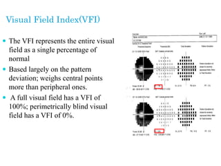
1
Dr. Md. Ashfakur Rahaman (Rayhan)
D.O Student
RpMC
Definition of Perimetry:
Defined as Numeric, Graphical and Symbolic
presentation of visual field.
Objectives:
 Identification and quantification of abnormal visual
fields
 Longitudinal assessment to detect glaucomatous
progression and measure rates of change.
The Visual Field:
The visual field can be represented as a three-
dimensional structure akin to a hill of increasing
sensitivity.
THE NORMAL VISUAL
FIELD :
50˚ SUPERIORLY
60˚ NASALLY
70˚ INFERIORLY
90˚ TEMPORALLY
Common terms used in V.F & Perimetry
 An isopter is a line connecting points of the same
sensitivity, and on a two-dimensional isopter plot
encloses an area within which a stimulus of a given
strength is visible.
 When the field is represented as a hill, isopters
resemble the contour lines on a map.
Cont….
 A scotoma is an area of reduced (‘relative’) or total
(‘absolute’) loss of vision surrounded by a seeing
area.
 Luminance is the intensity or ‘brightness’ of a light
stimulus, measured in apostilbs (asb).
 Scotoma
Cont…
Stimulus brightness:
 Apostilbe (Asb): Unit of intensity of light projected as
stimulus.
 Decibel (dB): Unit of neutral density filters used to make
stimulus dimmer.
0dB(withoutfilter)=10,000Asb
(Humphrey)
10dB = 1/10th = 1000Asb
20dB = 1/100th = 100Asb etc.
(10dB = 1Lu)
Cont…
 Threshold at a given location in the visual field is the
brightness of a stimulus at which it can be detected by
the subject.
 After the age of 20 years the sensitivity decreases by
about 1 dB per 10 years.
Threshold
Cont…
 Background luminance: The retinal sensitivity at
any location varies depending on background
luminance.
 Rod photoreceptors are more sensitive in dim light.
 Cone photoreceptors are more sensitive in Bright light
 Peripheral retina more sensitive in proportion to the
central retina.
Types of perimetry
 Kinetic
Goldmann
Lister
Bjerrum s Screen
 Static
Humphrey
Octopus
Henson
Automated Static Perimetry
 To find visual field sensitivity at pre-selected
locations
 Presenting stimulus of known size and brightness
 known background for known amount of time.
 We measure differential light sensitivity or contrast
sensitivity.
Testing algorithms
Threshold
 Used for detailed assessment of the hill of vision
 By plotting the threshold luminance value at various
locations
Cont…
Suprathreshold
 Testing with stimuli of luminance above the expected
normal threshold levels
 It enables testing to be carried out rapidly
 Reserved for screening.
Fast algorithms
 Introduced with shorter testing times,
 Providing efficiency benefits with little or no
detriment to testing accuracy.
 SITA-Standard and SITA-Fast versions are available
Stimulus inteansity
in decibel value
20
∞ Retinal Sensitivity
Testing
protocols
Testing Protocols
 Most common ---- central 30-2 (76 points) or 24-2 (54 points),
 Neurological disease – 30-2 or 24-2,
 Small central scotoma – 10-2 (68 points)
 Peripheral – 60-4
 Macula
30-1 30-2
SITA (Swedish interactive threshold algorithm)
 Dramatic reduction of test time
without sacrificing accuracy.
 More accurate choice of stimulus
brightness.
 Complete use of patient information.
 Test pace completely determined by
patient.
INTERPRETATIONS
Zone 1 Patient data & test data
Zone 2 Foveal Therashold & Reliable
indices
Zone 3 Gray Scale
Zone 4 Total Deviation Plot
Zone 5 Pattern Deviation Plot
Zone 6 Global Indices
Zone 7 Glaucoma Hemifield test
Zone 8 Raw data
 Zone 1 Patient data & test data
 Zone 2 Foveal Therashold &
Reliable indices
 Zone 3 Gray Scale
 Zone 4 Total Deviation Plot
 Zone 5 Pattern Deviation Plot
 Zone 6 Global Indices
 Zone 7 Glaucoma Hemifield
test
 Zone 8 Raw Data
RELIABILITY INDICES
Reliability indices
 B) False positive error rates :
 Should be < 33%
 Increased fixation loss value.
 C) False negative error rates :
 Should be < 33% ( No ‘xx’ for FN %
in newer SITA strategy).
Reliability indices –
a) Gaze stability –
 Heijl-Krakau blind spot
method - Fixation loss
should be <1/5th
 Gaze monitor
 Video monitoring
Single Field Printout:zone-3
 Interpolated gray
scale.(within green
ring)
Total Deviation plots-zone-4
I . Numeric value in upper plot - diff. in dB between the
patients test results and the age corrected normal values at each
tested point in the visual Field. Value is abnormal -> if 5 dB
less than normal .
 2. Gray -scale symbols in lower plot -> translates values in
upper plot, Darker the symbols, more the depth of defect.
 Total deviation (from
normal values for age) -
 Normal sensitivity
range – larger peripherally
than centrally and
superiorly than inferiorly.
Pattern Deviation
Is similar to total deviation
Except that any generalised depression in the field caused by
other factors such as lens opacities or miosis.
 Pattern deviation :zone-5
 Single most useful analysis
 For exposing localized
defect masked by
generalized depression or
elevation
 Early field defect appear
first in probability map
 Doesn’t come in very
advanced field defects
P- Value
 Probability (Chance) - Chances of occurrence by chance
expressed by 'P'. Ranges -> from '0' to I . P = 0, means no
chance of an event happening or its occurrence is impossible
e.g. Chances of survival after rabies are zero or nil .
 P = 1, means chances of an event happening are 100% e.g.
death of any living being.
 P value -> Significant in 5% level, (95% confidence limit)
40
Uniform total dev. plot
depression but normal pattern
dev. plot – Media opacity
Normal total dev. but
abnormal pattern dev.–
trigger happy pt.
Statistical Analysis: zone; 6
Global indices :
 Mean deviation (M.D.) –
 Denote average severity of field
loss.
 Pattern standard dev. (PSD) –
 It is the S.D. around M.D.
 ↑ PSD - ↑ focal defect.
 Global indices:
 Short term fluctuation (SF) -
 Index of intra test measurement variability.
 Between 1-2.5 dB.
 Significance limited by small no. of points tested.
 High in inconsistency and in diseased area (not done in
SITA).
 Corrected PSD (CPSD) -
 When SF removed from PSD.
 5 Zones in upper
field compared with
5 mirror-image zones
in lower field
In 30-2 and 24-2
tests only
Glaucoma hemifield test
(GHT):zone-7
Glaucoma Hemifield Test (GHT)
 Five possible messages :
Outside normal limits (one or more zone difference by
P < 0.01)
Borderline
General reduction of sensitivity
Abnormally high sensitivity
Within normal limits
Visual Field Index(VFI)
 The VFI represents the entire visual
field as a single percentage of
normal
 Based largely on the pattern
deviation; weighs central points
more than peripheral ones.
 A full visual field has a VFI of
100%; perimetrically blind visual
field has a VFI of 0%.
Overview -
Minimum 4 field
tests -- for evidence of
progression. 6 or
more -- for certainty.
Glaucoma Progression
Analysis(GPA)
 Like GCP, but smarter
 Adjusts for cataract
 Deterioration from baseline at
P<5% - ∆
 Deterioration at same location, 2
consec. tests –
 Deterioration at same location, 3
consec. tests –▲
Artifact
 Edge artifact:
 Eye brow
 Lens rim
Artifact due to
patient fatigue
(clover leaf
pattern)
53
Stimulus – Blue (440 nm),
Size V
Background – Intense
yellow
Can detect presence and
progression of glaucomatous
V. F. change several years
earlier than standard white
on white auto perimetry
(SAP).
FDP
(Frequency Doubling Perimetry)
55
Microperimetry
 Microperimetry is a visual field test that measures
retinal sensitivity and fixation behaviour in patients
with macular disease and focal glaucoma involving
the central visual field.
56
Cont...
 Goldmann size 3 stimuli are projected onto the central
9° of the fundus.
 A 4-2 staircase thresholding technique is used,
 Using this instrument normal retinal sensitivity is 18
dB.
Cont...
 Results are printed, together with reliability indices
(fixation loss), probabilities, and retinal sensitivity
values,
 Which are colour-coded.
 It can also be helpful in patients with early glaucoma,
particularly when there is subtle change close to
fixation.
Sources of error in perimetry
 Inexperienced or unskilled technician
 Incorrect patient details
 Miosis
 Lens opacities
 Uncorrected refractive error
 Spectacles rim artefact
 Ptosis
 Poor patient performance
 Inadequate retinal adaptation
62

Visual field Analysis .ppt

  • 1.
    1 Dr. Md. AshfakurRahaman (Rayhan) D.O Student RpMC
  • 2.
    Definition of Perimetry: Definedas Numeric, Graphical and Symbolic presentation of visual field.
  • 3.
    Objectives:  Identification andquantification of abnormal visual fields  Longitudinal assessment to detect glaucomatous progression and measure rates of change.
  • 4.
    The Visual Field: Thevisual field can be represented as a three- dimensional structure akin to a hill of increasing sensitivity.
  • 5.
    THE NORMAL VISUAL FIELD: 50˚ SUPERIORLY 60˚ NASALLY 70˚ INFERIORLY 90˚ TEMPORALLY
  • 7.
    Common terms usedin V.F & Perimetry  An isopter is a line connecting points of the same sensitivity, and on a two-dimensional isopter plot encloses an area within which a stimulus of a given strength is visible.  When the field is represented as a hill, isopters resemble the contour lines on a map.
  • 8.
    Cont….  A scotomais an area of reduced (‘relative’) or total (‘absolute’) loss of vision surrounded by a seeing area.  Luminance is the intensity or ‘brightness’ of a light stimulus, measured in apostilbs (asb).
  • 9.
  • 10.
    Cont… Stimulus brightness:  Apostilbe(Asb): Unit of intensity of light projected as stimulus.  Decibel (dB): Unit of neutral density filters used to make stimulus dimmer. 0dB(withoutfilter)=10,000Asb (Humphrey) 10dB = 1/10th = 1000Asb 20dB = 1/100th = 100Asb etc. (10dB = 1Lu)
  • 11.
    Cont…  Threshold ata given location in the visual field is the brightness of a stimulus at which it can be detected by the subject.  After the age of 20 years the sensitivity decreases by about 1 dB per 10 years.
  • 12.
  • 13.
    Cont…  Background luminance:The retinal sensitivity at any location varies depending on background luminance.  Rod photoreceptors are more sensitive in dim light.  Cone photoreceptors are more sensitive in Bright light  Peripheral retina more sensitive in proportion to the central retina.
  • 14.
    Types of perimetry Kinetic Goldmann Lister Bjerrum s Screen  Static Humphrey Octopus Henson
  • 15.
    Automated Static Perimetry To find visual field sensitivity at pre-selected locations  Presenting stimulus of known size and brightness  known background for known amount of time.  We measure differential light sensitivity or contrast sensitivity.
  • 16.
    Testing algorithms Threshold  Usedfor detailed assessment of the hill of vision  By plotting the threshold luminance value at various locations
  • 18.
    Cont… Suprathreshold  Testing withstimuli of luminance above the expected normal threshold levels  It enables testing to be carried out rapidly  Reserved for screening.
  • 19.
    Fast algorithms  Introducedwith shorter testing times,  Providing efficiency benefits with little or no detriment to testing accuracy.  SITA-Standard and SITA-Fast versions are available
  • 20.
    Stimulus inteansity in decibelvalue 20 ∞ Retinal Sensitivity
  • 21.
  • 22.
    Testing Protocols  Mostcommon ---- central 30-2 (76 points) or 24-2 (54 points),  Neurological disease – 30-2 or 24-2,  Small central scotoma – 10-2 (68 points)  Peripheral – 60-4  Macula
  • 23.
  • 24.
    SITA (Swedish interactivethreshold algorithm)  Dramatic reduction of test time without sacrificing accuracy.  More accurate choice of stimulus brightness.  Complete use of patient information.  Test pace completely determined by patient.
  • 26.
  • 27.
    Zone 1 Patientdata & test data Zone 2 Foveal Therashold & Reliable indices Zone 3 Gray Scale Zone 4 Total Deviation Plot Zone 5 Pattern Deviation Plot Zone 6 Global Indices Zone 7 Glaucoma Hemifield test Zone 8 Raw data
  • 28.
     Zone 1Patient data & test data  Zone 2 Foveal Therashold & Reliable indices  Zone 3 Gray Scale  Zone 4 Total Deviation Plot  Zone 5 Pattern Deviation Plot  Zone 6 Global Indices  Zone 7 Glaucoma Hemifield test  Zone 8 Raw Data
  • 30.
  • 31.
    Reliability indices  B)False positive error rates :  Should be < 33%  Increased fixation loss value.  C) False negative error rates :  Should be < 33% ( No ‘xx’ for FN % in newer SITA strategy).
  • 32.
    Reliability indices – a)Gaze stability –  Heijl-Krakau blind spot method - Fixation loss should be <1/5th  Gaze monitor  Video monitoring
  • 33.
    Single Field Printout:zone-3 Interpolated gray scale.(within green ring)
  • 34.
    Total Deviation plots-zone-4 I. Numeric value in upper plot - diff. in dB between the patients test results and the age corrected normal values at each tested point in the visual Field. Value is abnormal -> if 5 dB less than normal .  2. Gray -scale symbols in lower plot -> translates values in upper plot, Darker the symbols, more the depth of defect.
  • 35.
     Total deviation(from normal values for age) -  Normal sensitivity range – larger peripherally than centrally and superiorly than inferiorly.
  • 36.
    Pattern Deviation Is similarto total deviation Except that any generalised depression in the field caused by other factors such as lens opacities or miosis.
  • 37.
     Pattern deviation:zone-5  Single most useful analysis  For exposing localized defect masked by generalized depression or elevation  Early field defect appear first in probability map  Doesn’t come in very advanced field defects
  • 39.
    P- Value  Probability(Chance) - Chances of occurrence by chance expressed by 'P'. Ranges -> from '0' to I . P = 0, means no chance of an event happening or its occurrence is impossible e.g. Chances of survival after rabies are zero or nil .  P = 1, means chances of an event happening are 100% e.g. death of any living being.  P value -> Significant in 5% level, (95% confidence limit)
  • 40.
  • 41.
    Uniform total dev.plot depression but normal pattern dev. plot – Media opacity Normal total dev. but abnormal pattern dev.– trigger happy pt.
  • 42.
    Statistical Analysis: zone;6 Global indices :  Mean deviation (M.D.) –  Denote average severity of field loss.  Pattern standard dev. (PSD) –  It is the S.D. around M.D.  ↑ PSD - ↑ focal defect.
  • 43.
     Global indices: Short term fluctuation (SF) -  Index of intra test measurement variability.  Between 1-2.5 dB.  Significance limited by small no. of points tested.  High in inconsistency and in diseased area (not done in SITA).  Corrected PSD (CPSD) -  When SF removed from PSD.
  • 44.
     5 Zonesin upper field compared with 5 mirror-image zones in lower field In 30-2 and 24-2 tests only Glaucoma hemifield test (GHT):zone-7
  • 45.
    Glaucoma Hemifield Test(GHT)  Five possible messages : Outside normal limits (one or more zone difference by P < 0.01) Borderline General reduction of sensitivity Abnormally high sensitivity Within normal limits
  • 46.
    Visual Field Index(VFI) The VFI represents the entire visual field as a single percentage of normal  Based largely on the pattern deviation; weighs central points more than peripheral ones.  A full visual field has a VFI of 100%; perimetrically blind visual field has a VFI of 0%.
  • 48.
    Overview - Minimum 4field tests -- for evidence of progression. 6 or more -- for certainty.
  • 49.
    Glaucoma Progression Analysis(GPA)  LikeGCP, but smarter  Adjusts for cataract  Deterioration from baseline at P<5% - ∆  Deterioration at same location, 2 consec. tests –  Deterioration at same location, 3 consec. tests –▲
  • 50.
    Artifact  Edge artifact: Eye brow  Lens rim
  • 51.
    Artifact due to patientfatigue (clover leaf pattern)
  • 53.
    53 Stimulus – Blue(440 nm), Size V Background – Intense yellow Can detect presence and progression of glaucomatous V. F. change several years earlier than standard white on white auto perimetry (SAP).
  • 54.
  • 55.
  • 56.
    Microperimetry  Microperimetry isa visual field test that measures retinal sensitivity and fixation behaviour in patients with macular disease and focal glaucoma involving the central visual field. 56
  • 58.
    Cont...  Goldmann size3 stimuli are projected onto the central 9° of the fundus.  A 4-2 staircase thresholding technique is used,  Using this instrument normal retinal sensitivity is 18 dB.
  • 59.
    Cont...  Results areprinted, together with reliability indices (fixation loss), probabilities, and retinal sensitivity values,  Which are colour-coded.  It can also be helpful in patients with early glaucoma, particularly when there is subtle change close to fixation.
  • 61.
    Sources of errorin perimetry  Inexperienced or unskilled technician  Incorrect patient details  Miosis  Lens opacities  Uncorrected refractive error  Spectacles rim artefact  Ptosis  Poor patient performance  Inadequate retinal adaptation
  • 62.