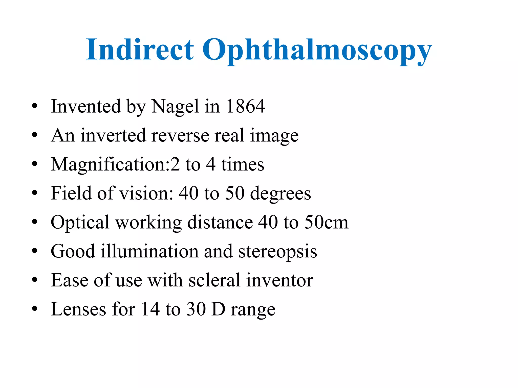
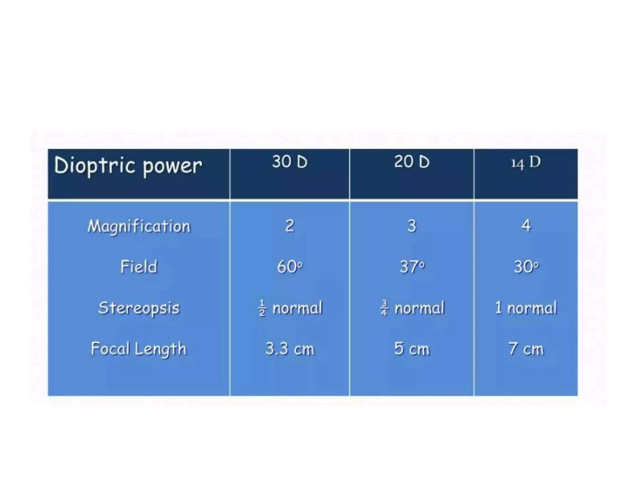
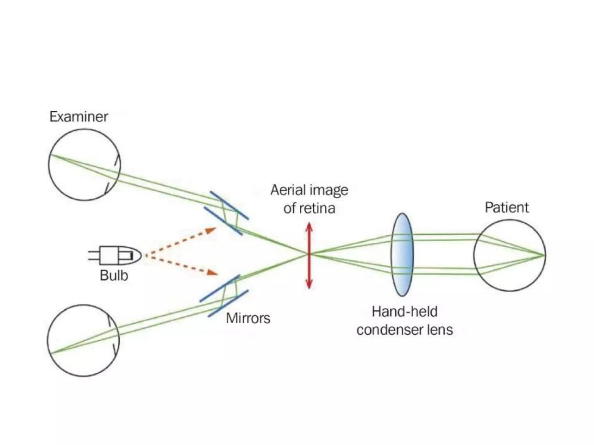
Indirect ophthalmoscopy allows examination of the retina using an inverted magnified image. A strong convex lens is placed near the eye to make the eye highly myopic. This brings light from the retina to a focused real image between the lens and observer. The image size and position depends on the eye's refractive error - it is at the lens focus for emmetropia, in front for myopia, and behind for hyperopia. Indirect ophthalmoscopy provides a large field of view useful for retinal pathology assessment and laser treatment. Though difficult to learn, it allows examination of peripheral retina and is useful in media opacity.