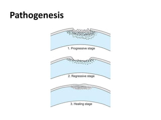
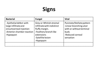
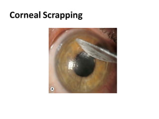
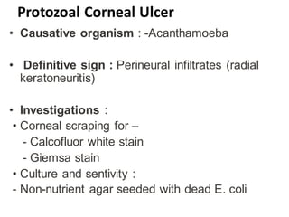
CORNEALULCER
Dr.Md.Ashfakur Rahaman
DO Student
Rangpur Medical College
Contents
• Applied anatomy of the cornea
• Definition of corneal ulcer
• Classification
• Predisposing factors
• Pathogenesis
• Clinical presentation
• Management
• Complications
Defination
Refers to corneal tissue excavation
associated with an epithelial defect, usually
with infiltration and necrosis.
Histology of Cornea
Classification Corneal ulcers
Corneal ulcers can be classified in three ways :
A. On the basis of Aetiology
B. On the basis of Location of ulcer
C. On the basis of Involvement of the corneal
layers
A.On the basis of Aetiology
A. Infective:
- Bacterial
- Viral
- Fungal
- Protozoal
B. Non-infective/sterile
-Neuroparalytic
-Neurotrophic
-Corneal ulcer due to Vit A
deficiency
- Mooren ulcer
B. On the basis of Location
C. On the basis of involvement of the
corneal layers
Predisposing Factors
Local Factors:-
• Contact lens wear
• Ocular trauma
• Dry eye
• Chronic dacryocystitis
• Exophthalmos
• Xerophthalmia
• Systemic Factors :
• Malnutrition
• DM
• Alcoholism
• Malignancy
• Immunosuppressive
drugs
Pathogenesis
Clinical feature
Symptoms
Bacterial Fungal Viral
Pain and Irritation
Blurred Vision
Redness
Photophobia
Watering
Discharge
Eyelid Swelling
Blurred Vision
Redness
Eyelid Swelling
Photophobia
Pain and Irritation
Discomfortand Gritty
feeling
Photophobia
Redness
Watering
Blurred vision
Signs
Bacterial Fungal Viral
-Epithelialdefect with
large infiltrateand
circumcorneal injection
-Anterior chamber reaction
-Hypopyon
Grey or Whitish stromal
infiltratewith indistinct
fluffy margin.
-Feathery branch like
extensions
-Satellitelesion
-Hypopyon
Punctate/Stellatepattern
-Linear branching ulcer
with or without terminal
buds
-Reduced corneal
sensation
BACTERIAL CORNEAL ULCER
FUNGAL CORNEAL ULCER
VIRAL CORNEAL ULCER
Investigations
• Corneal Scrapping for-
Bacterial Fungal Viral
Gram staining 10% KOH
Giemsa stain
PCR
Culture and
Sensitivity
-Blood agar media
-Chocolateagar
media
-McConkayagar
media
Culture and
Sensitivity
-Sabouraud
Dextrose agar
media
Corneal Scrapping
Culture Media
S.Aureus grows on blood agar media
N.Gonorrheagrows onChocolate agar media
Candida grows on Sabouraud dextrose agar media
Protozoal Corneal Ulcer
• Causative organism : -Acanthamoeba
• Definitive sign : Perineural infiltrates (radial
keratoneuritis)
• Investigations :
• Corneal scraping for –
- Calcofluor white stain
- Giemsa stain
• Culture and sentivity :
- Non-nutrient agar seeded with dead E. coli
Protozoal Corneal Ulcer
Treatment of Corneal Ulcer
Principles of treatment
1.Control of infection
2.Relief of symptoms
3.Promotion of heeling
4.Prevention of
complications
5.Treatment of
complications
A.Generally Measures
B.Medical treatment
C.Surgical treatment
General measures
• Hospital admission
• Discontinuation of contact lens wear
• Using eye shield/dark glass
• Improvement of nutrition (Vit–C )
Treatment Bacterial Corneal ulcer
Medical Treatment :-
Topical Antibiotic
•Antibiotic monotherapy
•Antibiotic duotherapy
Cycloplegics
Analgesic
Systemic Therapy
– Systemic antibiotic
Surgical treatment:- (if medical
treatment failed)
• Conjuctival hooding
• Tarsorrhaphy
• Amniotic membrane graft
• Keratoplasty
Treatment Fungal Corneal Ulcer
Topical Therapy :
• Topical antifungal
• Cycloplegics
• Subconjunctival injection
• Anterior chamber washout
• Removal of the epithelium
Systemic Therapy :
• Antifungals
• Tetracycline/ Doxicycline
Treatment Viral Corneal Ulcer
Local Therapy :-
• Topical Antiviral
• Antibiotic prophylaxis
• Topical steroids
• Cycloplegics
Systemic Therapy :
• Oral antiviral
• Interferon monotherapy
Treatment Cont...
Local care :
• Debridement of ulcer
• Care of skin lesions
- aciclovir cream five times daily
• IOP control
Treatment Protozoal Corneal Ulcer
• Topical amoebicides
• Topical antibiotic
• Topical steroid
• Control of pain
• Therapeutic Keratoplasty
• Debridement of involved epithelium
Management of apparent
treatment failure :
- Frequent instillation of fortified
aminoglycosides
- Review of antibiotic regimen
- Re-scraping and additional staining
- Corneal biopsy
Complication
Complications of corneal ulcer can be
described under two headings :
1.Before perforation
2.After perforation
Before perforation :
• Anterior uveitis
• Hypopyon
• Secondary glaucoma
• Descemetocele
• Corneal scarring - Nebula - Macula -
Leucoma
Beforeperforation
After perforation :
• Anterior synaechiae
• Iris prolapse
• Adherent leucoma
• Complicated cataract
• Sublaxation of the lens
• Anterior staphyloma
• Endophthalmitis
• Phthisis bulbi
After perforation
Treatment of Complication
Management of perforation :
-Tissue adhesive glue with bandage
contact lens.
-Corneal patch graft
- Occlusive surface repair techniques
- Penetrating keratoplasty
Treatment of Complication Cont..
• Management of endophthalmitis
• Visual rehabilitation :
-Keratoplasty
-Rigid contact lens
-Cataract surgery
Tissue adhesive glue
Amniotic membrane graft
Keratoplasty
Neurotrophic keratitis/
neurotrophic keratopathy
• Neurotrophic keratopathy is a corneal
degenerative disease characterized by a
reduction or absence of corneal
sensitivity.Usually associated with Herpes
keratitis,Topical anesthetic abuse,Corneal
surgery.
Clinical features
• A full cranial nerve
examination is
mandatory.
Symptoms:-
-Redness
-Swollen lid and defective
vison
-No pain,No tearing
• Sign:-
- Reduced Corneal
Sensation
- Cilliary Congestion
- Interpalpebral epithelial
irregularity
-Rolled and thickened
edges
-Stromal melting
Treatment
• Discontinuation
• Topical lubricant
• Anticollagenase agents
• Cenegermin
Exposure Keratopathy
• Is result of incomplete lid
closure(lagophthalmos),withdrying of cornea
despite normal tear production.
• Causes include neuroparalytic, Facial nerve
palsy,Parkinsonism,lid scaring,post
blepharoplasty,proptosis etc.
Clinical features
Symptoms :-
• Irritation
• Burning sensation
• Foreign body sensation
• Intolerance to drafts
and winds.
Signs:-
• Drying of cornea
• Punctate epithelial
defects
• Corneal ulceration
Treatment
• Artificial tears
• Instillationof ointment
• Soft bandage contact lens
• Tarsorrhaphy
• Treatment of cause
Peripheral Ulcerative Keratopathies
• Marginal keratitis (catarrhal ulcer)
• Mooren‘s ulcer
• Terrien marginal degeration
• Dellen
• Associated with systemic autoimmune
diseases
Marginal Keratitis
• Superficial ulcers situated near the limbus,
Common in old people usually cuased by
hypersensitivity reaction to Staphylococcal
toxin.
Clinical features
• Mild ocular irritation,pain,photophobia and
watering.
• Ulcer is shallow, slightly infiltrated and often
multiple.
• Vascularization
• Recurrence very common
Treatment
• Topical steroid
• Antibiotic
Mooren’s ulcer/chronic serpiginous
/rodent ulcer
• Rare autoimmune disease
• Progressive circumferential peripheral stromal
ulceration
• Often unilateral
• Mainly affect older people
Clinical features
• Symptoms
-Pain
-Photophobia
-Blurred Vision
• Signs
-Peripheral
ulceration(involve 1/3 of
stroma)
-Undermined and
Infiltrated edge
-Limbitis
-Vascularization
Mooren’s Ulcer
Treatment
Medical
• Topical steroid
• Topical Ciclosporin
• Tacrolimus ointment
• Artificial tears
• Collagenase inhibitors
• Systemic
immunosuppression
Surgical
• Conjuctival resection
• Lamellar keratectomy
• Visual rehabilitation
-Keratoplasty
Conclusion
Timely diagnosis and proper management of
corneal ulcer can save the valuable vision of
the patient
THANK YOU

Corneal Ulcer.pdf