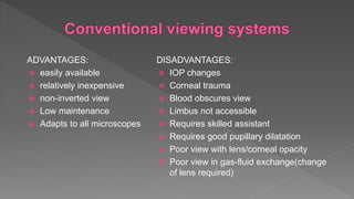
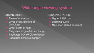
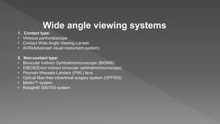
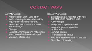
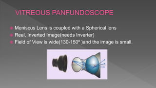
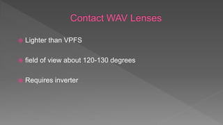
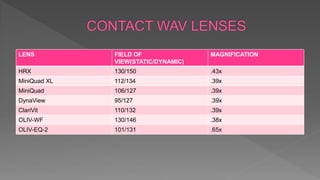
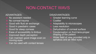
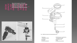
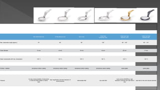
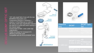
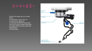
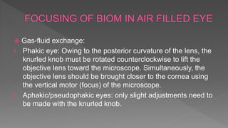
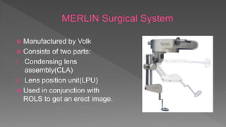
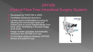
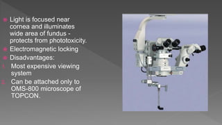
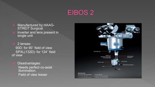
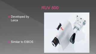
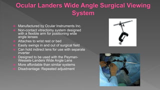
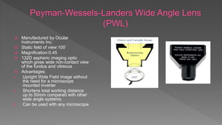
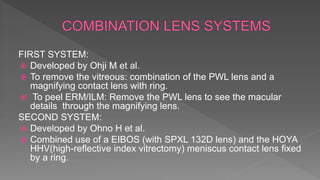
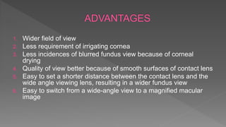
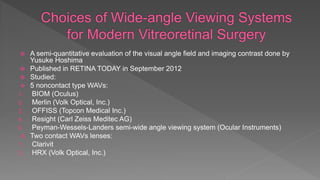
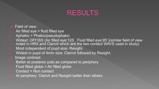
ADVANTAGES:
 easily available
 relatively inexpensive
 non-inverted view
 Low maintenance
 Adapts to all microscopes
DISADVANTAGES:
 IOP changes
 Corneal trauma
 Blood obscures view
 Limbus not accessible
 Requires skilled assistant
 Requires good pupillary dilatation
 Poor view with lens/corneal opacity
 Poor view in gas-fluid exchange(change
of lens required)
ADVANTAGES:
 Ease of operation
 Gives overall picture of
pathology
 Great depth of field
 Easy view in gas-fluid exchange
 Facilitates SOI-PFCL exchange
 Facilitates bimanual surgery
DISADVANTAGES:
 Higher initial cost
 Learning curve
 May need skilled assistant
Wide angle viewing systems
1. Contact type:
• Vitreous panfundoscope
• Contact Wide Angle Viewing Lenses
• AVIS(Advanced visual instrument system)
2. Non-contact type:
• Binocular indirect Ophthalmomicroscope (BIOM®)
• EIBOS(Erect indirect binocular ophthalmomicroscope)
• Peyman-Wessels-Landers (PWL) lens
• Optical fiber-free intravitreal surgery system (OFFISS)
• Merlin™ system
• Resight® 500/700 system
 Made of Prisms
 Uses:
1. Forms erect image
2. Corrects laterality
3. Stereopsis
 Examples:
 SDI
 ROLS
 AVIS
 Invertertube E
 Developed by Spitznas in 1987 along with BIOM.
 Permits the microscopic image to be vertically inverted and laterally
reversed, while retaining full stereopsis.
 Currently SDI4 available.
SDI 4m
The basic version, easy to
mount on nearly every
microscope type. No
voltage
supply or cables required.
The inverting function is
controlled via sterile
covered control knob on the
front side.
SDI 4e
Can be activated by an
optional foot switch.
Some microscope types also
offer the integration of the
switch control into the foot
pedal of the microscope
SDI4c
Most comfortable version.
It is required to control the
motorized focus of the
BIOM 4c. It also can
be used together with any
other version of the
BIOM.
Used with BIOM 4c, the
SDI 4c is activated
 Single-element re-inverter prism design
 Fundus image and surgical instrumentation in the eye presented in an
upright and correctly oriented position
 guarantees left/right eye image fusion, high efficiency light transmission and
high optical transparency
 Diode laser safety filter permanently installed
 Can be used with contact lens,non-contact lens systems(eg. Merlin)
ADVANTAGES:
 Wider field of view (upto 150⁰)
 Self-stabilising lenses (Volk,SSV)
require less skilled assistant.
 Better resolution and contrast of
fundus image
 Cheaper
 Corneal aberrations and reflections
from corneal surface eliminated
 Maintains stereopsis
DISADVANTAGES:
 Skilled assistant required with non-
self stabilising handheld lens.
 IOP changes
 Image lost if eye is rotated
 Lens and corneal opacities
problematic
 Corneal trauma
 Poor access to limbus
 Poor with steep corneal curvatures
 Fixed field of viewing
 Meniscus Lens is coupled with a Spherical lens
 Real, Inverted Image(needs Inverter)
 Field of View is wide(130-150⁰ )and the image is small.
 Lighter than VPFS
 field of view about 120-130 degrees
 Requires inverter
LENS FIELD OF
VIEW(STATIC/DYNAMIC)
MAGNIFICATION
HRX 130/150 .43x
MiniQuad XL 112/134 .39x
MiniQuad 106/127 .39x
DynaView 95/127 .39x
ClariVit 110/132 .39x
OLIV-WF 130/146 .38x
OLIV-EQ-2 101/131 .65x
OLIV-
WF
OLIV-EQ-2 HR
X
MINI
QUAD
MINI QUAD
XL
MINI QUAD
(Autoclavable)
DynaVie CLARIV
 Consists of following parts:
1. AVI stereo inverter-to invert the image
2. 2 miniature contact lenses-one for 68 degree field of view other for 130 degree field
of view.
3. lens handles
4. lens retaining ring
5. Dual mode cannula giving wide field illumination and infusion simultaneously that
fits through a single 20 gauge sclerotomy port
ADVANTAGES:
 No assistant needed
 No corneal trauma
 Good with fluid air exchange
 Good in paediatric cases
 Good for steep corneas
 Ease of accessibility to limbus
 Improved depth perception
 Can maintain good image even on
moving eyes
 Can be used with contact lenses
DISADVANTAGES:
 Greater learning curve
 Costlier
 Adaptabilty to microscopes
 Less resolution
 Dehydration of corneal surface
 Condensation on front lens-proper
draping of the patient
 Wider field of view provided only in
aphakics and air filled eyes
• Manufactured by OCULUS
• Developed by Spitznas in 1987
• Needs to be used with
Stereoscopic Diagonal Inverter
 Reduction lens - fixed focal distance
 Ophthalmoscope lens - Variable distance
 Focal length of reduction lens is chosen such that the system
would be kept in focus when changing from external to
internal observation.
 2 Key elements:
1. Distance between ophthalmoscope lens of BIOM and cornea
2. Distance between microscope and cornea
 To focus - both the distances need to be adjusted with foot pedal.
 At a low microscope magnification: Begin initial focusing of the BIOM image by turning
the BIOM focus adjusting wheel.
 Magnify the image section by actuating the focus foot switch at the microscope.
 Then use the microscope footswitch control to zoom in to maximum magnification.
 Now finely focus the image with the adjusting knob of the BIOM.
 The microscope magnification should then be reduced to the minimum required, in
order to achieve as wide a fundus view as possible.
 The use of the focusing function with the microscope foot pedal when the BIOM is in
use only changes the size of the image field ("keyhole effect").
BIOM 3m BIOM 4m BIOM 5m
Mini Wide Field Lens Hi Res Macula Lens 90 D Lens
Wide Field
Enhanced Lens
Wide Field High
Definition Lens
Wide Field High
Definition Mini
Max. observation angle (approx.) 70° 60° 90° 120° 60° - 130° 60° - 110°
Outer diameter 12 mm 19 mm 19 mm 19 mm 20 mm 17 mm
Steam autoclavable with max. temperature 134° C 134° C 134° C 134° C 134° C 134° C
Surface / material amorphous carbon coating amorphous carbon coating amorphous carbon coating amorphous carbon coating quartz glass quartz glass
Features
for easy maneuverability of instruments
in small and deep seated or infants´
eyes
high magnifcation and best resolution of
small structures
intermediate field very wide field
most common BIOM®lens, high
resolution combined with wide field of
view
wide field of view with reduced diameter
• 130° wide angle field of view with the new
BIOM®HD Disposable Lens
• Outstanding resolution in periphery –
whether in a fluid filled eye or under air
• HD clarity under high magnification reduces
the need for a contact lens when working in
the macula region
• Superb depth of field – even under high
magnification
• Single-use design for increased O.R.
efficiency and lower costs
• Compatible with all OCULUS BIOM® 3/4/5
Objective
lens
BIOM® BIOM® Optic
Set
f = 175 mm BIOM® 3 m/c,
4 m/c, 5 m/c
HD
Professional
for f = 175 mm
f = 200 mm
BIOM® 3 ml/cl,
4 ml/cl, 5 ml/cl
HD
Professional
for f = 200 mm
BIOM® m/c, 4
m/c, 5 m/c
HD Flex
• World’s first single use non-contact
WAVS.
• No sterilization “down time” for
increased O.R. efficiency.
• Incorporates BIOM®HD Disposable
Lens which gives excellent stereopsis
and High Definition clarity under high
magnification.
• Pre-assembled
 Gas-fluid exchange:
1. Phakic eye: Owing to the posterior curvature of the lens, the
knurled knob must be rotated counterclockwise to lift the
objective lens toward the microscope. Simultaneously, the
objective lens should be brought closer to the cornea using
the vertical motor (focus) of the microscope.
2. Aphakic/pseudophakic eyes: only slight adjustments need to
be made with the knurled knob.
 Manufactured by Volk
 Consists of two parts:
1. Condensing lens
assembly(CLA)
2. Lens position unit(LPU)
 Used in conjunction with
ROLS to get an erect image.
 Condensing lens Unit:
Recognizes the surgical lens
position to switch the internal
condensing lens into, and out of,
the optical pathway as needed.
 Lens Positioning Unit:
1. Allows for fine focus
adjustment of the vitreous and
retina.
2. Can be rotated 360° to provide
precise lens positioning or aid
in convenient positioning for
the surgeon or assistant.
 Manufactured by ZEISS.
 System automatically fold up if
unintended contact is made with
the patient's eye guaranteeing
optimal patient safety.
 RESIGHT 700-foot control panel to
focus while your hands remain in
the surgical field.
 RESIGHT 500-manual focusing
 Automatic inversion with
Invertertube® E allow a smooth
workflow with focus on the patient
1. Base and focusing unit
2. Lens support with lens
turret for 175 and 200 mm
lenses.
3. Re-sterilizable aspheric
lenses (60 D and 128 D)
Disadvantage:
Can only be used with Zeiss
microscopes.
 Developed by TOPCON in 2003
 Facilitates bimanual vitrectomy
 Lenses avoid complicated focusing by
allowing the microscope head and
indirect lens to move independently from
each other, facilitating a focused image
at all times.
 Image inverter activates automatically
whenever the OFFISS is in use
 Comfortable distance between OFFISS
lenses and patient’s eye.
 Light is focused near
cornea and illuminates
wide area of fundus -
protects from phototoxicity.
 Electromagnetic locking
 Disadvantages:
1. Most expensive viewing
system
2. Can be attached only to
OMS-800 microscope of
TOPCON.
 Manufactured by HAAG-
STREIT Surgical.
 Inverter and lens present in
single unit
 2 lenses:
90D: for 90˚ field of view
SPXL(132D): for 124˚ field
of view
 Disadvantages:
1. Needs perfect co-axial
illumination.
2. Field of view lesser
 Developed by
Leica
 Similar to EIBOS
 Manufactured by Ocular Instruments Inc.
 Non-contact vitrectomy system designed
with a flexible arm for positioning wide
angle lenses
 Attaches to wrist rest or bed
 Easily swings in and out of surgical field
 Can hold indirect lens for use with separate
inverter
 Designed to be used with the Peyman-
Wessels-Landers Wide Angle Lens
 More affordable than similar systems
 Disadvantage: Repeated adjustment
 Manufactured by Ocular
Instruments Inc.
 Static field of view:100˚
 Magnification:0.45
 132D aspheric imaging optic
which gives wide non-contact view
of the fundus and vitreous
 Advantages:
1. Upright Wide Field image without
the need for a microscope
mounted inverter
2. Shortens total working distance
up to 50mm compared with other
wide angle systems.
3. Can be used with any microscope
FIRST SYSTEM:
 Developed by Ohji M et al.
 To remove the vitreous: combination of the PWL lens and a
magnifying contact lens with ring.
 To peel ERM/ILM: Remove the PWL lens to see the macular
details through the magnifying lens.
SECOND SYSTEM:
 Developed by Ohno H et al.
 Combined use of a EIBOS (with SPXL 132D lens) and the HOYA
HHV(high-reflective index vitrectomy) meniscus contact lens fixed
by a ring.
1. Wider field of view
2. Less requirement of irrigating cornea
3. Less incidences of blurred fundus view because of corneal
drying
4. Quality of view better because of smooth surfaces of contact lens
5. Easy to set a shorter distance between the contact lens and the
wide angle viewing lens, resulting in a wider fundus view
6. Easy to switch from a wide-angle view to a magnified macular
image
 A semi-quantitative evaluation of the visual angle field and imaging contrast done by
Yusuke Hoshima
 Published in RETINA TODAY in September 2012
 Studied:
 5 noncontact type WAVs:
1. BIOM (Oculus)
2. Merlin (Volk Optical, Inc.)
3. OFFISS (Topcon Medical Inc.)
4. Resight (Carl Zeiss Meditec AG)
5. Peyman-Wessels-Landers semi-wide angle viewing system (Ocular Instruments)
 Two contact WAVs lenses:
1. Clarivit
2. HRX (Volk Optical, Inc.)
 Field of view:
1. Air filled eye > fluid filled eye
2. Aphakic > Phakic/pseudophakic
3. Widest: OFFISS (Air filled eye:125˚, Fluid filled eye:95˚)(similar field of view
noted in HRX and Clarivit which are the two contact WAVS used in study)
4. Most independent of pupil size: Resight
5. Widest in pupil of 4mm size: Clarivit followed by Resight.
 Image contrast:
1. Better at posterior pole as compared to periphery
2. Fluid filled globe > Air filled globe
3. Contact > Non contact
4. At periphery: Clarivit and Resight better than others

Wide angle viewing systems

  • 2.
    ADVANTAGES:  easily available relatively inexpensive  non-inverted view  Low maintenance  Adapts to all microscopes DISADVANTAGES:  IOP changes  Corneal trauma  Blood obscures view  Limbus not accessible  Requires skilled assistant  Requires good pupillary dilatation  Poor view with lens/corneal opacity  Poor view in gas-fluid exchange(change of lens required)
  • 3.
    ADVANTAGES:  Ease ofoperation  Gives overall picture of pathology  Great depth of field  Easy view in gas-fluid exchange  Facilitates SOI-PFCL exchange  Facilitates bimanual surgery DISADVANTAGES:  Higher initial cost  Learning curve  May need skilled assistant
  • 4.
    Wide angle viewingsystems 1. Contact type: • Vitreous panfundoscope • Contact Wide Angle Viewing Lenses • AVIS(Advanced visual instrument system) 2. Non-contact type: • Binocular indirect Ophthalmomicroscope (BIOM®) • EIBOS(Erect indirect binocular ophthalmomicroscope) • Peyman-Wessels-Landers (PWL) lens • Optical fiber-free intravitreal surgery system (OFFISS) • Merlin™ system • Resight® 500/700 system
  • 5.
     Made ofPrisms  Uses: 1. Forms erect image 2. Corrects laterality 3. Stereopsis  Examples:  SDI  ROLS  AVIS  Invertertube E
  • 6.
     Developed bySpitznas in 1987 along with BIOM.  Permits the microscopic image to be vertically inverted and laterally reversed, while retaining full stereopsis.  Currently SDI4 available.
  • 7.
    SDI 4m The basicversion, easy to mount on nearly every microscope type. No voltage supply or cables required. The inverting function is controlled via sterile covered control knob on the front side. SDI 4e Can be activated by an optional foot switch. Some microscope types also offer the integration of the switch control into the foot pedal of the microscope SDI4c Most comfortable version. It is required to control the motorized focus of the BIOM 4c. It also can be used together with any other version of the BIOM. Used with BIOM 4c, the SDI 4c is activated
  • 8.
     Single-element re-inverterprism design  Fundus image and surgical instrumentation in the eye presented in an upright and correctly oriented position  guarantees left/right eye image fusion, high efficiency light transmission and high optical transparency  Diode laser safety filter permanently installed  Can be used with contact lens,non-contact lens systems(eg. Merlin)
  • 9.
    ADVANTAGES:  Wider fieldof view (upto 150⁰)  Self-stabilising lenses (Volk,SSV) require less skilled assistant.  Better resolution and contrast of fundus image  Cheaper  Corneal aberrations and reflections from corneal surface eliminated  Maintains stereopsis DISADVANTAGES:  Skilled assistant required with non- self stabilising handheld lens.  IOP changes  Image lost if eye is rotated  Lens and corneal opacities problematic  Corneal trauma  Poor access to limbus  Poor with steep corneal curvatures  Fixed field of viewing
  • 10.
     Meniscus Lensis coupled with a Spherical lens  Real, Inverted Image(needs Inverter)  Field of View is wide(130-150⁰ )and the image is small.
  • 11.
     Lighter thanVPFS  field of view about 120-130 degrees  Requires inverter
  • 12.
    LENS FIELD OF VIEW(STATIC/DYNAMIC) MAGNIFICATION HRX130/150 .43x MiniQuad XL 112/134 .39x MiniQuad 106/127 .39x DynaView 95/127 .39x ClariVit 110/132 .39x OLIV-WF 130/146 .38x OLIV-EQ-2 101/131 .65x
  • 13.
    OLIV- WF OLIV-EQ-2 HR X MINI QUAD MINI QUAD XL MINIQUAD (Autoclavable) DynaVie CLARIV
  • 14.
     Consists offollowing parts: 1. AVI stereo inverter-to invert the image 2. 2 miniature contact lenses-one for 68 degree field of view other for 130 degree field of view. 3. lens handles 4. lens retaining ring 5. Dual mode cannula giving wide field illumination and infusion simultaneously that fits through a single 20 gauge sclerotomy port
  • 15.
    ADVANTAGES:  No assistantneeded  No corneal trauma  Good with fluid air exchange  Good in paediatric cases  Good for steep corneas  Ease of accessibility to limbus  Improved depth perception  Can maintain good image even on moving eyes  Can be used with contact lenses DISADVANTAGES:  Greater learning curve  Costlier  Adaptabilty to microscopes  Less resolution  Dehydration of corneal surface  Condensation on front lens-proper draping of the patient  Wider field of view provided only in aphakics and air filled eyes
  • 16.
    • Manufactured byOCULUS • Developed by Spitznas in 1987 • Needs to be used with Stereoscopic Diagonal Inverter
  • 17.
     Reduction lens- fixed focal distance  Ophthalmoscope lens - Variable distance  Focal length of reduction lens is chosen such that the system would be kept in focus when changing from external to internal observation.
  • 18.
     2 Keyelements: 1. Distance between ophthalmoscope lens of BIOM and cornea 2. Distance between microscope and cornea  To focus - both the distances need to be adjusted with foot pedal.  At a low microscope magnification: Begin initial focusing of the BIOM image by turning the BIOM focus adjusting wheel.  Magnify the image section by actuating the focus foot switch at the microscope.  Then use the microscope footswitch control to zoom in to maximum magnification.  Now finely focus the image with the adjusting knob of the BIOM.  The microscope magnification should then be reduced to the minimum required, in order to achieve as wide a fundus view as possible.  The use of the focusing function with the microscope foot pedal when the BIOM is in use only changes the size of the image field ("keyhole effect").
  • 19.
    BIOM 3m BIOM4m BIOM 5m
  • 20.
    Mini Wide FieldLens Hi Res Macula Lens 90 D Lens Wide Field Enhanced Lens Wide Field High Definition Lens Wide Field High Definition Mini Max. observation angle (approx.) 70° 60° 90° 120° 60° - 130° 60° - 110° Outer diameter 12 mm 19 mm 19 mm 19 mm 20 mm 17 mm Steam autoclavable with max. temperature 134° C 134° C 134° C 134° C 134° C 134° C Surface / material amorphous carbon coating amorphous carbon coating amorphous carbon coating amorphous carbon coating quartz glass quartz glass Features for easy maneuverability of instruments in small and deep seated or infants´ eyes high magnifcation and best resolution of small structures intermediate field very wide field most common BIOM®lens, high resolution combined with wide field of view wide field of view with reduced diameter
  • 21.
    • 130° wideangle field of view with the new BIOM®HD Disposable Lens • Outstanding resolution in periphery – whether in a fluid filled eye or under air • HD clarity under high magnification reduces the need for a contact lens when working in the macula region • Superb depth of field – even under high magnification • Single-use design for increased O.R. efficiency and lower costs • Compatible with all OCULUS BIOM® 3/4/5 Objective lens BIOM® BIOM® Optic Set f = 175 mm BIOM® 3 m/c, 4 m/c, 5 m/c HD Professional for f = 175 mm f = 200 mm BIOM® 3 ml/cl, 4 ml/cl, 5 ml/cl HD Professional for f = 200 mm BIOM® m/c, 4 m/c, 5 m/c HD Flex
  • 22.
    • World’s firstsingle use non-contact WAVS. • No sterilization “down time” for increased O.R. efficiency. • Incorporates BIOM®HD Disposable Lens which gives excellent stereopsis and High Definition clarity under high magnification. • Pre-assembled
  • 23.
     Gas-fluid exchange: 1.Phakic eye: Owing to the posterior curvature of the lens, the knurled knob must be rotated counterclockwise to lift the objective lens toward the microscope. Simultaneously, the objective lens should be brought closer to the cornea using the vertical motor (focus) of the microscope. 2. Aphakic/pseudophakic eyes: only slight adjustments need to be made with the knurled knob.
  • 24.
     Manufactured byVolk  Consists of two parts: 1. Condensing lens assembly(CLA) 2. Lens position unit(LPU)  Used in conjunction with ROLS to get an erect image.
  • 25.
     Condensing lensUnit: Recognizes the surgical lens position to switch the internal condensing lens into, and out of, the optical pathway as needed.  Lens Positioning Unit: 1. Allows for fine focus adjustment of the vitreous and retina. 2. Can be rotated 360° to provide precise lens positioning or aid in convenient positioning for the surgeon or assistant.
  • 27.
     Manufactured byZEISS.  System automatically fold up if unintended contact is made with the patient's eye guaranteeing optimal patient safety.  RESIGHT 700-foot control panel to focus while your hands remain in the surgical field.  RESIGHT 500-manual focusing  Automatic inversion with Invertertube® E allow a smooth workflow with focus on the patient
  • 28.
    1. Base andfocusing unit 2. Lens support with lens turret for 175 and 200 mm lenses. 3. Re-sterilizable aspheric lenses (60 D and 128 D) Disadvantage: Can only be used with Zeiss microscopes.
  • 29.
     Developed byTOPCON in 2003  Facilitates bimanual vitrectomy  Lenses avoid complicated focusing by allowing the microscope head and indirect lens to move independently from each other, facilitating a focused image at all times.  Image inverter activates automatically whenever the OFFISS is in use  Comfortable distance between OFFISS lenses and patient’s eye.
  • 30.
     Light isfocused near cornea and illuminates wide area of fundus - protects from phototoxicity.  Electromagnetic locking  Disadvantages: 1. Most expensive viewing system 2. Can be attached only to OMS-800 microscope of TOPCON.
  • 31.
     Manufactured byHAAG- STREIT Surgical.  Inverter and lens present in single unit  2 lenses: 90D: for 90˚ field of view SPXL(132D): for 124˚ field of view  Disadvantages: 1. Needs perfect co-axial illumination. 2. Field of view lesser
  • 32.
  • 33.
     Manufactured byOcular Instruments Inc.  Non-contact vitrectomy system designed with a flexible arm for positioning wide angle lenses  Attaches to wrist rest or bed  Easily swings in and out of surgical field  Can hold indirect lens for use with separate inverter  Designed to be used with the Peyman- Wessels-Landers Wide Angle Lens  More affordable than similar systems  Disadvantage: Repeated adjustment
  • 34.
     Manufactured byOcular Instruments Inc.  Static field of view:100˚  Magnification:0.45  132D aspheric imaging optic which gives wide non-contact view of the fundus and vitreous  Advantages: 1. Upright Wide Field image without the need for a microscope mounted inverter 2. Shortens total working distance up to 50mm compared with other wide angle systems. 3. Can be used with any microscope
  • 35.
    FIRST SYSTEM:  Developedby Ohji M et al.  To remove the vitreous: combination of the PWL lens and a magnifying contact lens with ring.  To peel ERM/ILM: Remove the PWL lens to see the macular details through the magnifying lens. SECOND SYSTEM:  Developed by Ohno H et al.  Combined use of a EIBOS (with SPXL 132D lens) and the HOYA HHV(high-reflective index vitrectomy) meniscus contact lens fixed by a ring.
  • 36.
    1. Wider fieldof view 2. Less requirement of irrigating cornea 3. Less incidences of blurred fundus view because of corneal drying 4. Quality of view better because of smooth surfaces of contact lens 5. Easy to set a shorter distance between the contact lens and the wide angle viewing lens, resulting in a wider fundus view 6. Easy to switch from a wide-angle view to a magnified macular image
  • 37.
     A semi-quantitativeevaluation of the visual angle field and imaging contrast done by Yusuke Hoshima  Published in RETINA TODAY in September 2012  Studied:  5 noncontact type WAVs: 1. BIOM (Oculus) 2. Merlin (Volk Optical, Inc.) 3. OFFISS (Topcon Medical Inc.) 4. Resight (Carl Zeiss Meditec AG) 5. Peyman-Wessels-Landers semi-wide angle viewing system (Ocular Instruments)  Two contact WAVs lenses: 1. Clarivit 2. HRX (Volk Optical, Inc.)
  • 38.
     Field ofview: 1. Air filled eye > fluid filled eye 2. Aphakic > Phakic/pseudophakic 3. Widest: OFFISS (Air filled eye:125˚, Fluid filled eye:95˚)(similar field of view noted in HRX and Clarivit which are the two contact WAVS used in study) 4. Most independent of pupil size: Resight 5. Widest in pupil of 4mm size: Clarivit followed by Resight.  Image contrast: 1. Better at posterior pole as compared to periphery 2. Fluid filled globe > Air filled globe 3. Contact > Non contact 4. At periphery: Clarivit and Resight better than others