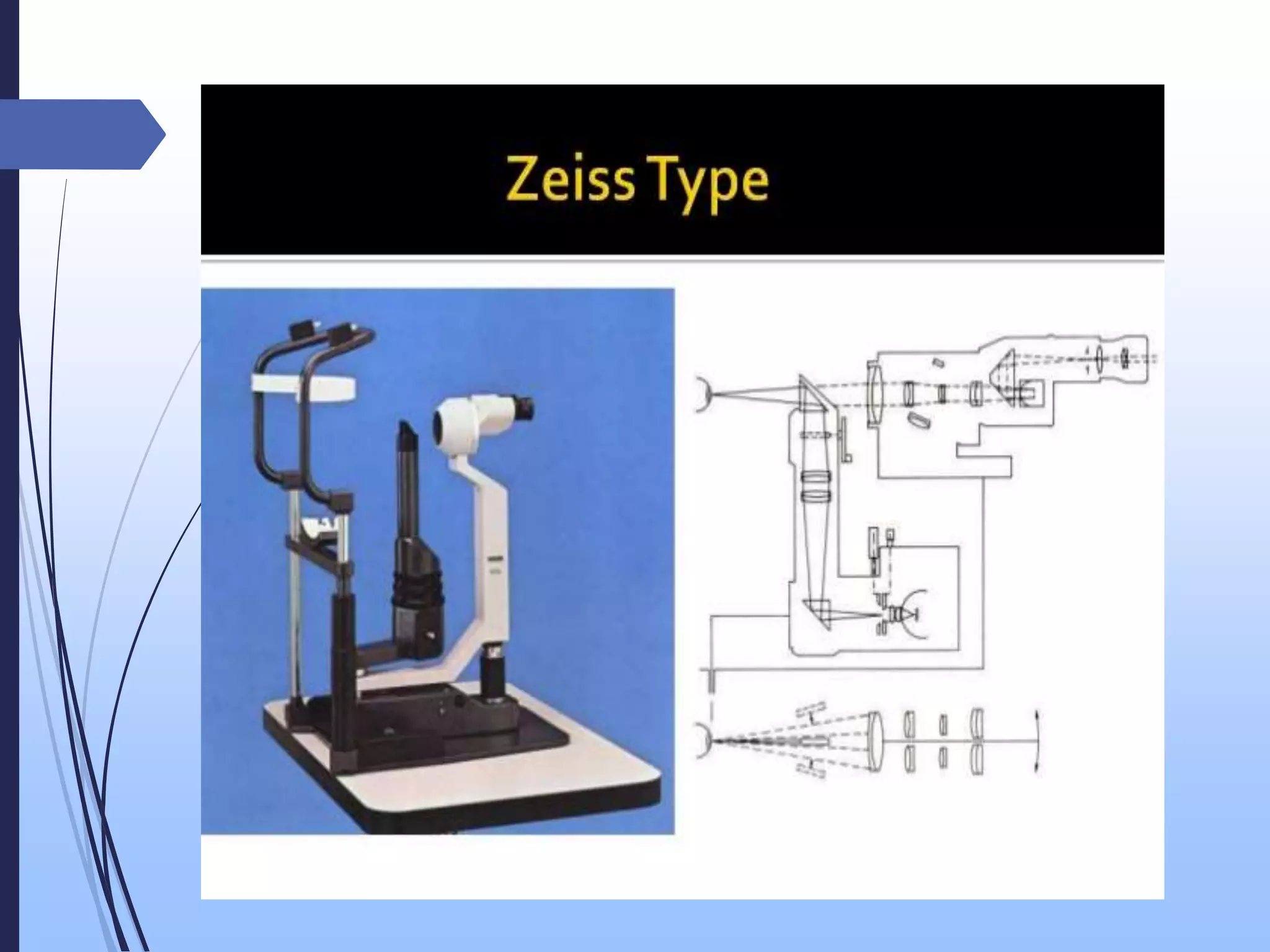
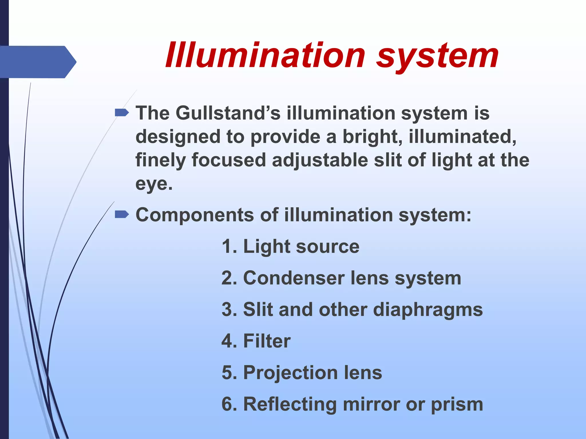
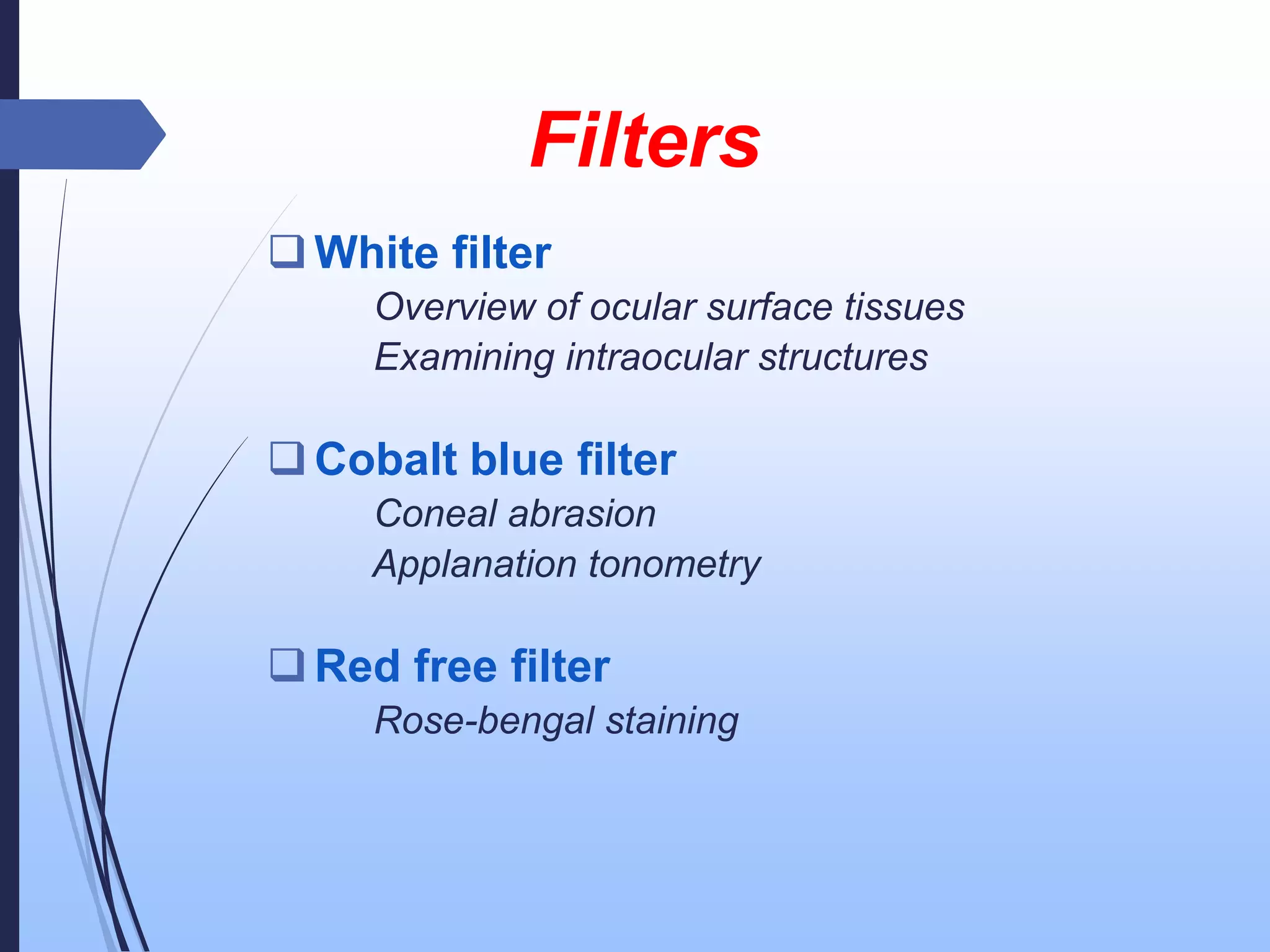
Presented by
Dr. Md.Ashfakur Rahaman
D.O Student, RpMC
SLIT LAMP
Slit lamp biomicroscope is a
relatively low powered compound
microscope with a adjustable bright
light source, specially designed to
view the different optically
transparent layers of the eye.
The instrument is known as
slit lamp because the
illumination is arranged in a
pattern that a narrow
vertical slit of light is
projected onto the eye.
Criteria of a modern slit lamp
 Binocular stereoscopic view.
 Wide range of magnification from
cornea to retina.
 Illumination of variable shape and
intensities.
 Quantitative measurement by its
auxiliary devices like IOP, endothelial
cell count, pupil size, corneal thickness,
anterior chamber depth etc.
 Photography of every part is possible.
Types
Parts of a slit lamp
Mechanical support system
Observation system
Illumination system
Mechanical System
Joy stick arrangement
Up and down movement
arrangement
Patient support arrangement
Fixation target
Mechanical coupling
Observation system
 It is a compound microscope which is
composed of two convex lenses-
The objective lens (+22D)
The eye piece lens (+10D)
 To provide good stereopsis the tubes are
converged at an angle of 10-15 degree.
Illumination system
 The Gullstand’s illumination system is
designed to provide a bright, illuminated,
finely focused adjustable slit of light at the
eye.
 Components of illumination system:
1. Light source
2. Condenser lens system
3. Slit and other diaphragms
4. Filter
5. Projection lens
6. Reflecting mirror or prism
Methods of illumination
1. Direct illumination:
a) Diffuse illumination
b)Slit /direct focal
c) Specular reflection
2. Indirect illumination:
a) Retro illumination
- fundus retroillumination
- iris retroillumination
b) Sclerotic scatter
Direct diffuse illumination
 Full height
 Broad beam
 Directed obliquely
 Lower illumination level
 Filters can be used
Used for overview of anterior segment of eye.
Direct focal/ slit illumination :
 Full height & medium width
 Medium-bright beam
 Obliquely directed
 An optical cross-section (slit) is created
by narrowing to a very thin beam < 1 mm
wide
Uses:
Cornea in detail
Anterior chamber
 Crystalline lens
Anterior part of vitreous
Grading cell and flare in anterior
chamber
Specular reflection
 The angle of incidence is equal to the
angle of reflection.
 The light reflected from the anterior or
posterior corneal surface
 Method is monocular
 Shows abnormalities of the endothelium
such as reduced cell density and guttata.
Indirect illumination:
 Slit beam focused on a position just
beside the areas to be examined
 Beam width moderate
 Illumination low, medium or high
 Angle 30-40°
Uses:
Corneal infiltrates
Micro-cysts
Vacuoles
Epithelial cells
Retroillumination:
 Light reflected on iris or fundus
 Microscope focused on cornea
 For better visualization of:
Epithelial cysts
Keratic precipitates
Small blood vessels
Scleral Scatter
 Light incident on the limbus
 The microscope focused centrally
 Light transmitted to cornea by total internal
reflection
 Useful to detect subtle stromal haze and
cellular or lipid infiltration
Filters
White filter
Overview of ocular surface tissues
Examining intraocular structures
Cobalt blue filter
Coneal abrasion
Applanation tonometry
Red free filter
Rose-bengal staining
Accessory devices
Specialized examinations can be perform by slit
lamp biomicroscope with the help of some
accessory devices. These are:
Gonioscopy
Pachymetry
Applanation tonometry
Ophthalmo dynamometry
Slit lamp photography
Slit lamp videography
Laser interferometry
Slit lamp as delivery system for
argon, diode and YAG laser
Ophthalmologic uses of
slit lamp biomicroscope
Diagnostic uses
Therapeutic uses
Procedures
Diagnostic uses
Anterior segment
1. Eye lid- to determine blepharitis, stye
2. Eyelashes- pediculosis, distichiasis
3. Conjunctiva- scarring, conjunctivitis, pattern of
congestion
4. Cornea-thickness, bullae, K.P, infiltratre
5. Anterior chamber- regularity, any pathology
6. Iris and pupil- appearance, colour, synechiae,
neovascularization, iridodonesis, coloboma
7. Lens- grading and staging of cataract,
phacodonesis, position of lens
Posterior segment
1. To see the vitreous, media, optic disc,
macula, background retina
2. To differentiate position of pathology
3. Any macular degeneration
4. Retinal detachment
Procedures by slit lamp
1. Applanation tonometry
2. Gonioscopy
3. Contact lens fitting
Therapeutic uses
1. Removal of corneal F.B
2. Epilation
SLIT LAMP BIOMICROSCOPY.pptx

SLIT LAMP BIOMICROSCOPY.pptx

  • 1.
    Presented by Dr. Md.AshfakurRahaman D.O Student, RpMC
  • 2.
    SLIT LAMP Slit lampbiomicroscope is a relatively low powered compound microscope with a adjustable bright light source, specially designed to view the different optically transparent layers of the eye.
  • 4.
    The instrument isknown as slit lamp because the illumination is arranged in a pattern that a narrow vertical slit of light is projected onto the eye.
  • 7.
    Criteria of amodern slit lamp  Binocular stereoscopic view.  Wide range of magnification from cornea to retina.  Illumination of variable shape and intensities.  Quantitative measurement by its auxiliary devices like IOP, endothelial cell count, pupil size, corneal thickness, anterior chamber depth etc.  Photography of every part is possible.
  • 8.
  • 11.
    Parts of aslit lamp Mechanical support system Observation system Illumination system
  • 12.
    Mechanical System Joy stickarrangement Up and down movement arrangement Patient support arrangement Fixation target Mechanical coupling
  • 14.
    Observation system  Itis a compound microscope which is composed of two convex lenses- The objective lens (+22D) The eye piece lens (+10D)  To provide good stereopsis the tubes are converged at an angle of 10-15 degree.
  • 17.
    Illumination system  TheGullstand’s illumination system is designed to provide a bright, illuminated, finely focused adjustable slit of light at the eye.  Components of illumination system: 1. Light source 2. Condenser lens system 3. Slit and other diaphragms 4. Filter 5. Projection lens 6. Reflecting mirror or prism
  • 19.
    Methods of illumination 1.Direct illumination: a) Diffuse illumination b)Slit /direct focal c) Specular reflection 2. Indirect illumination: a) Retro illumination - fundus retroillumination - iris retroillumination b) Sclerotic scatter
  • 20.
    Direct diffuse illumination Full height  Broad beam  Directed obliquely  Lower illumination level  Filters can be used Used for overview of anterior segment of eye.
  • 22.
    Direct focal/ slitillumination :  Full height & medium width  Medium-bright beam  Obliquely directed  An optical cross-section (slit) is created by narrowing to a very thin beam < 1 mm wide
  • 23.
    Uses: Cornea in detail Anteriorchamber  Crystalline lens Anterior part of vitreous Grading cell and flare in anterior chamber
  • 24.
    Specular reflection  Theangle of incidence is equal to the angle of reflection.  The light reflected from the anterior or posterior corneal surface  Method is monocular  Shows abnormalities of the endothelium such as reduced cell density and guttata.
  • 26.
    Indirect illumination:  Slitbeam focused on a position just beside the areas to be examined  Beam width moderate  Illumination low, medium or high  Angle 30-40° Uses: Corneal infiltrates Micro-cysts Vacuoles Epithelial cells
  • 27.
    Retroillumination:  Light reflectedon iris or fundus  Microscope focused on cornea  For better visualization of: Epithelial cysts Keratic precipitates Small blood vessels
  • 28.
    Scleral Scatter  Lightincident on the limbus  The microscope focused centrally  Light transmitted to cornea by total internal reflection  Useful to detect subtle stromal haze and cellular or lipid infiltration
  • 30.
    Filters White filter Overview ofocular surface tissues Examining intraocular structures Cobalt blue filter Coneal abrasion Applanation tonometry Red free filter Rose-bengal staining
  • 38.
    Accessory devices Specialized examinationscan be perform by slit lamp biomicroscope with the help of some accessory devices. These are: Gonioscopy Pachymetry Applanation tonometry Ophthalmo dynamometry Slit lamp photography Slit lamp videography Laser interferometry Slit lamp as delivery system for argon, diode and YAG laser
  • 39.
    Ophthalmologic uses of slitlamp biomicroscope Diagnostic uses Therapeutic uses Procedures
  • 40.
    Diagnostic uses Anterior segment 1.Eye lid- to determine blepharitis, stye 2. Eyelashes- pediculosis, distichiasis 3. Conjunctiva- scarring, conjunctivitis, pattern of congestion 4. Cornea-thickness, bullae, K.P, infiltratre 5. Anterior chamber- regularity, any pathology 6. Iris and pupil- appearance, colour, synechiae, neovascularization, iridodonesis, coloboma 7. Lens- grading and staging of cataract, phacodonesis, position of lens
  • 41.
    Posterior segment 1. Tosee the vitreous, media, optic disc, macula, background retina 2. To differentiate position of pathology 3. Any macular degeneration 4. Retinal detachment
  • 42.
    Procedures by slitlamp 1. Applanation tonometry 2. Gonioscopy 3. Contact lens fitting
  • 43.
    Therapeutic uses 1. Removalof corneal F.B 2. Epilation