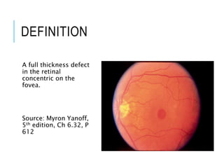
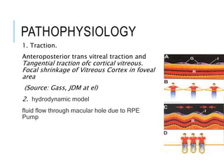
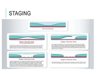
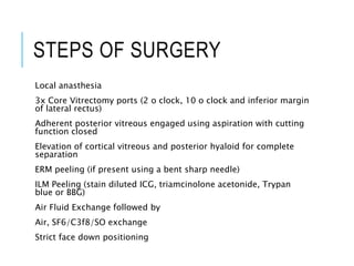
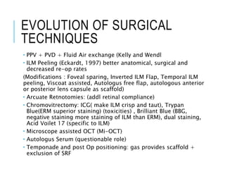
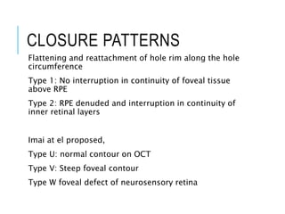
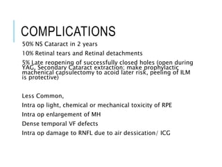
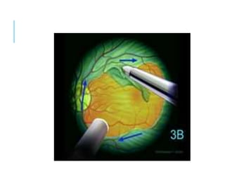
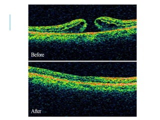
MACULAR HOLE
DEFINITION
A full thickness defect
in the retinal
concentric on the
fovea.
Source: Myron Yanoff,
5th edition, Ch 6.32, P
612
MOST FULL THICKNESS MH
ARE IDIOPATHIC
1 in 500 patients
6th to 7th decade
Predominantly female 2:1 (67%-91%)
Younger age in myopes
10-20% bilateral (not simultaneous)
PATHOPHYSIOLOGY
1. Traction.
Anteroposterior trans vitreal traction and
Tangential traction ofc cortical vitreous.
Focal shrinkage of Vitreous Cortex in foveal
area
(Source: Gass, JDM at el)
2. hydrodynamic model
fluid flow through macular hole due to RPE
Pump
NATURAL HISTORY OF
MACULAR HOLE
deterioration over a few years
stabilization of both visual acuity and hole size.
198 patients with untreated macular hole,
about 1/3rd of patients had an increase in size of the hole
almost half experienced a decrease in visual acuity of at least
two lines. Approximately 7 percent of patients developed a
macular hole in the fellow eye
Chew EY, Sperduto RD, Hiller R, et al. Clinical course of macular holes: The Eye Disease
Case-Control Study. Arch Ophthalmol 1999;117:242-6
OCULAR SIGNS
Hallmark complaint is painless central visual distortion or
blur of acute/subacute nature. (May go unnoticed)
Gass Classification explains clinically observed
appearance in 4 stages.
ASSOCIATIONS
A macular hole is mostly idiopathic or related to vitreomacular traction syndrome.
The macular hole may be associated with various forms of macular pathology
including
High Myopia
After laser treatment
Intraocular surgical intervention
Epiretinal membrane
Polypoidal choroidal vasculopathy (PCV)
Hypertensive retinopathy (HTNR)
Diabetic retinopathy (DR)
Vitelliform dystrophy
Ruptured retinal arterial macroaneurysm (RAM)
STAGING
Old/ Clinical Staging system by Gass
New International Vitreomacular Traction Study (IVTS)
Group:
based on OCT findings
STAGING
STAGE 0 MACULAR HOLE
(IVTS: VMA)
OCT finding of oblique foveal VR traction before Clinical changes
VMA: Residual attachment of Vitreous within 3mm radius of
central macula in the presence of perifoveal vitreous separation
No distortion of foveal contour or secondary changes
 Focal VMA <1500um
 Broad VMA >1500um
STAGE 1
(IVTS: VMT)
Typically asymptomatic
Loss of foveal depression
1A impending hole: Yellow spot
1B occult hole: yellow ring
50% resolved spontaneously
50% progress to stage 2
VMT: Perifoveal PVD (3mm) with retinal
changes on OCT
Associatons: AMD, RVO,DR
No true retinal defect present, PR
layer is intact,
No vitreofoveal separation
Oblique Vitreofoveal traction is
responsible
Source of image: AAO, Text Yanoff,
STAGE 2-3
IVTS 2: SMALL OR MEDIUM
FTMH WITH VMT
3: MEDIUM ORLARGE FTMH
WITH VMT
STAGE 4
IVTS: SMALL MEDIUM OF LARGE
FTMH WITHOUT VMT
Complete PVD (weiss ring often present)
Significant cases are associated with ERM
FTMH: foveal lesion featuring interruption of all layers of retina
from ILM to RPE. Small (<250um) Medium (250 to 400um) &
large (>400um)
DDX Stage 1a: Adult Vitilliform Macular dystrophy,
Solar & Laser retinopathy, CME
Stage 2 – 4 : Lamellar hole, Macular ERM with
pseudohole, VMT without full thickness macular
hole, Foveal drusen , Choroidal neovascular
membrane, Central areolar pigment
epitheliopathy, Pattern Dystrophy, CSCR
INVESTIGATIONS
Amsler Grid: Non specific distortion
> Scotoma
Watzke-Allen test
OCT (IVTS Classification)
FAF:
2: punctate fluorescence
3-4: marked fluorescent foveolar
spot
FA:
Not routinely indicated except in
cases of any other association CMO,
DR, PCV
Circular transmission defect(stage 2-
3-4)
MACULAR INDICES
MHI, THI: inc values indicate better post Op functional
outcome
BD, MLD: associated with better anatomical outcomes
MHI: ELM restoration
MANAGEMENT
1. Observation: suitable for Stage 1 MH with good VA
2. Pharmacological Vitreolysis:
 Suitable for small early stage holes, ERM is a poor prognostic factor
 Intra Vitreal Orciplasmin (human recombinant plasmin) releases VMT
in 25% eyes
 Pneumatic IV Injection
3. Surgery : Suitable for Stage 2 and above (stage 1 if
persistent decrease VA and prolonged period)
STEPS OF SURGERY
Local anasthesia
3x Core Vitrectomy ports (2 o clock, 10 o clock and inferior margin
of lateral rectus)
Adherent posterior vitreous engaged using aspiration with cutting
function closed
Elevation of cortical vitreous and posterior hyaloid for complete
separation
ERM peeling (if present using a bent sharp needle)
ILM Peeling (stain diluted ICG, triamcinolone acetonide, Trypan
blue or BBG)
Air Fluid Exchange followed by
Air, SF6/C3f8/SO exchange
Strict face down positioning
EVOLUTION OF SURGICAL
TECHNIQUES
 PPV + PVD + Fluid Air exchange (Kelly and Wendl
 ILM Peeling (Eckardt, 1997) better anatomical, surgical and
decreased re-op rates
(Modifications : Foveal sparing, Inverted ILM Flap, Temporal ILM
peeling, Viscoat assisted, Autologus free flap, autologous anterior
or posterior lens capsule as scaffold)
 Arcuate Retnotomies: (addl retinal compliance)
 Chromovitrectomy: ICG( make ILM crisp and taut), Trypan
Blue(ERM superior staining) (toxicities) , Brilliant Blue (BBG,
negative staining more staining of ILM than ERM), dual staining,
Acid Voilet 17 (specific to ILM)
 Microscope assisted OCT (Mi-OCT)
 Autologus Serum (questionable role)
 Temponade and post Op positioning: gas provides scaffold +
exclusion of SRF
CLOSURE PATTERNS
Flattening and reattachment of hole rim along the hole
circumference
Type 1: No interruption in continuity of foveal tissue
above RPE
Type 2: RPE denuded and interruption in continuity of
inner retinal layers
Imai at el proposed,
Type U: normal contour on OCT
Type V: Steep foveal contour
Type W foveal defect of neurosensory retina
COMPLICATIONS
50% NS Cataract in 2 years
10% Retinal tears and Retinal detachments
5% Late reopening of successfully closed holes (open during
YAG, Secondary Cataract extraction; make prophylactic
machenical capsulectomy to acoid later risk, peeling of ILM
is protective)
Less Common,
Intra op light, chemical or mechanical toxicity of RPE
Intra op enlargement of MH
Dense temporal VF defects
Intra op damage to RNFL due to air dessication/ ICG
macular hole.pptx
macular hole.pptx
macular hole.pptx
macular hole.pptx
macular hole.pptx
macular hole.pptx
macular hole.pptx

macular hole.pptx

  • 1.
  • 2.
    DEFINITION A full thicknessdefect in the retinal concentric on the fovea. Source: Myron Yanoff, 5th edition, Ch 6.32, P 612
  • 3.
    MOST FULL THICKNESSMH ARE IDIOPATHIC 1 in 500 patients 6th to 7th decade Predominantly female 2:1 (67%-91%) Younger age in myopes 10-20% bilateral (not simultaneous)
  • 4.
    PATHOPHYSIOLOGY 1. Traction. Anteroposterior transvitreal traction and Tangential traction ofc cortical vitreous. Focal shrinkage of Vitreous Cortex in foveal area (Source: Gass, JDM at el) 2. hydrodynamic model fluid flow through macular hole due to RPE Pump
  • 5.
    NATURAL HISTORY OF MACULARHOLE deterioration over a few years stabilization of both visual acuity and hole size. 198 patients with untreated macular hole, about 1/3rd of patients had an increase in size of the hole almost half experienced a decrease in visual acuity of at least two lines. Approximately 7 percent of patients developed a macular hole in the fellow eye Chew EY, Sperduto RD, Hiller R, et al. Clinical course of macular holes: The Eye Disease Case-Control Study. Arch Ophthalmol 1999;117:242-6
  • 6.
    OCULAR SIGNS Hallmark complaintis painless central visual distortion or blur of acute/subacute nature. (May go unnoticed) Gass Classification explains clinically observed appearance in 4 stages.
  • 7.
    ASSOCIATIONS A macular holeis mostly idiopathic or related to vitreomacular traction syndrome. The macular hole may be associated with various forms of macular pathology including High Myopia After laser treatment Intraocular surgical intervention Epiretinal membrane Polypoidal choroidal vasculopathy (PCV) Hypertensive retinopathy (HTNR) Diabetic retinopathy (DR) Vitelliform dystrophy Ruptured retinal arterial macroaneurysm (RAM)
  • 8.
    STAGING Old/ Clinical Stagingsystem by Gass New International Vitreomacular Traction Study (IVTS) Group: based on OCT findings
  • 9.
  • 10.
    STAGE 0 MACULARHOLE (IVTS: VMA) OCT finding of oblique foveal VR traction before Clinical changes VMA: Residual attachment of Vitreous within 3mm radius of central macula in the presence of perifoveal vitreous separation No distortion of foveal contour or secondary changes  Focal VMA <1500um  Broad VMA >1500um
  • 11.
    STAGE 1 (IVTS: VMT) Typicallyasymptomatic Loss of foveal depression 1A impending hole: Yellow spot 1B occult hole: yellow ring 50% resolved spontaneously 50% progress to stage 2 VMT: Perifoveal PVD (3mm) with retinal changes on OCT Associatons: AMD, RVO,DR No true retinal defect present, PR layer is intact, No vitreofoveal separation Oblique Vitreofoveal traction is responsible Source of image: AAO, Text Yanoff,
  • 12.
    STAGE 2-3 IVTS 2:SMALL OR MEDIUM FTMH WITH VMT 3: MEDIUM ORLARGE FTMH WITH VMT
  • 13.
    STAGE 4 IVTS: SMALLMEDIUM OF LARGE FTMH WITHOUT VMT Complete PVD (weiss ring often present) Significant cases are associated with ERM FTMH: foveal lesion featuring interruption of all layers of retina from ILM to RPE. Small (<250um) Medium (250 to 400um) & large (>400um)
  • 14.
    DDX Stage 1a:Adult Vitilliform Macular dystrophy, Solar & Laser retinopathy, CME Stage 2 – 4 : Lamellar hole, Macular ERM with pseudohole, VMT without full thickness macular hole, Foveal drusen , Choroidal neovascular membrane, Central areolar pigment epitheliopathy, Pattern Dystrophy, CSCR
  • 16.
    INVESTIGATIONS Amsler Grid: Nonspecific distortion > Scotoma Watzke-Allen test OCT (IVTS Classification) FAF: 2: punctate fluorescence 3-4: marked fluorescent foveolar spot FA: Not routinely indicated except in cases of any other association CMO, DR, PCV Circular transmission defect(stage 2- 3-4)
  • 17.
    MACULAR INDICES MHI, THI:inc values indicate better post Op functional outcome BD, MLD: associated with better anatomical outcomes MHI: ELM restoration
  • 18.
    MANAGEMENT 1. Observation: suitablefor Stage 1 MH with good VA 2. Pharmacological Vitreolysis:  Suitable for small early stage holes, ERM is a poor prognostic factor  Intra Vitreal Orciplasmin (human recombinant plasmin) releases VMT in 25% eyes  Pneumatic IV Injection 3. Surgery : Suitable for Stage 2 and above (stage 1 if persistent decrease VA and prolonged period)
  • 19.
    STEPS OF SURGERY Localanasthesia 3x Core Vitrectomy ports (2 o clock, 10 o clock and inferior margin of lateral rectus) Adherent posterior vitreous engaged using aspiration with cutting function closed Elevation of cortical vitreous and posterior hyaloid for complete separation ERM peeling (if present using a bent sharp needle) ILM Peeling (stain diluted ICG, triamcinolone acetonide, Trypan blue or BBG) Air Fluid Exchange followed by Air, SF6/C3f8/SO exchange Strict face down positioning
  • 20.
    EVOLUTION OF SURGICAL TECHNIQUES PPV + PVD + Fluid Air exchange (Kelly and Wendl  ILM Peeling (Eckardt, 1997) better anatomical, surgical and decreased re-op rates (Modifications : Foveal sparing, Inverted ILM Flap, Temporal ILM peeling, Viscoat assisted, Autologus free flap, autologous anterior or posterior lens capsule as scaffold)  Arcuate Retnotomies: (addl retinal compliance)  Chromovitrectomy: ICG( make ILM crisp and taut), Trypan Blue(ERM superior staining) (toxicities) , Brilliant Blue (BBG, negative staining more staining of ILM than ERM), dual staining, Acid Voilet 17 (specific to ILM)  Microscope assisted OCT (Mi-OCT)  Autologus Serum (questionable role)  Temponade and post Op positioning: gas provides scaffold + exclusion of SRF
  • 21.
    CLOSURE PATTERNS Flattening andreattachment of hole rim along the hole circumference Type 1: No interruption in continuity of foveal tissue above RPE Type 2: RPE denuded and interruption in continuity of inner retinal layers Imai at el proposed, Type U: normal contour on OCT Type V: Steep foveal contour Type W foveal defect of neurosensory retina
  • 22.
    COMPLICATIONS 50% NS Cataractin 2 years 10% Retinal tears and Retinal detachments 5% Late reopening of successfully closed holes (open during YAG, Secondary Cataract extraction; make prophylactic machenical capsulectomy to acoid later risk, peeling of ILM is protective) Less Common, Intra op light, chemical or mechanical toxicity of RPE Intra op enlargement of MH Dense temporal VF defects Intra op damage to RNFL due to air dessication/ ICG