Download as PDF, PPTX


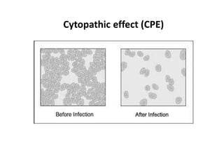


Viruses are obligate intracellular parasites that lack cell organelles and exist in two phases - an extracellular phase where they possess few enzymes, and an intracellular phase where they induce host cells to synthesize viral components. They have a nucleocapsid containing either DNA or RNA surrounded by a protein capsid. Viruses replicate through either lytic or lysogenic cycles, with the lytic cycle involving adsorption to and penetration of host cells, synthesis of viral components, assembly of new virus particles, and cell lysis. Viruses can be grown in tissue culture or through plaque assays using bacterial hosts.


![Classification Of Virus[Summary]](https://cdn.slidesharecdn.com/ss_thumbnails/classificationofvirussummary-100325002939-phpapp01-thumbnail.jpg?width=640&height=640&fit=bounds)


