Downloaded 1,189 times


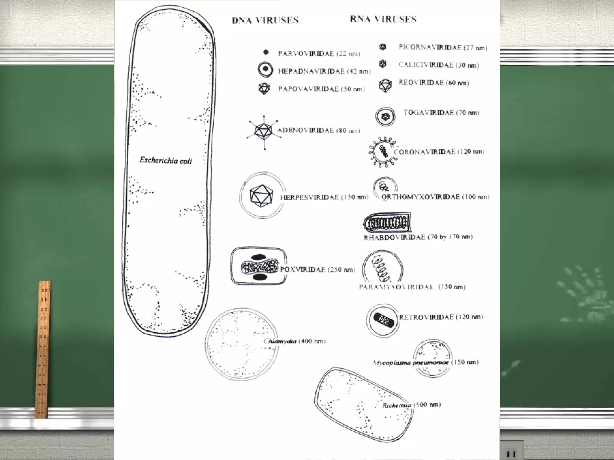


The document summarizes key aspects of virology. It describes that viruses are small infectious agents that contain either DNA or RNA and use the machinery of host cells to replicate. Viruses infect cells and program them to produce new viral components for assembly of new virus particles. The document then discusses viral structure, morphology, replication cycles involving attachment, entry, uncoating, production of components, assembly and release. It also covers pathogenesis, diagnosis, cultivation, and methods for prevention and treatment of viral infections including vaccines, interferons and antiviral drugs.