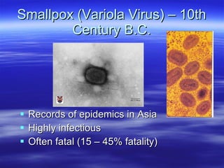
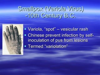
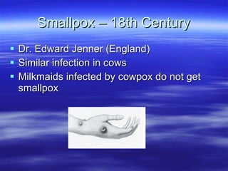
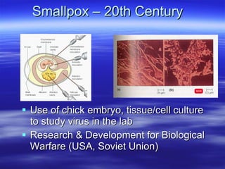
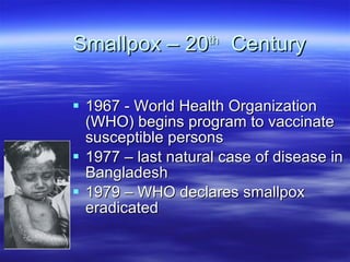
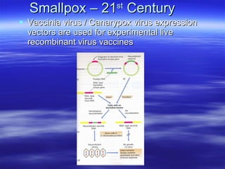
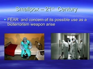
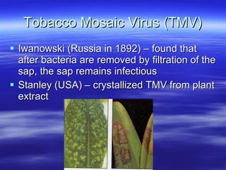
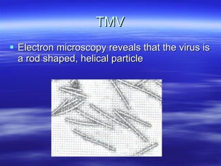
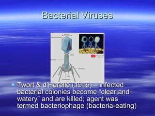
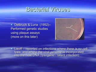
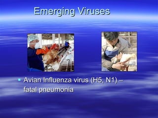
Introduction to Virology Brief History, Viral Structure and Classification, and Role of Host Cells in Viral Infections
Smallpox (Variola Virus) – 10th Century B.C. Records of epidemics in Asia Highly infectious Often fatal (15 – 45% fatality)
Smallpox (Variola Virus) -10th Century B.C. Variola, “spot” – vesicular rash Chinese prevent infection by self-inoculation of pus from lesions Termed “variolation”
Smallpox – 18th Century Dr. Edward Jenner (England) Similar infection in cows Milkmaids infected by cowpox do not get smallpox
Used the pus from cowpox lesions to inoculate and prevent smallpox Termed “vaccination” (vaccinus, cow)
Smallpox – 20th Century Use of chick embryo, tissue/cell culture to study virus in the lab Research & Development for Biological Warfare (USA, Soviet Union)
Smallpox – 20 th   Century 1967 - World Health Organization (WHO) begins program to vaccinate susceptible persons 1977 – last natural case of disease in Bangladesh 1979 – WHO declares smallpox eradicated
Smallpox – 21 st  Century Vaccinia virus / Canarypox virus expression vectors are used for experimental live recombinant virus vaccines
Smallpox – 21 st   Century FEAR  and concern of its possible use as a bioterrorism weapon arise
Tobacco Mosaic Virus (TMV) Iwanowski (Russia in 1892) – found that after bacteria are removed by filtration of the sap, the sap remains infectious Stanley (USA) – crystallized TMV from plant extract
TMV Electron microscopy reveals that the virus is a rod shaped, helical particle
Bacterial Viruses Twort & d’Herelle (1915) – infected bacterial colonies become “clear,and watery” and are killed; agent was termed bacteriophage (bacteria-eating)
Bacterial Viruses Delbruck & Luria  (1952)– Performed genetic studies  using plaque assays (more on this later) Lwoff – reported on infections where there is no cell lysis, and where the viral genome is incorporated into the host DNA (lysogenic / latent infection)
RNA Tumor Viruses Rous (USA, 1911) – reported  that a virus infection of  chickens resulted in sarcoma Related viruses were  shown to cause leukemia   in cats, mice, and cows
RNA Tumor Viruses Identified as Retroviruses (RNA to DNA)
DNA Tumor Viruses Shope (USA) – shows that a DNA virus is  responsible for papilloma in rabbits
DNA Tumor Viruses Related viruses were found in mice, cows, horses, and primates The Virus:Host interactions of these viruses were used as a model to study cell regulation Human Papilloma  Virus
Emerging Infections
Emerging Viruses Ebola Virus -  fatal hemorrhagic  fever HIV –  chronic infection,   immune deficiency
Emerging Viruses Hantavirus –  hemorrhagic fever  + pulmonary infection Prion – proteinaeous infectious particle causing subacute spongiform encephalopathy
Emerging Viruses West Nile Virus – asymptomatic, encephalitis (1%) Severe Acute  Respiratory Syndrome  (SARS)
Emerging Viruses Avian Influenza virus (H5, N1) –  fatal pneumonia
Introduction to Virology What is a virus? Extremely small Submicroscopic Must use an electron microscope to “see” Passes through filters used to “sterilize” solutions Obligate, intracellular parasites Can’t be cultured on artificial media
Variation in virus size
What is a virus? Biochemically viruses are similar to, but different from “living organisms” Both viruses and other living organisms contain proteins and glycoproteins While other living organisms contain both RNA and DNA, viruses contain  either DNA or RNA , but not both DNA viruses  may be linear with open or closed ends, circular (closed or nicked), single stranded, or  double stranded RNA viruses may be linear single stranded,  segmented single stranded, or  segmented double stranded  If single stranded, the strand may be of either the plus or the minus sense (more later on this)
Types of viral nucleic acid
What is a virus? Unlike other living organisms, viruses contain no polysaccharides, small molecules or ions Other living organisms contain lipids. Lipids, if found in viruses, are only found in enveloped viruses (more on this later on) Viruses lack the genetic information that encodes the apparatus necessary for the generation of metabolic energy or for protein synthesis The growth curves of viruses are very different from those of other organisms:
Growth curves of bacteria (A) versus bacteriophages (B)
Differences in growth curves: Virus particles are produced from the assembly of pre-formed components: other organisms grow from an increase in the integrated sum of their components and reproduce by cell division. Viruses don’t “grow” or undergo division There are six basic phases in the multiplication cycle of all viruses Attachment Penetration Uncoating Biosynthesis Assembly Release
The multiplication cycle of a virus
One step growth curve A one-step growth curve of bacteriophage λ following infection of susceptible bacteria (Escherichia coli). During the eclipse phase(1), the infectivity of the cell-associated, infecting virus is lost as it uncoats; during the maturation phase(2) infectious virus is assembled inside cells  (cell-associated virus), but not yet released; and the latent phase(3) measures the period before infectious virus is released from cells into the medium. Total virus is the sum of cell-associated virus +released virus. Cell-associated virus decreases as cells are lysed. This classic experiment shows that phages develop intracellularly.
What is a virion? A  virion  is a structurally complete virus that is capable of infecting new cells
Structure of viruses: Composed of nucleic acid (either DNA or RNA) Surrounding the nucleic acid is a protein outer coat (a  capsid ) which is composed of units called  capsomers  which are formed by the association of individual proteins called  protomers.  The capsid : Functions to protect the delicate inner nucleic acid from physical, chemical or enzymatic damage. May function in attachment of the virus to the host cell. May provide enzymes essential for virus entry Functions to ensure that the virus genome is released only at the appropriate time and location
Capsomers, continued The arrangement of the capsomers determines the architecture of the virus or the  nucleocapsid  (composed of nucleic acid and the capsid).  There are two basic types of capsomer arrangements: Helix Looks like a hollow tube or cylinder with the nucleic acid inside.  The proteins are arranged around the circumference of a circle to form a disc.  Multiple discs are stacked on top of each other.  The helix may be rigid or flexible.
Helical Structure
Helical structure
Capsid architecture continued Icosahedral Looks like a sphere, but it actually has  20 triangular faces and 12 corners  made by the intersection of 5 faces.  For small viruses, each face (capsomer) consists of three structural subunits (protomers).  The faces of larger viruses are made from  multiples of three subunits .
Icosahedral architecture
Capsid architecture continued Some viruses are more complex and don’t fit into either architectural type.
Poxvirus
Reovirus
Structure of Viruses, continued The nucleocapsid of many viruses is surrounded by an envelope.  The envelope is derived from host cell membranes.  Viruses differ as to which host cell membrane is used for their envelope, i.e., plasma membrane, Golgi, endoplasmic reticulum (E.R.), or nuclear membranes may be used.
Enveloped viruses Sindbis virus: an enveloped icosahedron
Enveloped viruses Influenza A virus (an orthomyxovirus) and vesicular stomatitis virus (a rhabdovirus): viruses with enveloped helical structures. Although their morphology is different, these viruses are constructedin the same way.
Structure of Viruses, continued Viruses without an envelope are called  naked viruses.  Enveloped viruses  have an advantage in that they may exit the host cell without destroying it.  For example, viruses that use the host cell plasma membrane as the envelope, may take part of the host cell plasma membrane as the viruses exit the cell (a process called  budding ) and the host cell membrane reseals itself. (More detail will be provided on this process later in the quarter)
Budding
Enveloped viruses Enveloped viruses modify their lipid envelopes by directing the synthesis of different classes of virally encoded proteins that are specifically transported to and associated with the membrane that eventually becomes their envelope. Matrix proteins  – bind to the inner surface of the membrane to link the nucleocapsid to the membrane in the assembly process. Glycoproteins  – are transmembrane proteins. External glycoproteins have large ectodomains and small endodomains.  Monomers of these proteins often associate to form multimers and may function in:
Enveloped viruses Receptor binding Fusion, a process that takes place during entry of the virus into the host cell (more on this later) Transport channel proteins  May span the membrane several times.  May be important for modifying the internal environment of the virus by altering membrane permeability.
Types of viral proteins associated with the envelope Influenza   virus
Structural Roles of Host Cells in Viral Infections In order for a virus to successfully infect a host cell, the cell must contain the  receptor  that the virus binds to in the process of initiating an infection. Receptors on animal cells are found on the plasma membrane.  They may be proteins, glycoproteins, or glycolipids. Many viruses bind to the carbohydrate side chains of glycoproteins.
Receptors on animal cells
Structural Roles of Host Cells in Viral Infections In order for a virus to successfully infect a host cell, the host cell must: Contain the receptor for the virus,  It must also have the  cellular machinery  that the virus needs for replication.  Differences in the organization of the cell’s genome and how it carries out the processes of replication, transcription and translation play an important role in virus replication.
Eukaryotic cells Several linear chromosomes; diploid No operons; each gene is regulated by its own controlling elememts; monocistronic mRNA (rare IRES sites) Post-trascriptional modification of RNA Splicing Addition of 5’cap Addition of 3’ poly A tail
Eukaryotic cells, continued Internal compartmentalization Nucleus for replication and transcription Cytoplasm for translation Once cells differentiate, they exit the cell cycle.
Classification of Viruses There are various ways to classify viruses: On the basis of disease On basis of the host organism On basis of virus particle morphology On the basis of viral nucleic acid The most commonly used classification scheme is the Baltimore scheme. This scheme is based on the relationship between the viral genome and the mRNA used for translation during expression of the viral genome: Class I = double stranded DNA viruses Class 2a= single stranded + sense DNA (DNA has the same sequence as the mRNA, except T replaces U) Class 2b= single stranded – sense DNA (The DNA is complementary to the mRNA)
The Baltimore Classification of Viruses Class 3= double stranded RNA Class 4= single stranded + sense RNA (the genome sequence is the same as the mRNA and can often be directly translated into protein product) Class 5= single stranded – sense RNA (the RNA is complementary to the mRNA and thus, the genomic RNA cannot be directly translated into a protein product) Class 6= single stranded + sense RNA that requires synthesis of a double stranded DNA molecule for the expression of the genome (retroviruses) Class 7 = this is a new class for viruses termed reversiviruses which replicate their double stranded DNA genome via a positive sense single stranded RNA intermediate
Baltimore Classification of Viruses

33. introduction to virology

  • 1.
    Introduction to VirologyBrief History, Viral Structure and Classification, and Role of Host Cells in Viral Infections
  • 2.
    Smallpox (Variola Virus)– 10th Century B.C. Records of epidemics in Asia Highly infectious Often fatal (15 – 45% fatality)
  • 3.
    Smallpox (Variola Virus)-10th Century B.C. Variola, “spot” – vesicular rash Chinese prevent infection by self-inoculation of pus from lesions Termed “variolation”
  • 4.
    Smallpox – 18thCentury Dr. Edward Jenner (England) Similar infection in cows Milkmaids infected by cowpox do not get smallpox
  • 5.
    Used the pusfrom cowpox lesions to inoculate and prevent smallpox Termed “vaccination” (vaccinus, cow)
  • 6.
    Smallpox – 20thCentury Use of chick embryo, tissue/cell culture to study virus in the lab Research & Development for Biological Warfare (USA, Soviet Union)
  • 7.
    Smallpox – 20th Century 1967 - World Health Organization (WHO) begins program to vaccinate susceptible persons 1977 – last natural case of disease in Bangladesh 1979 – WHO declares smallpox eradicated
  • 8.
    Smallpox – 21st Century Vaccinia virus / Canarypox virus expression vectors are used for experimental live recombinant virus vaccines
  • 9.
    Smallpox – 21st Century FEAR and concern of its possible use as a bioterrorism weapon arise
  • 10.
    Tobacco Mosaic Virus(TMV) Iwanowski (Russia in 1892) – found that after bacteria are removed by filtration of the sap, the sap remains infectious Stanley (USA) – crystallized TMV from plant extract
  • 11.
    TMV Electron microscopyreveals that the virus is a rod shaped, helical particle
  • 12.
    Bacterial Viruses Twort& d’Herelle (1915) – infected bacterial colonies become “clear,and watery” and are killed; agent was termed bacteriophage (bacteria-eating)
  • 13.
    Bacterial Viruses Delbruck& Luria (1952)– Performed genetic studies using plaque assays (more on this later) Lwoff – reported on infections where there is no cell lysis, and where the viral genome is incorporated into the host DNA (lysogenic / latent infection)
  • 14.
    RNA Tumor VirusesRous (USA, 1911) – reported that a virus infection of chickens resulted in sarcoma Related viruses were shown to cause leukemia in cats, mice, and cows
  • 15.
    RNA Tumor VirusesIdentified as Retroviruses (RNA to DNA)
  • 16.
    DNA Tumor VirusesShope (USA) – shows that a DNA virus is responsible for papilloma in rabbits
  • 17.
    DNA Tumor VirusesRelated viruses were found in mice, cows, horses, and primates The Virus:Host interactions of these viruses were used as a model to study cell regulation Human Papilloma Virus
  • 18.
  • 19.
    Emerging Viruses EbolaVirus - fatal hemorrhagic fever HIV – chronic infection, immune deficiency
  • 20.
    Emerging Viruses Hantavirus– hemorrhagic fever + pulmonary infection Prion – proteinaeous infectious particle causing subacute spongiform encephalopathy
  • 21.
    Emerging Viruses WestNile Virus – asymptomatic, encephalitis (1%) Severe Acute Respiratory Syndrome (SARS)
  • 22.
    Emerging Viruses AvianInfluenza virus (H5, N1) – fatal pneumonia
  • 23.
    Introduction to VirologyWhat is a virus? Extremely small Submicroscopic Must use an electron microscope to “see” Passes through filters used to “sterilize” solutions Obligate, intracellular parasites Can’t be cultured on artificial media
  • 24.
  • 25.
    What is avirus? Biochemically viruses are similar to, but different from “living organisms” Both viruses and other living organisms contain proteins and glycoproteins While other living organisms contain both RNA and DNA, viruses contain either DNA or RNA , but not both DNA viruses may be linear with open or closed ends, circular (closed or nicked), single stranded, or double stranded RNA viruses may be linear single stranded, segmented single stranded, or segmented double stranded If single stranded, the strand may be of either the plus or the minus sense (more later on this)
  • 26.
    Types of viralnucleic acid
  • 27.
    What is avirus? Unlike other living organisms, viruses contain no polysaccharides, small molecules or ions Other living organisms contain lipids. Lipids, if found in viruses, are only found in enveloped viruses (more on this later on) Viruses lack the genetic information that encodes the apparatus necessary for the generation of metabolic energy or for protein synthesis The growth curves of viruses are very different from those of other organisms:
  • 28.
    Growth curves ofbacteria (A) versus bacteriophages (B)
  • 29.
    Differences in growthcurves: Virus particles are produced from the assembly of pre-formed components: other organisms grow from an increase in the integrated sum of their components and reproduce by cell division. Viruses don’t “grow” or undergo division There are six basic phases in the multiplication cycle of all viruses Attachment Penetration Uncoating Biosynthesis Assembly Release
  • 30.
  • 31.
    One step growthcurve A one-step growth curve of bacteriophage λ following infection of susceptible bacteria (Escherichia coli). During the eclipse phase(1), the infectivity of the cell-associated, infecting virus is lost as it uncoats; during the maturation phase(2) infectious virus is assembled inside cells (cell-associated virus), but not yet released; and the latent phase(3) measures the period before infectious virus is released from cells into the medium. Total virus is the sum of cell-associated virus +released virus. Cell-associated virus decreases as cells are lysed. This classic experiment shows that phages develop intracellularly.
  • 32.
    What is avirion? A virion is a structurally complete virus that is capable of infecting new cells
  • 33.
    Structure of viruses:Composed of nucleic acid (either DNA or RNA) Surrounding the nucleic acid is a protein outer coat (a capsid ) which is composed of units called capsomers which are formed by the association of individual proteins called protomers. The capsid : Functions to protect the delicate inner nucleic acid from physical, chemical or enzymatic damage. May function in attachment of the virus to the host cell. May provide enzymes essential for virus entry Functions to ensure that the virus genome is released only at the appropriate time and location
  • 34.
    Capsomers, continued Thearrangement of the capsomers determines the architecture of the virus or the nucleocapsid (composed of nucleic acid and the capsid). There are two basic types of capsomer arrangements: Helix Looks like a hollow tube or cylinder with the nucleic acid inside. The proteins are arranged around the circumference of a circle to form a disc. Multiple discs are stacked on top of each other. The helix may be rigid or flexible.
  • 35.
  • 36.
  • 37.
    Capsid architecture continuedIcosahedral Looks like a sphere, but it actually has 20 triangular faces and 12 corners made by the intersection of 5 faces. For small viruses, each face (capsomer) consists of three structural subunits (protomers). The faces of larger viruses are made from multiples of three subunits .
  • 38.
  • 39.
  • 40.
    Capsid architecture continuedSome viruses are more complex and don’t fit into either architectural type.
  • 41.
  • 42.
  • 43.
    Structure of Viruses,continued The nucleocapsid of many viruses is surrounded by an envelope. The envelope is derived from host cell membranes. Viruses differ as to which host cell membrane is used for their envelope, i.e., plasma membrane, Golgi, endoplasmic reticulum (E.R.), or nuclear membranes may be used.
  • 44.
    Enveloped viruses Sindbisvirus: an enveloped icosahedron
  • 45.
    Enveloped viruses InfluenzaA virus (an orthomyxovirus) and vesicular stomatitis virus (a rhabdovirus): viruses with enveloped helical structures. Although their morphology is different, these viruses are constructedin the same way.
  • 46.
    Structure of Viruses,continued Viruses without an envelope are called naked viruses. Enveloped viruses have an advantage in that they may exit the host cell without destroying it. For example, viruses that use the host cell plasma membrane as the envelope, may take part of the host cell plasma membrane as the viruses exit the cell (a process called budding ) and the host cell membrane reseals itself. (More detail will be provided on this process later in the quarter)
  • 47.
  • 48.
    Enveloped viruses Envelopedviruses modify their lipid envelopes by directing the synthesis of different classes of virally encoded proteins that are specifically transported to and associated with the membrane that eventually becomes their envelope. Matrix proteins – bind to the inner surface of the membrane to link the nucleocapsid to the membrane in the assembly process. Glycoproteins – are transmembrane proteins. External glycoproteins have large ectodomains and small endodomains. Monomers of these proteins often associate to form multimers and may function in:
  • 49.
    Enveloped viruses Receptorbinding Fusion, a process that takes place during entry of the virus into the host cell (more on this later) Transport channel proteins May span the membrane several times. May be important for modifying the internal environment of the virus by altering membrane permeability.
  • 50.
    Types of viralproteins associated with the envelope Influenza virus
  • 51.
    Structural Roles ofHost Cells in Viral Infections In order for a virus to successfully infect a host cell, the cell must contain the receptor that the virus binds to in the process of initiating an infection. Receptors on animal cells are found on the plasma membrane. They may be proteins, glycoproteins, or glycolipids. Many viruses bind to the carbohydrate side chains of glycoproteins.
  • 52.
  • 53.
    Structural Roles ofHost Cells in Viral Infections In order for a virus to successfully infect a host cell, the host cell must: Contain the receptor for the virus, It must also have the cellular machinery that the virus needs for replication. Differences in the organization of the cell’s genome and how it carries out the processes of replication, transcription and translation play an important role in virus replication.
  • 54.
    Eukaryotic cells Severallinear chromosomes; diploid No operons; each gene is regulated by its own controlling elememts; monocistronic mRNA (rare IRES sites) Post-trascriptional modification of RNA Splicing Addition of 5’cap Addition of 3’ poly A tail
  • 55.
    Eukaryotic cells, continuedInternal compartmentalization Nucleus for replication and transcription Cytoplasm for translation Once cells differentiate, they exit the cell cycle.
  • 56.
    Classification of VirusesThere are various ways to classify viruses: On the basis of disease On basis of the host organism On basis of virus particle morphology On the basis of viral nucleic acid The most commonly used classification scheme is the Baltimore scheme. This scheme is based on the relationship between the viral genome and the mRNA used for translation during expression of the viral genome: Class I = double stranded DNA viruses Class 2a= single stranded + sense DNA (DNA has the same sequence as the mRNA, except T replaces U) Class 2b= single stranded – sense DNA (The DNA is complementary to the mRNA)
  • 57.
    The Baltimore Classificationof Viruses Class 3= double stranded RNA Class 4= single stranded + sense RNA (the genome sequence is the same as the mRNA and can often be directly translated into protein product) Class 5= single stranded – sense RNA (the RNA is complementary to the mRNA and thus, the genomic RNA cannot be directly translated into a protein product) Class 6= single stranded + sense RNA that requires synthesis of a double stranded DNA molecule for the expression of the genome (retroviruses) Class 7 = this is a new class for viruses termed reversiviruses which replicate their double stranded DNA genome via a positive sense single stranded RNA intermediate
  • 58.