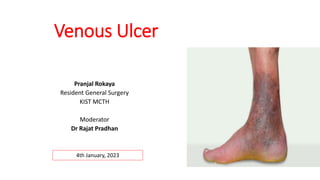
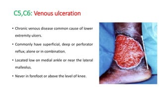
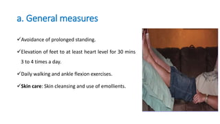
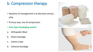
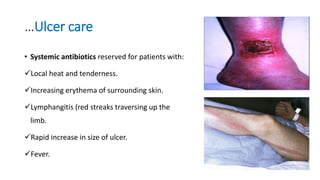
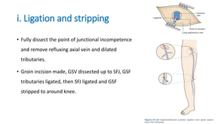
Venous Ulcer
Pranjal Rokaya
Resident General Surgery
KIST MCTH
Moderator
Dr Rajat Pradhan
4th January, 2023
Outline
1. Ulcer: Introduction
2. The venous system of lower limb
3. Lower limb venous disorders
4. Pathophysiology
5. Risk factors
6. Classification
7. Clinical features
8. Venous ulcer
9. Comparison of ulcers
10. Management of venous ulcer
Ulcer
• Break in the continuity of covering epithelium(skin or mucous membrane)
following molecular death of surface epithelium or its traumatic removal.
Classification
Clinically
1. Spreading Ulcer
2. Healing Ulcer
3. Callous/Chronic ulcer
Pathologically
1. Nonspecific Ulcers
 Traumatic
 Arterial
 Venous
 Neurogenic (trophic)
 A/w malnutrition
2. Specific ulcers
3. Malignant ulcers
Venous system of lower limb
Venous drainage of foot Deep venous system
Saphenous vein axis: Superficial system Perforator system
Lower extremity venous disorders
• A large spectrum of morphological and functional abnormalities.
• Can be congenital or acquired.
• May or may not be symptomatic.
• Ranges from minimal dilatation of veins to massive varicosities and ulceration.
Venous pathophysiology
Inadequate
muscle pump
function
Incompetent
venous valves
Venous
thrombosis
Non thrombotic
obstruction
Venous hypertension
Risk factors
Advancing age
Family history of venous disease
Ligamentous laxity
Prolonged standing
Increased BMI
Smoking
Lower extremity trauma
Prior venous thrombosis
Pregnancy
High estrogen states
Some hereditary conditions
Classification: CEAP system
C : Clinical
E : Etiological
A : Anatomic
P : Pathophysiology
Clinical Etiological
C0 No visible or palpable sign of venous disease Ep Primary
C1 Telangiectasia, reticular veins Es Secondary
C2 Varicose veins Ec Congenital
C3 Edema En No cause identified
C4 Changes in skin and subcutaneous tissue
C5 Healed
C6 Active venous ulcer
S With symptoms
A Without symptoms
Anatomical Pathophysiology
As Superficial veins Pr Reflux
Ad Deep veins Po Obstruction
Ap Perforator veins Pr,o Reflux and obstruction
An No venous anatomic
location identified
Pn No venous pathology
identifiable
Clinical features: Symptoms
Limb discomfort including leg heaviness or aching.
Throbbing or burning pain: Worse on standing/ sitting with feet dependent for a
prolonged period.
Itching.
Limb swelling.
Skin changes
Clinical signs by CEAP category
Co
20% of patients with clinical symptoms have no visible signs.
C1: Telangiectasia / reticular veins
Most frequently encountered sign.
Telangiectasia: Confluence of dilated
intradermal venules/ capillaries less than 1
mm in diameter.
Reticular veins: Dilated, bluish subdermal
veins, 1-3 mm in diameter.
C2: Varicose veins
Subcutaneous, dilated, tortuous veins more
than 3 mm in diameter.
Often a/w superficial axial venous reflux.
C3: Edema
Dependent ankle edema may progress to
include the calf region.
In the early stage, present only at end of the
day.
Clues:
a. Edema limited to lower extremity, often unilateral, a/w other signs.
b. Typically subsides with recumbency. (d/d from lymphatic obstruction).
c. CVP normal.
d. Nonresponsive to diuretics; may develop signs of hypoperfusion
C4a: Skin pigmentation/ dermatitis
• Initially prominent at medial ankle but later
encroach upon foot and lower leg.
• Pigmentation due to hemosiderin deposition.
• Venous reflux predisposes to stasis dermatitis
• Presents as eczematous rash.
C4b: Lipodermatosclerosis
Fibrosing panniculitis of subcutaneous tissue.
Characterized by firm are of induration,
initially located at medial ankle.
In late stage, entire legs can be
circumferentially involved.
Fibrosis may be extensive enough to strangle
lower leg.
Prone to repeated bouts of cellulitis.
C4c: Corona phlebectatita
Crown of numerous small, intradermal veins on
medial or lateral aspect of ankle or foot.
Indicator of venous hypertension due to saphenous
or perforator venous insufficiency.
C5,C6: Venous ulceration
• Chronic venous disease common cause of lower
extremity ulcers.
• Commonly have superficial, deep or perforator
reflux; alone or in combination.
• Located low on medial ankle or near the lateral
malleolus.
• Never in forefoot or above the level of knee.
Venous leg ulcer: Epidemiology
• Responsible for 85% of all chronic lower limb ulcers in resource rich countries.
• Prevalence: 0.1-0.3 % in adults (2-4% in elderly).
• 15-30% of patients have concomitant arterial occlusive disease
Venous ulcer: Pathology
Static blood flow in veins Hypoxia Tissue death
Not confirmed: Venous oxygen saturation found higher in ulcerated limbs.
Ambulatory
venous
hypertension
Incompetence of perforating
veins
Valve incompetence of saphenous veins
Incompetence or obstruction of
deep veins
Venous ulcer: Pathology
High venous pressure
Pericapillary infiltrate (
fibrin)
Fibrosis
Acts as impediment to
diffusion of nutrients.
Ulcer
environment
Growth factors inhibited
Increased ROS:
generate free
radicals; cause tissue
damage
Increased no. of
mast cells
monocytes and
lymphocytes
Venous ulcer: Morphology
Distribution : Medial ankle (gaiter region).
Shape : Shallow and flat
Margin : Thin and blue of growing epithelium.
Edge : Sloping; pale purple in color.
Floor : Pale granulation tissue
Base : Never penetrates deep fascia
Discharge : Seropurulent with an occasional trace of
blood.
History
• Complete medical history should be obtained.
• Diabetes, hypertension, CKD.
• Smoking history.
• Occupation history.
• Past history of DVT.
• Family history. Increased JVP: Sign of RHF
Examination
• General and systemic examination.
• Assessment of signs of venous
disease.
• Ulcer: Size, shape, number,
position, edge, base, margin,
discharge.
• Pulse examination.
• Neurological assessment
Types of ulcer edges
ABI to r/o PAD/mixed ulcer
Comparison of major types of leg ulcer
Venous Arterial Neuropathic
History • Prior DVT, stroke
• Obesity, multiple
pregnancies
• Pain upon prolonged
standing
• Cigarette smoking
• Diabetes
• Dyslipidemia
• Intermittent claudication
• Diabetes
• Other causes of
peripheral neuropathy
Location • Gaiter distribution
• Malleolar regions (M>L)
• Pressure sites
• Distal points (toes)
• Pressure sites
Pain • Mild to moderate • Severe • Painless
Morphology • Irregular borders
• Shallow
• Necrotic eschar
• Punched out
• Punched out
Comparison of major types of leg ulcer
Venous Arterial Neuropathic
Other findings • Varicosities
• Peripheral edema
• Stasis dermatitis
• Weak/absent pulses
• Prolonged CRT
• Pallor on leg
elevation (45 deg for
1 min)
• Peripheral
neuropathy with
decreased
sensation.
Relevant studies • Duplex
ultrasonography
• Ankle-brachial index
• CT angiography
• MR angiography
Investigations
• All new patients: CBC, RBS, ESR, CRP; Sickle cell test
a. Duplex scan
Presence of reflux in the deep and superficial venous
system.
Extent and distribution of reflux
Presence of obstruction in the deep venous system.
Presence of thrombus in the superficial system.
Investigations
b. Venography
Ascending: Dye injected into a superficial pedal vein.
Descending: Dye into the deep venous system at the groin or popliteal vein.
 Observation of flow defines regions of thrombus or obstruction.
 Adjunct to clinical exam and duplex scanning to identify specific valvular
incompetence.
Management
General measures
Compression therapy
Ulcer care
Pharmacotherapy
Surgical therapy
a. General measures
Avoidance of prolonged standing.
Elevation of feet to at least heart level for 30 mins
3 to 4 times a day.
Daily walking and ankle flexion exercises.
Skin care: Skin cleansing and use of emollients.
• Skin care
a. Stasis dermatitis
• For chronic dryness, petroleum-based
emollient.
• If oozing or vesiculation: Mid potency topical
steroid.
b. Contact dermatitis
• Avoid use of topical moisturizers/ antibiotics.
b. Compression therapy
• Keystone of management is to decrease venous
HTN.
• Primary way: Use of compression
• Four-layer bandaging system
a. Orthopedic Wool
b. Elastic bandage
c. Cotton crepe
d. Cohesive bandage
…Bandaging
The ideal interface pressure is 35-40 mm Hg.
The interval between bandage applications is based on the amount of exudate
and speed of healing.
Compression in mixed ulcer controversial.
For ABPI >0.5, a pressure of 30 mm Hg is safe and effective.
For ABPI <0.5, must undergo revascularisation before any compression.
Compression stockings
Different commercially available stockings with graded pressure
c. Ulcer care
• Basic wound care techniques are required.
i. Role of antibiotics
• Only in those with acute cellulitis or clinically infected ulcer.
• Routine use in uncomplicated ulcers doesn’t reduce bacterial colonization or
improve the healing rate; can cause emergence of resistance.
…Ulcer care
• Systemic antibiotics reserved for patients with:
Local heat and tenderness.
Increasing erythema of surrounding skin.
Lymphangitis (red streaks traversing up the
limb.
Rapid increase in size of ulcer.
Fever.
Ulcer care: Debridement
• Removal of necrotic tissue and fibrinous debris aids in the formation of granulation
tissue and enhances re-epithelialization.
• Regular dressing to control exudate, maintain moisture balance, and control odor
and pain.
• Options include simple nonadhesive dressing, paraffin gauze, hydrogels,
hydrocolloids, and silver-impregnated dressings.
• Few trials support a role of skin grafting for very large ulcers or ulcers present >12
months
Ulcer healing and recurrence
• Continued use of compression bandaging after healing reduces recurrence.
• Patient offered the strongest compression with which they can comply.
• Ulcers that persist beyond 6 months or recurrent ulcers should undergo a venous
duplex ultrasound to identify segments of venous incompetence amenable to
venous ablation.
d. Pharmacotherapy
• Variety of agents that affect venous tone used.
• Increase tone by a mechanism related to NE pathway.
• Other actions include reduction of capillary hyperpermeability, improved
lymphatic drainage, anti-inflammatory effects, and decreased blood viscosity.
…Pharmacotherapy
a. Flavonoids
Hydroxyethylrutoside (HR)
Escin (horse chestnut seed extract: HCE)
Micronized purified flavonoid fraction
 Meta-analysis of 15 RCTs showed HR has a higher response in pain and swelling
control vs placebo.
 Two meta-analyses: HCE improved symptoms related to chronic venous
insufficiency vs placebo
..Pharmacotherapy
 Aspirin: Insufficient evidence to detect beneficial effect or even harm.
 Stanazolol: Several RCTs have noted improvement in lipodermatosclerosis areas
and possibly faster ulcer healing rates.
Pentoxifylline: Metanalaysis of 11 trials: Significantly more effective for complete
or partial ulcer healing than placebo (800 mg TDS).
 Sulodexide, calcium dobesilate: low-quality evidence.
e. Surgical therapy
 Speciality referral in case of:
a. Arterial insufficiency.
b. Nonhealing ulcer.
c. Ulcer recurrence.
d. Suspected contact dermatitis.
e. Resistant or recurrent cellulitis.
Surgical therapy
Treat superficial
reflux
Ligation and stripping
Saphenofemoral
ligation and great
saphenous stripping
Saphenopopliteal
junction ligation and
small saphenous
stripping
Endothermal ablation
Laser ablation
Radiofrequency
ablation
Ultrasound guided
sclerotherapy
i. Ligation and stripping
• Fully dissect the point of junctional incompetence
and remove refluxing axial vein and dilated
tributaries.
• Groin incision made, GSV dissected up to SFJ, GSF
tributaries ligated, then SFJ ligated and GSF
stripped to around knee.
ii. Endothermal ablation
• Safer, faster recovery, cost effective.
• Concept:
A device inserted into incompetent axial vein p/c.
Vein surrounded by LA solution; compresses the vein emptying it of blood.
Device produces thermal energy that destroys structure of vein , resulting in
permanent occlusion.
Endothermal ablation
• Laser ablation
Laser energy at wavelength 1470 nm
transmitted to blood itself.
Steam bubbles are generated by laser
energy, coagulation occurs after
energy delivery.
• Radiofrequency ablation
 Radiofrequency heat is delivered at a
temperature of 120 C.
Directly injures vein wall endothelium,
resulting in collagen contraction and
thrombosis of treated vein.
iii. Ultrasound guided sclerotherapy
• Used for treatment of incompetent
perforators and large venous tributaries
causes by neovascularisation.
• Injection of sclerosing agent directly into
veins.
• Most commn agent: Sodium tetradecyl
sulphate.
• Causes cellular death resulting in thrombosis,
fibrosis and sclerosis.
Prognosis
• Nearly all venous ulcers can be healed.
• Even with successful ablation and/or compression; a reulceration rate of 20-30%
by five years.
• Reulceration is greatest in the post-thrombotic leg.
Summary
• Venous leg ulcer a/w profound impairment in quality of life.
• High chances of recurrence.
• Treatment a/w high cost to the healthcare system and patient.
• The mainstay of t/t: reduction in venous hypertension with compression and
superficial venous ablation.
References
• Bailey and Love textbook of surgery, 27th edition.
• Sabiston textbook of Surgery, 11th edition.
• Swartz textbook of surgery.
Thank You.

Venous ulcer

  • 1.
    Venous Ulcer Pranjal Rokaya ResidentGeneral Surgery KIST MCTH Moderator Dr Rajat Pradhan 4th January, 2023
  • 2.
    Outline 1. Ulcer: Introduction 2.The venous system of lower limb 3. Lower limb venous disorders 4. Pathophysiology 5. Risk factors 6. Classification 7. Clinical features 8. Venous ulcer 9. Comparison of ulcers 10. Management of venous ulcer
  • 3.
    Ulcer • Break inthe continuity of covering epithelium(skin or mucous membrane) following molecular death of surface epithelium or its traumatic removal. Classification Clinically 1. Spreading Ulcer 2. Healing Ulcer 3. Callous/Chronic ulcer Pathologically 1. Nonspecific Ulcers  Traumatic  Arterial  Venous  Neurogenic (trophic)  A/w malnutrition 2. Specific ulcers 3. Malignant ulcers
  • 4.
    Venous system oflower limb Venous drainage of foot Deep venous system
  • 5.
    Saphenous vein axis:Superficial system Perforator system
  • 6.
    Lower extremity venousdisorders • A large spectrum of morphological and functional abnormalities. • Can be congenital or acquired. • May or may not be symptomatic. • Ranges from minimal dilatation of veins to massive varicosities and ulceration.
  • 7.
    Venous pathophysiology Inadequate muscle pump function Incompetent venousvalves Venous thrombosis Non thrombotic obstruction Venous hypertension
  • 8.
    Risk factors Advancing age Familyhistory of venous disease Ligamentous laxity Prolonged standing Increased BMI Smoking Lower extremity trauma Prior venous thrombosis Pregnancy High estrogen states Some hereditary conditions
  • 9.
    Classification: CEAP system C: Clinical E : Etiological A : Anatomic P : Pathophysiology
  • 10.
    Clinical Etiological C0 Novisible or palpable sign of venous disease Ep Primary C1 Telangiectasia, reticular veins Es Secondary C2 Varicose veins Ec Congenital C3 Edema En No cause identified C4 Changes in skin and subcutaneous tissue C5 Healed C6 Active venous ulcer S With symptoms A Without symptoms
  • 11.
    Anatomical Pathophysiology As Superficialveins Pr Reflux Ad Deep veins Po Obstruction Ap Perforator veins Pr,o Reflux and obstruction An No venous anatomic location identified Pn No venous pathology identifiable
  • 12.
    Clinical features: Symptoms Limbdiscomfort including leg heaviness or aching. Throbbing or burning pain: Worse on standing/ sitting with feet dependent for a prolonged period. Itching. Limb swelling. Skin changes
  • 13.
    Clinical signs byCEAP category Co 20% of patients with clinical symptoms have no visible signs.
  • 14.
    C1: Telangiectasia /reticular veins Most frequently encountered sign. Telangiectasia: Confluence of dilated intradermal venules/ capillaries less than 1 mm in diameter. Reticular veins: Dilated, bluish subdermal veins, 1-3 mm in diameter.
  • 15.
    C2: Varicose veins Subcutaneous,dilated, tortuous veins more than 3 mm in diameter. Often a/w superficial axial venous reflux.
  • 16.
    C3: Edema Dependent ankleedema may progress to include the calf region. In the early stage, present only at end of the day. Clues: a. Edema limited to lower extremity, often unilateral, a/w other signs. b. Typically subsides with recumbency. (d/d from lymphatic obstruction). c. CVP normal. d. Nonresponsive to diuretics; may develop signs of hypoperfusion
  • 17.
    C4a: Skin pigmentation/dermatitis • Initially prominent at medial ankle but later encroach upon foot and lower leg. • Pigmentation due to hemosiderin deposition. • Venous reflux predisposes to stasis dermatitis • Presents as eczematous rash.
  • 18.
    C4b: Lipodermatosclerosis Fibrosing panniculitisof subcutaneous tissue. Characterized by firm are of induration, initially located at medial ankle. In late stage, entire legs can be circumferentially involved. Fibrosis may be extensive enough to strangle lower leg. Prone to repeated bouts of cellulitis.
  • 19.
    C4c: Corona phlebectatita Crownof numerous small, intradermal veins on medial or lateral aspect of ankle or foot. Indicator of venous hypertension due to saphenous or perforator venous insufficiency.
  • 20.
    C5,C6: Venous ulceration •Chronic venous disease common cause of lower extremity ulcers. • Commonly have superficial, deep or perforator reflux; alone or in combination. • Located low on medial ankle or near the lateral malleolus. • Never in forefoot or above the level of knee.
  • 21.
    Venous leg ulcer:Epidemiology • Responsible for 85% of all chronic lower limb ulcers in resource rich countries. • Prevalence: 0.1-0.3 % in adults (2-4% in elderly). • 15-30% of patients have concomitant arterial occlusive disease
  • 22.
    Venous ulcer: Pathology Staticblood flow in veins Hypoxia Tissue death Not confirmed: Venous oxygen saturation found higher in ulcerated limbs. Ambulatory venous hypertension Incompetence of perforating veins Valve incompetence of saphenous veins Incompetence or obstruction of deep veins
  • 23.
    Venous ulcer: Pathology Highvenous pressure Pericapillary infiltrate ( fibrin) Fibrosis Acts as impediment to diffusion of nutrients. Ulcer environment Growth factors inhibited Increased ROS: generate free radicals; cause tissue damage Increased no. of mast cells monocytes and lymphocytes
  • 24.
    Venous ulcer: Morphology Distribution: Medial ankle (gaiter region). Shape : Shallow and flat Margin : Thin and blue of growing epithelium. Edge : Sloping; pale purple in color. Floor : Pale granulation tissue Base : Never penetrates deep fascia Discharge : Seropurulent with an occasional trace of blood.
  • 25.
    History • Complete medicalhistory should be obtained. • Diabetes, hypertension, CKD. • Smoking history. • Occupation history. • Past history of DVT. • Family history. Increased JVP: Sign of RHF
  • 26.
    Examination • General andsystemic examination. • Assessment of signs of venous disease. • Ulcer: Size, shape, number, position, edge, base, margin, discharge. • Pulse examination. • Neurological assessment Types of ulcer edges
  • 27.
    ABI to r/oPAD/mixed ulcer
  • 28.
    Comparison of majortypes of leg ulcer Venous Arterial Neuropathic History • Prior DVT, stroke • Obesity, multiple pregnancies • Pain upon prolonged standing • Cigarette smoking • Diabetes • Dyslipidemia • Intermittent claudication • Diabetes • Other causes of peripheral neuropathy Location • Gaiter distribution • Malleolar regions (M>L) • Pressure sites • Distal points (toes) • Pressure sites Pain • Mild to moderate • Severe • Painless Morphology • Irregular borders • Shallow • Necrotic eschar • Punched out • Punched out
  • 29.
    Comparison of majortypes of leg ulcer Venous Arterial Neuropathic Other findings • Varicosities • Peripheral edema • Stasis dermatitis • Weak/absent pulses • Prolonged CRT • Pallor on leg elevation (45 deg for 1 min) • Peripheral neuropathy with decreased sensation. Relevant studies • Duplex ultrasonography • Ankle-brachial index • CT angiography • MR angiography
  • 30.
    Investigations • All newpatients: CBC, RBS, ESR, CRP; Sickle cell test a. Duplex scan Presence of reflux in the deep and superficial venous system. Extent and distribution of reflux Presence of obstruction in the deep venous system. Presence of thrombus in the superficial system.
  • 31.
    Investigations b. Venography Ascending: Dyeinjected into a superficial pedal vein. Descending: Dye into the deep venous system at the groin or popliteal vein.  Observation of flow defines regions of thrombus or obstruction.  Adjunct to clinical exam and duplex scanning to identify specific valvular incompetence.
  • 32.
    Management General measures Compression therapy Ulcercare Pharmacotherapy Surgical therapy
  • 33.
    a. General measures Avoidanceof prolonged standing. Elevation of feet to at least heart level for 30 mins 3 to 4 times a day. Daily walking and ankle flexion exercises. Skin care: Skin cleansing and use of emollients.
  • 34.
    • Skin care a.Stasis dermatitis • For chronic dryness, petroleum-based emollient. • If oozing or vesiculation: Mid potency topical steroid. b. Contact dermatitis • Avoid use of topical moisturizers/ antibiotics.
  • 35.
    b. Compression therapy •Keystone of management is to decrease venous HTN. • Primary way: Use of compression • Four-layer bandaging system a. Orthopedic Wool b. Elastic bandage c. Cotton crepe d. Cohesive bandage
  • 36.
    …Bandaging The ideal interfacepressure is 35-40 mm Hg. The interval between bandage applications is based on the amount of exudate and speed of healing. Compression in mixed ulcer controversial. For ABPI >0.5, a pressure of 30 mm Hg is safe and effective. For ABPI <0.5, must undergo revascularisation before any compression.
  • 37.
    Compression stockings Different commerciallyavailable stockings with graded pressure
  • 38.
    c. Ulcer care •Basic wound care techniques are required. i. Role of antibiotics • Only in those with acute cellulitis or clinically infected ulcer. • Routine use in uncomplicated ulcers doesn’t reduce bacterial colonization or improve the healing rate; can cause emergence of resistance.
  • 39.
    …Ulcer care • Systemicantibiotics reserved for patients with: Local heat and tenderness. Increasing erythema of surrounding skin. Lymphangitis (red streaks traversing up the limb. Rapid increase in size of ulcer. Fever.
  • 40.
    Ulcer care: Debridement •Removal of necrotic tissue and fibrinous debris aids in the formation of granulation tissue and enhances re-epithelialization. • Regular dressing to control exudate, maintain moisture balance, and control odor and pain. • Options include simple nonadhesive dressing, paraffin gauze, hydrogels, hydrocolloids, and silver-impregnated dressings. • Few trials support a role of skin grafting for very large ulcers or ulcers present >12 months
  • 41.
    Ulcer healing andrecurrence • Continued use of compression bandaging after healing reduces recurrence. • Patient offered the strongest compression with which they can comply. • Ulcers that persist beyond 6 months or recurrent ulcers should undergo a venous duplex ultrasound to identify segments of venous incompetence amenable to venous ablation.
  • 42.
    d. Pharmacotherapy • Varietyof agents that affect venous tone used. • Increase tone by a mechanism related to NE pathway. • Other actions include reduction of capillary hyperpermeability, improved lymphatic drainage, anti-inflammatory effects, and decreased blood viscosity.
  • 43.
    …Pharmacotherapy a. Flavonoids Hydroxyethylrutoside (HR) Escin(horse chestnut seed extract: HCE) Micronized purified flavonoid fraction  Meta-analysis of 15 RCTs showed HR has a higher response in pain and swelling control vs placebo.  Two meta-analyses: HCE improved symptoms related to chronic venous insufficiency vs placebo
  • 44.
    ..Pharmacotherapy  Aspirin: Insufficientevidence to detect beneficial effect or even harm.  Stanazolol: Several RCTs have noted improvement in lipodermatosclerosis areas and possibly faster ulcer healing rates. Pentoxifylline: Metanalaysis of 11 trials: Significantly more effective for complete or partial ulcer healing than placebo (800 mg TDS).  Sulodexide, calcium dobesilate: low-quality evidence.
  • 45.
    e. Surgical therapy Speciality referral in case of: a. Arterial insufficiency. b. Nonhealing ulcer. c. Ulcer recurrence. d. Suspected contact dermatitis. e. Resistant or recurrent cellulitis.
  • 46.
    Surgical therapy Treat superficial reflux Ligationand stripping Saphenofemoral ligation and great saphenous stripping Saphenopopliteal junction ligation and small saphenous stripping Endothermal ablation Laser ablation Radiofrequency ablation Ultrasound guided sclerotherapy
  • 47.
    i. Ligation andstripping • Fully dissect the point of junctional incompetence and remove refluxing axial vein and dilated tributaries. • Groin incision made, GSV dissected up to SFJ, GSF tributaries ligated, then SFJ ligated and GSF stripped to around knee.
  • 48.
    ii. Endothermal ablation •Safer, faster recovery, cost effective. • Concept: A device inserted into incompetent axial vein p/c. Vein surrounded by LA solution; compresses the vein emptying it of blood. Device produces thermal energy that destroys structure of vein , resulting in permanent occlusion.
  • 49.
    Endothermal ablation • Laserablation Laser energy at wavelength 1470 nm transmitted to blood itself. Steam bubbles are generated by laser energy, coagulation occurs after energy delivery. • Radiofrequency ablation  Radiofrequency heat is delivered at a temperature of 120 C. Directly injures vein wall endothelium, resulting in collagen contraction and thrombosis of treated vein.
  • 50.
    iii. Ultrasound guidedsclerotherapy • Used for treatment of incompetent perforators and large venous tributaries causes by neovascularisation. • Injection of sclerosing agent directly into veins. • Most commn agent: Sodium tetradecyl sulphate. • Causes cellular death resulting in thrombosis, fibrosis and sclerosis.
  • 51.
    Prognosis • Nearly allvenous ulcers can be healed. • Even with successful ablation and/or compression; a reulceration rate of 20-30% by five years. • Reulceration is greatest in the post-thrombotic leg.
  • 52.
    Summary • Venous legulcer a/w profound impairment in quality of life. • High chances of recurrence. • Treatment a/w high cost to the healthcare system and patient. • The mainstay of t/t: reduction in venous hypertension with compression and superficial venous ablation.
  • 53.
    References • Bailey andLove textbook of surgery, 27th edition. • Sabiston textbook of Surgery, 11th edition. • Swartz textbook of surgery.
  • 54.