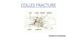
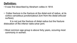
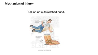
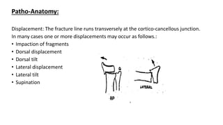
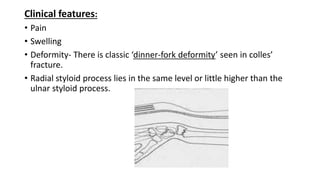
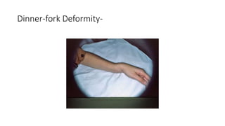
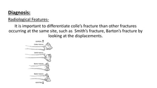
COLLES FRACTURE
-MONICA.N.DHANANI
Definition:
• It was first described by Abraham colles in 1814.
• Colles fracture is the fracture at the distal end of radius, at its
cortico cancellous junction(about 2cm from the distal articular
surface).
It is not just the fracture of distal radius but the fracture
dislocation of the inferior radio-ulnar joint.
• Most common age group is above forty years, occuring most
commonly in women.
Mechanism of Injury-
Fall on an outstretched hand.
Patho-Anatomy:
Displacement: The fracture line runs transversely at the cortico-cancellous junction.
In many cases one or more displacements may occur as follows.:
• Impaction of fragments
• Dorsal displacement
• Dorsal tilt
• Lateral displacement
• Lateral tilt
• Supination
Clinical features:
• Pain
• Swelling
• Deformity- There is classic ‘dinner-fork deformity’ seen in colles’
fracture.
• Radial styloid process lies in the same level or little higher than the
ulnar styloid process.
Dinner-fork Deformity-
Diagnosis:
Radiological Features-
It is important to differentiate colle’s fracture than other fractures
occurring at the same site, such as Smith’s fracture, Barton’s fracture by
looking at the displacements.
X-RAY:
Lateral view
• Dorsal tilt- It can be detected by looking at the direction of distal
articular surface
AP view
• Lateral tilt- similarly it can be detected by looking at the articular
surface if it faces medially it is normal,if it becomes horizontal or faces
laterally ,a lateral tilt is present.
Treatment-
Conservative Method:
For Undisplaced fracture-immobilisation in a below-elbow plaster cast
for six weeks
For Displaced fractures- Manipulative reduction followed by
immobilization in a colles cast.
Manipulative reduction :
The muscles of the forearm are to be relaxed either by general or regional
anaesthsia.
• Step-1: the first step is to disimpact the fragments this can be achieved
by firm longitudinal traction against the counter-traction by an assistant.
• Step-2:Press the distal fragment into palmar flexion and ulnar deviation
by using thumb of other hand.
• Step-3:Now hand is drawn into pronation, palmar flexion and ulnar
deviation. A plaster cast is applied extending from below the elbow to
the metacarpal heads,maintaining the wrist in palmar flexion and ulnar
deviation. After six weeks, plaster cast is removed and joint mobilising
and strengthening exercises are started.
Manipulative reduction technique
Surgical method:
• 1 . Closed reduction and percutaneous pinning with k – wires
• 2 . Open reduction and plate fixation.
Complications-
1. Stiffness of joints
2. Mal-union
3. Subluxation of the inferior radio-ulnar joint
4. Carpel –tunnel syndrome
5. Sudecks osteodystrophy
6. Rupture of the extensor pollicis longus tendon
THANKYOU

Colles fracture

  • 1.
  • 2.
    Definition: • It wasfirst described by Abraham colles in 1814. • Colles fracture is the fracture at the distal end of radius, at its cortico cancellous junction(about 2cm from the distal articular surface). It is not just the fracture of distal radius but the fracture dislocation of the inferior radio-ulnar joint. • Most common age group is above forty years, occuring most commonly in women.
  • 3.
    Mechanism of Injury- Fallon an outstretched hand.
  • 4.
    Patho-Anatomy: Displacement: The fractureline runs transversely at the cortico-cancellous junction. In many cases one or more displacements may occur as follows.: • Impaction of fragments • Dorsal displacement • Dorsal tilt • Lateral displacement • Lateral tilt • Supination
  • 5.
    Clinical features: • Pain •Swelling • Deformity- There is classic ‘dinner-fork deformity’ seen in colles’ fracture. • Radial styloid process lies in the same level or little higher than the ulnar styloid process.
  • 6.
  • 7.
    Diagnosis: Radiological Features- It isimportant to differentiate colle’s fracture than other fractures occurring at the same site, such as Smith’s fracture, Barton’s fracture by looking at the displacements.
  • 8.
    X-RAY: Lateral view • Dorsaltilt- It can be detected by looking at the direction of distal articular surface AP view • Lateral tilt- similarly it can be detected by looking at the articular surface if it faces medially it is normal,if it becomes horizontal or faces laterally ,a lateral tilt is present.
  • 9.
    Treatment- Conservative Method: For Undisplacedfracture-immobilisation in a below-elbow plaster cast for six weeks For Displaced fractures- Manipulative reduction followed by immobilization in a colles cast.
  • 10.
    Manipulative reduction : Themuscles of the forearm are to be relaxed either by general or regional anaesthsia. • Step-1: the first step is to disimpact the fragments this can be achieved by firm longitudinal traction against the counter-traction by an assistant. • Step-2:Press the distal fragment into palmar flexion and ulnar deviation by using thumb of other hand. • Step-3:Now hand is drawn into pronation, palmar flexion and ulnar deviation. A plaster cast is applied extending from below the elbow to the metacarpal heads,maintaining the wrist in palmar flexion and ulnar deviation. After six weeks, plaster cast is removed and joint mobilising and strengthening exercises are started.
  • 11.
  • 12.
    Surgical method: • 1. Closed reduction and percutaneous pinning with k – wires • 2 . Open reduction and plate fixation.
  • 13.
    Complications- 1. Stiffness ofjoints 2. Mal-union 3. Subluxation of the inferior radio-ulnar joint 4. Carpel –tunnel syndrome 5. Sudecks osteodystrophy 6. Rupture of the extensor pollicis longus tendon
  • 14.