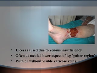
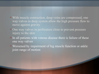
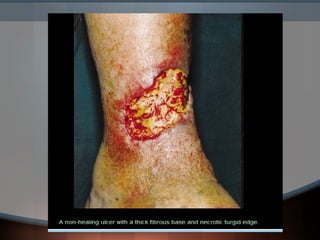
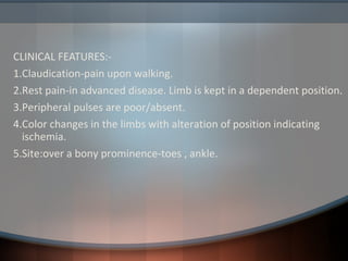
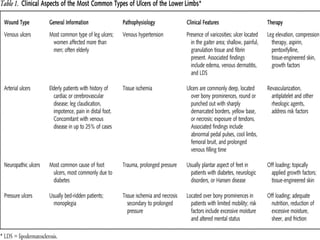
This document discusses different types of leg ulcers including venous, arterial, diabetic neuropathic, and hypertensive ulcers. It provides information on:
1. The causes, risk factors, signs and symptoms, investigations and management for each type of ulcer.
2. Venous ulcers are the most common type, caused by venous insufficiency and reflux, and are typically treated with compression therapy and dressings.
3. Arterial ulcers are caused by peripheral arterial disease and present with dry necrotic wounds, often over bony prominences of the feet. Revascularization may be required for healing.
4. Diabetic and neuropathic ulcers occur due to loss of sensation from
Venous ulcer is one of the commonest complication of varicose veins. It may also occur in a condition called post phlebitic limb which is a sequelae to acute deep vein thronbosis. Hurry in surgical treatment of this condition before the ulcer heals could lead to a failure. Good conservative treatment for healing of the ulcer followed by surgical intervention gives the best results.
Made by Ranjith R Thampi. A surgery powerpoint I made during internship for Management of Varicose Veins. Tried to cover as much as possible on the topic. Kindly comment before you download. Thanks!
Types, Investigation, complication and treatment of Incisional herniaimraxid
It is herniation through a weak abdominal scar (scar of previous surgery).
It is common in old age and obese individuals.
Predisposing Factors:
..> Vertical scar, midline scar, lower abdominal scar— may injure the nerves of the abdominal muscles.
...> Scar of major surgeries (biliary, pancreatic).
...> Scar of emergency surgeries (peritonitis, acute abdomen).
For Health Tips: http://MedicoPk.com/
NECROTISING FASCIITIS- the flesh eating infection
#surgicaleducator #necrotisingfasciitis #surgicaltutor #babysurgeon #usmle
· Dear Viewers
· Greetings from “Surgical Educator”
· Today in this episode I have discussed about Necrotising Fasciitis- the flesh eating infection
· It is common in immunocompromised patients even after trivial trauma.
· I have discussed about the overview,etiology,types,clinical features,complications and treatment of Necrotising Fasciitis
· I hope this video is interesting and also useful to all of you
· You can watch the video in the following links:
· surgicaleducator.blogspot.com youtube.com/c/surgicaleducator
Thank you for watching the video
Lecture on haemorrhoids for medical students. Encompasses basic sciences, classifications, principles and tips of management of this very common yet potentially complicated disorder.
Detailed presentation on Varicose veins, examination and management
Detailed presentation on Deep Vein Thrombosis, categories, staging and scoring systems and management.
Management also includes Endovascular and Surgical techniques.
Short notes made on IVC filters
NVBDCP.pptx Nation vector borne disease control programSapna Thakur
NVBDCP was launched in 2003-2004 . Vector-Borne Disease: Disease that results from an infection transmitted to humans and other animals by blood-feeding arthropods, such as mosquitoes, ticks, and fleas. Examples of vector-borne diseases include Dengue fever, West Nile Virus, Lyme disease, and malaria.
Ethanol (CH3CH2OH), or beverage alcohol, is a two-carbon alcohol
that is rapidly distributed in the body and brain. Ethanol alters many
neurochemical systems and has rewarding and addictive properties. It
is the oldest recreational drug and likely contributes to more morbidity,
mortality, and public health costs than all illicit drugs combined. The
5th edition of the Diagnostic and Statistical Manual of Mental Disorders
(DSM-5) integrates alcohol abuse and alcohol dependence into a single
disorder called alcohol use disorder (AUD), with mild, moderate,
and severe subclassifications (American Psychiatric Association, 2013).
In the DSM-5, all types of substance abuse and dependence have been
combined into a single substance use disorder (SUD) on a continuum
from mild to severe. A diagnosis of AUD requires that at least two of
the 11 DSM-5 behaviors be present within a 12-month period (mild
AUD: 2–3 criteria; moderate AUD: 4–5 criteria; severe AUD: 6–11 criteria).
The four main behavioral effects of AUD are impaired control over
drinking, negative social consequences, risky use, and altered physiological
effects (tolerance, withdrawal). This chapter presents an overview
of the prevalence and harmful consequences of AUD in the U.S.,
the systemic nature of the disease, neurocircuitry and stages of AUD,
comorbidities, fetal alcohol spectrum disorders, genetic risk factors, and
pharmacotherapies for AUD.
Pulmonary Thromboembolism - etilogy, types, medical- Surgical and nursing man...VarunMahajani
Disruption of blood supply to lung alveoli due to blockage of one or more pulmonary blood vessels is called as Pulmonary thromboembolism. In this presentation we will discuss its causes, types and its management in depth.
New Drug Discovery and Development .....NEHA GUPTA
The "New Drug Discovery and Development" process involves the identification, design, testing, and manufacturing of novel pharmaceutical compounds with the aim of introducing new and improved treatments for various medical conditions. This comprehensive endeavor encompasses various stages, including target identification, preclinical studies, clinical trials, regulatory approval, and post-market surveillance. It involves multidisciplinary collaboration among scientists, researchers, clinicians, regulatory experts, and pharmaceutical companies to bring innovative therapies to market and address unmet medical needs.
Acute scrotum is a general term referring to an emergency condition affecting the contents or the wall of the scrotum.
There are a number of conditions that present acutely, predominantly with pain and/or swelling
A careful and detailed history and examination, and in some cases, investigations allow differentiation between these diagnoses. A prompt diagnosis is essential as the patient may require urgent surgical intervention
Testicular torsion refers to twisting of the spermatic cord, causing ischaemia of the testicle.
Testicular torsion results from inadequate fixation of the testis to the tunica vaginalis producing ischemia from reduced arterial inflow and venous outflow obstruction.
The prevalence of testicular torsion in adult patients hospitalized with acute scrotal pain is approximately 25 to 50 percent
Ozempic: Preoperative Management of Patients on GLP-1 Receptor Agonists Saeid Safari
Preoperative Management of Patients on GLP-1 Receptor Agonists like Ozempic and Semiglutide
ASA GUIDELINE
NYSORA Guideline
2 Case Reports of Gastric Ultrasound
Lung Cancer: Artificial Intelligence, Synergetics, Complex System Analysis, S...Oleg Kshivets
RESULTS: Overall life span (LS) was 2252.1±1742.5 days and cumulative 5-year survival (5YS) reached 73.2%, 10 years – 64.8%, 20 years – 42.5%. 513 LCP lived more than 5 years (LS=3124.6±1525.6 days), 148 LCP – more than 10 years (LS=5054.4±1504.1 days).199 LCP died because of LC (LS=562.7±374.5 days). 5YS of LCP after bi/lobectomies was significantly superior in comparison with LCP after pneumonectomies (78.1% vs.63.7%, P=0.00001 by log-rank test). AT significantly improved 5YS (66.3% vs. 34.8%) (P=0.00000 by log-rank test) only for LCP with N1-2. Cox modeling displayed that 5YS of LCP significantly depended on: phase transition (PT) early-invasive LC in terms of synergetics, PT N0—N12, cell ratio factors (ratio between cancer cells- CC and blood cells subpopulations), G1-3, histology, glucose, AT, blood cell circuit, prothrombin index, heparin tolerance, recalcification time (P=0.000-0.038). Neural networks, genetic algorithm selection and bootstrap simulation revealed relationships between 5YS and PT early-invasive LC (rank=1), PT N0—N12 (rank=2), thrombocytes/CC (3), erythrocytes/CC (4), eosinophils/CC (5), healthy cells/CC (6), lymphocytes/CC (7), segmented neutrophils/CC (8), stick neutrophils/CC (9), monocytes/CC (10); leucocytes/CC (11). Correct prediction of 5YS was 100% by neural networks computing (area under ROC curve=1.0; error=0.0).
CONCLUSIONS: 5YS of LCP after radical procedures significantly depended on: 1) PT early-invasive cancer; 2) PT N0--N12; 3) cell ratio factors; 4) blood cell circuit; 5) biochemical factors; 6) hemostasis system; 7) AT; 8) LC characteristics; 9) LC cell dynamics; 10) surgery type: lobectomy/pneumonectomy; 11) anthropometric data. Optimal diagnosis and treatment strategies for LC are: 1) screening and early detection of LC; 2) availability of experienced thoracic surgeons because of complexity of radical procedures; 3) aggressive en block surgery and adequate lymph node dissection for completeness; 4) precise prediction; 5) adjuvant chemoimmunoradiotherapy for LCP with unfavorable prognosis.
TEST BANK for Operations Management, 14th Edition by William J. Stevenson, Ve...kevinkariuki227
TEST BANK for Operations Management, 14th Edition by William J. Stevenson, Verified Chapters 1 - 19, Complete Newest Version.pdf
These simplified slides by Dr. Sidra Arshad present an overview of the non-respiratory functions of the respiratory tract.
Learning objectives:
1. Enlist the non-respiratory functions of the respiratory tract
2. Briefly explain how these functions are carried out
3. Discuss the significance of dead space
4. Differentiate between minute ventilation and alveolar ventilation
5. Describe the cough and sneeze reflexes
Study Resources:
1. Chapter 39, Guyton and Hall Textbook of Medical Physiology, 14th edition
2. Chapter 34, Ganong’s Review of Medical Physiology, 26th edition
3. Chapter 17, Human Physiology by Lauralee Sherwood, 9th edition
4. Non-respiratory functions of the lungs https://academic.oup.com/bjaed/article/13/3/98/278874
Recomendações da OMS sobre cuidados maternos e neonatais para uma experiência pós-natal positiva.
Em consonância com os ODS – Objetivos do Desenvolvimento Sustentável e a Estratégia Global para a Saúde das Mulheres, Crianças e Adolescentes, e aplicando uma abordagem baseada nos direitos humanos, os esforços de cuidados pós-natais devem expandir-se para além da cobertura e da simples sobrevivência, de modo a incluir cuidados de qualidade.
Estas diretrizes visam melhorar a qualidade dos cuidados pós-natais essenciais e de rotina prestados às mulheres e aos recém-nascidos, com o objetivo final de melhorar a saúde e o bem-estar materno e neonatal.
Uma “experiência pós-natal positiva” é um resultado importante para todas as mulheres que dão à luz e para os seus recém-nascidos, estabelecendo as bases para a melhoria da saúde e do bem-estar a curto e longo prazo. Uma experiência pós-natal positiva é definida como aquela em que as mulheres, pessoas que gestam, os recém-nascidos, os casais, os pais, os cuidadores e as famílias recebem informação consistente, garantia e apoio de profissionais de saúde motivados; e onde um sistema de saúde flexível e com recursos reconheça as necessidades das mulheres e dos bebês e respeite o seu contexto cultural.
Estas diretrizes consolidadas apresentam algumas recomendações novas e já bem fundamentadas sobre cuidados pós-natais de rotina para mulheres e neonatos que recebem cuidados no pós-parto em unidades de saúde ou na comunidade, independentemente dos recursos disponíveis.
É fornecido um conjunto abrangente de recomendações para cuidados durante o período puerperal, com ênfase nos cuidados essenciais que todas as mulheres e recém-nascidos devem receber, e com a devida atenção à qualidade dos cuidados; isto é, a entrega e a experiência do cuidado recebido. Estas diretrizes atualizam e ampliam as recomendações da OMS de 2014 sobre cuidados pós-natais da mãe e do recém-nascido e complementam as atuais diretrizes da OMS sobre a gestão de complicações pós-natais.
O estabelecimento da amamentação e o manejo das principais intercorrências é contemplada.
Recomendamos muito.
Vamos discutir essas recomendações no nosso curso de pós-graduação em Aleitamento no Instituto Ciclos.
Esta publicação só está disponível em inglês até o momento.
Prof. Marcus Renato de Carvalho
www.agostodourado.com
Report Back from SGO 2024: What’s the Latest in Cervical Cancer?bkling
Are you curious about what’s new in cervical cancer research or unsure what the findings mean? Join Dr. Emily Ko, a gynecologic oncologist at Penn Medicine, to learn about the latest updates from the Society of Gynecologic Oncology (SGO) 2024 Annual Meeting on Women’s Cancer. Dr. Ko will discuss what the research presented at the conference means for you and answer your questions about the new developments.
263778731218 Abortion Clinic /Pills In Harare ,sisternakatoto
263778731218 Abortion Clinic /Pills In Harare ,ABORTION WOMEN’S CLINIC +27730423979 IN women clinic we believe that every woman should be able to make choices in her pregnancy. Our job is to provide compassionate care, safety,affordable and confidential services. That’s why we have won the trust from all generations of women all over the world. we use non surgical method(Abortion pills) to terminate…Dr.LISA +27730423979women Clinic is committed to providing the highest quality of obstetrical and gynecological care to women of all ages. Our dedicated staff aim to treat each patient and her health concerns with compassion and respect.Our dedicated group ABORTION WOMEN’S CLINIC +27730423979 IN women clinic we believe that every woman should be able to make choices in her pregnancy. Our job is to provide compassionate care, safety,affordable and confidential services. That’s why we have won the trust from all generations of women all over the world. we use non surgical method(Abortion pills) to terminate…Dr.LISA +27730423979women Clinic is committed to providing the highest quality of obstetrical and gynecological care to women of all ages. Our dedicated staff aim to treat each patient and her health concerns with compassion and respect.Our dedicated group of receptionists, nurses, and physicians have worked together as a teamof receptionists, nurses, and physicians have worked together as a team wwww.lisywomensclinic.co.za/
7. With muscle contraction, deep veins are compressed, one
way valves in deep system allow the high pressure flow to
move against gravity
One way valves in perforators close to prevent pressure
injury to the skin
In all patients with venous disease there is failure of these
one way valves
Worsened by impairment of leg muscle function or ankle
joint range of motion
8. Incompetent perforators and refluxIncompetent perforators and reflux
• Failure of venous valve (REFLUX)
and poor calf muscle function leads
to ambulatory venous hypertension
and sustained capillary hypertension
• 60% of venous ulcer patients have
isolated superficial vein insufficiency
9. PathogenesisPathogenesis
1. Capillary stasis: `Homans` postulated that stasis of venous
blood in post-thrombotic syndrome gave rise to anoxia and hence
venous ulcers
2. Fibrin cuff theory: `Browse and Bermand` postulated that
venous ulcer could be result of deposition of pericapillary fibrin due
to leakage of fibrinogen through pericapillary spaces. fibrinogen
polymerizes to form fibrin
Oxygen diffusion barrier
Increase in collagen IV in and around capillary
3. White cell trapping: `Coleridge and Smith` suggested that
leukocytes may become trapped in capillaries in static blood,
obstructing the flow
10. 4. Trapping growth factors: `Felanga and Eaglestein`
hypothesized that pericapillary fibrincuff , α-2 macroglobulin interfare
with growth factor transport
5. Multicausal model/ Maastricht model:
Elevated pressures on vascular side of capillaries lead to structural
changes in capillary wall
Interendothelial space broadens
Collagen IV layer disintegrates
Capillary wall becomes thicker
Water diffusion is effected- oedema
Larger molecules (fibrin ) escape- fibrin cuff formation
α-2 macroglobulin escapes and traps TGF-β
13. Signs and symptomsSigns and symptoms
• Usually preceded by patchy erythema or discoloration of an
intense bluish red colour (capillary congestion)
• Ischemia of the skin finally leads, to necrosis, often following
a minor episode of trauma (scratching, small knocks,
dermatitis)
• Ulcer is typically painless. Patients develop typical ischemic
pain on elevation of the ulcerated leg, which is edematous .
Associated with arterial disease can ( 1/3 of cases).
• Ulcer is covered with yellowish exudate over granulation
tissue
• Healing ulcers have a shallow sloping edge with healthy
granulation tissue in their base and little slough. Pink lip of
epithelium at the edge of ulcer is uniform and supplied by
relatively uncongested capillaries
14.
15.
16. • Signs:-
Signs of venous hypertension are present- LDS, varicose
veins, varicose eczema or oedema
An oedematous leg not responding to diuretics is a strong
clue to the diagnosis
18. DiagnosisDiagnosis
• Clinical:
Gaiter area
Signs of venous hypertension
Past history of venous thrombosis
Past history of treatment for varicose veins
Family history of venous disease
• Colour Doppler duplex USG: usually, after ulcer
heals
Confirm venous reflux, superficial venous incompitance
Confirm deep venous compliance
19. • Plethysmography:
To investigate calf muscle pump function when Colour
Duplex is normal
• Skin biopsy: not indicated in venous ulcer
Skin malignancy or vasculitis suspected
• Arterial disease must be excluded;
ABPI
Arterial duplex doppler scanning
arteriography
20. ManagementManagement
• Is done by the following means:-
1. CONSERVATIVE TREATMENT
2. SYSTEMIC MEDICATIONS
3. SURGICAL OPTIONS
• The goals of treatment are to reduce edema, improve ulcer
healing, and prevent recurrence.
21. COMPRESSION THERAPYCOMPRESSION THERAPY
• Compression therapy is the standard of care for venous ulcers
and chronic venous insufficiency
• Compression therapy reduces edema, improves venous reflux,
enhances healing of ulcers, and reduces pain.
• After an ulcer has healed, lifelong maintenance of compression
therapy may reduce the risk of recurrence
• Contraindications to compression therapy include clinically
significant arterial disease and uncompensated heart failure.
• Methods include
1. inelastic,
2. elastic,
3. intermittent pneumatic compression
22. 1. INELASTIC COMPRESSION :-
• Inelastic compression therapy provides high working pressure
during ambulation and muscle contraction, but no resting
pressure.
• The most common method of inelastic compression therapy is
the Unna boot, a zinc oxide–impregnated, moist bandage that
hardens after application.
• Disadvantages:- because of its inelasticity, the Unna boot does
not conform to changes in leg size and may be uncomfortable to
wear.
• The Unna boot may lead to a foul smell from the accumulation
of exudate from the ulcer, requiring frequent reapplications
23.
24. 2. ELASTIC COMPRESSION :-
• Elastic compression therapy methods conform to changes in leg
size and sustain compression during both rest and activity
• Stockings or bandages can be used; however, elastic wraps are
not recommended because they do not provide enough pressure
• Compression stockings are removed at night, and should be
replaced every six months because they lose pressure with regular
washing
• Elastic bandages are alternatives to compression stockings
(multilayer bandages are more effective than single layer.)
Disadvantages:- multilayer compression bandages require skilled
application in the physician’s office one or two times per week,
depending on drainage.
25.
26.
27. 3. INTERMITTENT PNEUMATIC COMPRESSION:-
• comprises a pump that delivers air to inflatable and deflatable
sleeves that embrace extremities, providing intermittent
compression.
• generally reserved for bedridden patients who cannot tolerate
continuous compression therapy
Disadvantages :- expensive and requires immobilization of the
patient
28. LEG ELEVATION
• Leg elevation requires raising lower extremities above the level
of the heart, with the aim of reducing edema, improving
microcirculation and oxygen delivery, and hastening ulcer healing
DRESSINGS
• Dressings are often used under compression bandages to promote
faster healing and prevent adherence of the bandage to the ulcer
29. • Treatment of underlying cause of eczema:
Varicose
Contact allergy
Contact irritant
Emollients , Steroids if inflammed
• Cleansing and debridement:
1. Irrigation of ulcer with warm tap water, sterile saline
2. Debridement improves wound healing
30. Systemic therapySystemic therapy
• Antibiotics:- only used if there is clear evidence of
infection
• Pentoxyphylline:-
Fibrinolytic
Reduction in leukocyte adhesion
Dose of 400 to 800mgs TDS
• Ortal enteric coated aspirin:- 300mg
• Flavonoid drugs (e.g. oxerutins)
• Daflon 500
• Stanazolol:-androgenic steroid with fibrinolytic property.
Improves LDS
• Iloprost infusion (vasodilator that inhibits platelet
aggregation)
32. TYPES OF DEBRIDEMENT:
I. SHARP DEBRIDEMENT
II. MECHANICAL DEBRIDEMENT
III.AUTOLYTIC DEBRIDEMENT
IV.ENZYMATIC DEBRIEMENT
V. BIOLOGICAL DEBRIDEMENT
33. Dressing and topical therapiesDressing and topical therapies
Should keep ulcer moist not wet
Simple ,low adherent
Left undisturbed as long as possible
`strike through` of exudate to outside of the bandage is indication
for change
1. Knitted viscose primary dressings + superimposed
absorbent pad (secondary dressing)
2. Hydrocolloid dressing- dry sloughy wounds to reduce pain
3. Absorptive dressing (alginate, foam, hydrofibre) – highly
exuding wound
4. Zinc paste bandage (unna boot)
34.
35. Surgical treatmentSurgical treatment
• Between 50-70 % of ulcers heal at 3 months, 80-90% by
12 months
• 50% overall recurrence rate by 5-7 yrs , mostly in post-
thrombotic limbs
Surgical ligation of saphenous vein and incompetent
communicating veins is `no better than` stanozolol and
stockings in preventing ulcer
Various procedures used:
I. Ligation and stripping of saphenous veins
II. Compressive sclerotherapy
III.Complete extirpation of the communicating veins
`feeding` the ulcer
36. IV- deep vein bypass
V – valvuloplasty
VI- brachial valve transplant
Shave therapy:
•Excision of ulcer and surrounding LDS followed by meshed
split skin graft. Heals 88% of ulcers
Skin grafting
•Punch grafting
•Split skin grafts
•Mesh grafts
38. Lifelong:Lifelong: CompressionCompression
TherapyTherapy
• After healing of ulcer,
fit for custom stockings
• Remove and bathe each evening, apply
moisturizer
• Each morning put on
to prevent edema
• Pt should purchase in pairs of two,
replace every 6 months
39. Associations and Complication ofAssociations and Complication of
venous leg ulcervenous leg ulcer
1. General disease
1. Obesity
2. Hypertension
3. Cardiovascular diseases
2. Anaemia, hypoproteinemia
3. Depression
4. Inverted foot, equinus ,calf muscle atrophy
5. Zinc depletion
6. Infections: staph. aureus, groupA β- haemolytic
streptococci, pseudomonas, candida albicans
7. Contact dermatitis
8. haemorrage
45. CLINICAL FEATURES:-
1.Claudication-pain upon walking.
2.Rest pain-in advanced disease. Limb is kept in a dependent position.
3.Peripheral pulses are poor/absent.
4.Color changes in the limbs with alteration of position indicating
ischemia.
5.Site:over a bony prominence-toes , ankle.
46. Clinical featuresClinical features
• Ulcer: typically round with sharply demarcated borders.
• Base-dry &covered with necrotic debris.
• No granulation tissue is seen.
• Exposure of tendons/deep tissues.
• Surrounding skin-normal /
dry, cold, shiny &hair less.
• Lossof S/C tissue; Muscle wasting & atrophic skin of
lower calf and foot.
• Toe nails-thickened.
• Severe pain is present.
• Audible bruits present.
49. MANAGEMENT:
• Aim is to establish adequate arterial supply.
• General-Low cholesterol diet;
Reduction of smoking;
weight reduction;
Control of HTN; DM; Hyperlipidemia.
• Drugs:
• Lipid lowering drugs-HMG-COA Reductase inhibitors.
• Antiplatelet drugs-Aspirin.
Clopidogrel
Cilastozol
• Others-Exercise-dev.of collateral circulation.
Elevation of head of bed
Adequate pain control
Keeping the limbs warm.
50. TreatmentTreatment
Treatment:-assess by arterial duplex USG or Angiography
Condition is often indolent, healing only when blood supply
is improved + ulcer base is excised and grafted
i.ABPI <0.5 :-immediate revascularization
ii.ABPI >0.5 but <0.85 :-modified compression, short stretch
bandages with low resting pressures, intermittent pneumatic
compression ?
Revascularization should be considered in ulcer that do not
heal
Amputation
54. b)Autonomic neuropathy hypohydrosis of
foot fissures
& calluses ulceration
c)Atherosclerotic changes in LL vessels
thickening of capillary BM & endothelial gapsinc.vascular
permeabilityulcer.
55. C/F:
• Burning,numbness,itching,paresthesias of distal extremeties.
• claudication history present.
• Location –at pressure sites.
a.Plantar surface overlying the I & V metatarsal heads.
b.Plantar surface of great toe& the heel.
• Ulcer-”punched out” with thick rim of callus surrounding the
ulcer.
• Dryness &fissuring of surrounding skin.
62. Hypertensive ulcer/martorell`s ulcerHypertensive ulcer/martorell`s ulcer
• Associated with longstanding, poorly controlled
hypertension
• Female to male preponderance
• Often initiated by trauma and ischemia, failure to meet
demand of repair
• CLINICAL FEATURES
Preceded by small macular cyanotic lesion
Bilateral superficial ulcerations over ant. aspect of leg b/w
middle and lower 1/3. livid edge is characteristic
Extremely painful, alleviated by holding leg in dependent
position
Normal peripheral pulses
63. • Pain and ulceration with livid reticulate edge but no LDS.
• Usually more proximal than venous ulcer
64. • BIOPSY:-
• Increased thickness of arteriolar wall with luminal
narrowing by subendothelial hyaline degeneration
• Smooth muscle hyperplasia, most marked in media. Later
replaced by collagen fibers
• TREATMENT:-
• Anti-hypertensives (ß-blockers avoided)
• Firm non-elastic (short-stretch) support bandage
• Leg should be placed in position to prevent oedema, not so
high to promote ischemia
• smoking is to be stopped
• Excision of ulcer area with grafting advised
65. VASCULITIS/VASCULVASCULITIS/VASCUL
OPATHYOPATHY
Mostly acute, sometimes subacute and chronic
Usually multiple
Palpable purpura is characteristic, may be
polymorphous, even pustular
An irregular border, black necrosis, erythema,
bluish or purple discoloration of adjacent skin are
suggestive of vasculitis
66. Cutaneous ulcerations is usually caused by medium sized
to small vessel leucocytoclastic vasculitis
Small vessel vasculitis include
I. Cutaneous small vessel vasculitis
II. HSP
III. Wegener`s granulomatosis
IV. Essential mixed cryoglobulinemia
V. Rheumatoid nodules
Medium vessel vasculitis include
I. Classical PAN
I. Cutaneous form
II. Kawasaki disease
67. Lab screening tests for vasculitisLab screening tests for vasculitis
i. Urine routine for proteinuria, haematuria, cylindruria
ii. Routine and immunohistopathology of skin
iii. ESR, haemoglobin, differential blood count, LFT, RFT
iv. ANA, RF
v. Complement C4, circulating immune complexes
vi. Paraproteins, immunoglobulin fractions
vii. ANCA
viii. Serological tests and cultures for underlying infections
72. LIVEDOID VASCULOPATHYLIVEDOID VASCULOPATHY
• Occlusive vasculopathy limited to gaiter region extending
down to the dorsum of foot
• Hyalinization and thrombosis of the microvasculature with
scarce inflammatory infiltrate
• Ulcers are small painful, heal with ivory white scars
(Atrophie Blanche)
• TREATMENT
• Immunosupression not effective, treatment of
hypercoagulable state
73. Pyoderma gangrenosumPyoderma gangrenosum
• Rare, non infectious, neutrophilic dermatosis commonly
associated with underlying disease
• Classic/Ulcerative PG variant usually presents with painful
ulcers ,commonly on legs (70%)
• Clinical features:
presents as solitory or multiple small, tender, red-blue
papules, plaques or pustules that evolve into painful ulcers
with charesteristic violaceous undermined edges
There may be granulation tissue, necrosis or purulent
exudate at the ulcer base
Pathergy occurs in 25%
Healing usually occurs with atrophic cribriform scar
79. treatmenttreatment
For early or mild lesions:-
Wet compresses, hydrophilic occlusive dressings, antimicrobial agents
and topical corticosteroid
Topical tacrolimus- mild early lesions
Intralesional corticosteroids
Benzoyl peroxide, 5-aminosalicylic acid, nitrogen mustard,
cromoglycate, PDGF, intralesional ciclosporin
For more severe/resistant to topical therapy
Corticosteroids are mainstay of treatment
Dapsone , low dose colchicine
Ciclosporine, cyclophosphamide, chlorambucil, intravenous
tacrolimus, mycophenolate mofetil and thalidomide
TNF-α inhibitors:- etanercept, adalimumab, infliximab
Plasmapharesis, IVIg
80. Necrobiosis lipoidicaNecrobiosis lipoidica
• Age of onset is around 30 years, women three times more
cases
• Prevalence of 0.3 to 3.0 % in patients with diabetes
• Other than Diabetes (75%) and GA other reported
associations include rheumatologic, endocrine, and
haematologic disorders
• Presents as one to several sharply demarcated yellow-
brown plaques on the anterior pretibial region, have
violaceous, irregular border that may be raised and
indurated.
• Clinical course is indolent spontaneous remission in 20%
• Ulceration is most severe complication (13 to 35%) ,
84. • Marjolin's ulcer is a rare and often aggressive cutaneous
malignancy (scc 75-96%) that arises in previously
traumatized or chronically inflamed skin, particularly after
burns
• Several theories including the toxin, chronic irritation,
traumatic epithelial elements implantation, heredity,
immunologic privileged site, co-carcinogen, ultraviolet
rays, initiation and promotion and environmental and
genetic interaction theories have been reported to explain
the malignant transformation
• The latency period from the time of injury to the onset of
malignant transformation averages 36 years
85. • Marjolin's ulcers are very aggressive tumors that necessitate
a well thought out treatment plan to optimize care and
assure patient survival. Early diagnosis and prompt surgical
intervention is mandatory
86. HAEMATOLOGICAL DISORDERSHAEMATOLOGICAL DISORDERS
• CAUSES
o Sickle cell anaemia
o Hereditary spherocytosis
o Other haemolytic diseases
o Myloproliferative diseases
o Cell size compromises capillary perfusion
o If thrombosis develops
o Treatment (hydroxycarbamide)
87. HYDROXYCARBAMIDEHYDROXYCARBAMIDE
• Leg ulceration was reported in 9% of patients taking this
drug in a prospective study
• Indicated for CML, CLL , polycythemia vera, essential
thrombocythaemia, sickle cell anemia
• Ulcers develop after at least 1 year of treatment in dose
dependent fashion
• Ulcers are located on or near the malleoli in ¾ of cases. Are
very painful, resemble atrophie blanche
• Treatment:- after withdrawal of hydroxycarbamide 85% of
ulcers heal spontaneously in 1-9 months
• Debridement, f/b split skin graft in non-healing ulcers
88. STEROID ULCERSTEROID ULCER
CAUSES:-
• I/L steroid injections particularly over areas with
impoverished blood supply
• Strong topical corticosteroid applied to venous or other
ulcer of lower leg
CLINICAL FEATURE:-
• Indolent ulcer with characteristic
greyish slough
90. Primary pyococcal ulcerationPrimary pyococcal ulceration
• Some microorganisms like β-haemolytic Streptococcus
pyogens can cause tissue necrosis
• Causes wide range of clinical symptoms
• erysipelas
• punched out ulcers (Ecthyma)
• Deep cellulitis
• Fascitis necroticans, sepsis and multiorgan failure
• Treatment:-
• Immediate high dose antibiotics
• Special attention to possibility of combined infection with Staph.
aureus and anaerobic species
91. Bazin`s disease/Tuberculous ulcerBazin`s disease/Tuberculous ulcer
• Usually present as subcutaneous nodules and plaques on
calves in young adult female
• Gradually involve overlying skin, often with ulceration
Ulcer has irregular bluish and friable undermined edges.
Generally multiple ,bilaterally symmetrical
• There is generally evidence of pulmonary or skeletal
tuberculosis
• Develop in cold weather initially
• TREATMENT:-
• ATT
92. Tropical/phagedenic ulcerTropical/phagedenic ulcer
• Synergistic bacterial infection by at least two organisms
fusobacterium species (F. ulcerans) and spirochete or some
anaerobe
• Common in hot and humid tropical regions, clustering of
cases
CLINICAL FEATURES
• Most lesions follow minor trauma on exposed parts such
as legs
• Papule rapidly breaks down to form sharply defined ulcer,
most often with undermined edges
• Painful , constitutional symptoms are usual
• No lymphadenopathy
94. PROLIDASE DEFICIENCYPROLIDASE DEFICIENCY
• Autosomal Recessive condition caused by defect in
prolidase gene on chromosome. 19q13.11
• Prolidase/iminodipeptidase degrades collagen .
• Cleaves dipeptides with hydroxyproline/proline at C-
terminus
• Deficiency results in impaired recycling of proline residues
CLINICAL FEATURES:-
• Presents before 12yrs, multisystem disorder, primarily
involves skin (85%)
95. • Recurrent, multiple ulcers of
lower extremities are seen in over 50%
• Fragility of skin, easy breakdown, usually
preceded by purpura or bruising
• Fine scarring, telangiectasis and
eczematous lesions may be feature
96. • Characteristic facies:- hypertelorism, saddle nose in
mentally retarded
• Other associations: dental caries, splenomegaly,
hyperextensibility of ligaments, osteoporosis, respiratory
infections, corneal opacities, amblyopia and optic atrophy
DIAGNOSIS:
Iminodipeptiduria >5 mmol/24 hour
Decreased prolidase activity in blood
Thrombocytopenia, hypergammaglobulinemia, iron
deficiency anaemia
97. TREATMENT:
Refractory to all forms of treatment, including graft
rejection
Oral and intravenous antibiotics in case of secondary
infection
Ascorbic acid and manganese (cofactors of prolidase)
Diphenylhydantoin
5% glycine and 5% proline ointment
Apheresis exchange
Topical and systemic growth hormone