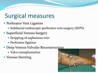
Dr.Devasheesh
Introduction
 Ulceration is an established defect in an epithelial
surface
 On basis of etiology they can be
 Neuropathic (most common)
 Venous
 Arterial
 Mixed (10%) (e.g. neuropathic–arterial, venous–arterial, etc)
 Venous ulcers are the most severe and debilitating sequlae
of chronic venous insufficiency (CVI) and venous
hypertension (HTN). They account for nearly 80% of all
lower extremity ulcers.
 Approximately 35% of patients afflicted with CVI will
develop a venous ulcer before the age of 40 and nearly two
thirds before the age of 65
Deep Veins
 Accompany axial arteries.
 Run within the muscles
deep to the muscle fascia.
Superficial veins
• Lie in the subcutaneous
tissue superficial to the
muscle fascia.
• They have their own, well-
developed muscle coat
• Long & short saphenous
veins and their tributaries
Comunicating Veins “Perforators”
 Perforate the fascia
connecting the
superficial & deep
veins at certain
points
Physiology
 Venous pressure in a foot vein on standing is approximately
100 mmHg
 To enable blood to be returned against gravity in the
standing position, an auxiliary pump in form of calf muscle
pump is present
 On calf muscles contraction, pressure within the calf
compartment rises to 200–300 mmHg which compress the
veins and the valves only allow blood to pass in the
direction of the heart.
 During muscle relaxation the pressure falls and blood from
the superficial veins enters the deep veins through the
perforating veins thus decreasing the pressure in
superficial veins to around 30 mmHg.
Risk factors
 Advancing age
 Family history of venous disease
 Prolonged standing
 Increased body mass index
 Smoking
 Sedentary lifestyle
 Lower extremity trauma
 Prior venous thrombosis (superficial or deep)
 Arteriovenous shunt
 Hereditary conditions
 Pregnancy 2.
Etiology
• AMBULATORY VENOUS HYPERTENSION
• Occurs when the vein valves become dysfunctional and impairs
venous blood return.
• Superficial venous reflux (long saphenous or short saphenous vein
reflux)
• Deep vein reflux (Primary or secondary to deep venous thrombosis
• Deep venous occlusion.
• Perforating vein reflux.
• Abnormal calf pump
(Neurological/musculoskeletal)
• Congenital absence of deep veins (e.g. Klippel-Trenaunay
syndrome)
• Venous trauma (ligation of proximal vein).
Pathology
 Fibrin cuff theory:
 widening of the pores between endothelial cells
allows the passage of larger molecules out of the
intravascular compartment into the tissues.
This allows the accumulation of fibrin around the
capillaries of the dermis to form a cuff which acts as a
barrier to oxygen and nutrient diffusion to interstitial
tissues and cutaneous skin cells resulting in local
tissue ischaemia and cell death which produces
ulceration.
White cell trapping theory:
Raised venous pressure reduces the capillary perfusion
pressure which causes trapping of white cells.
Venous hypertension results in expression of
leukocyte adhesion molecules that permit adherence
of leukocytes to the capillary endothelial cells.
The trapped cells become activated releasing
proteolytic enzymes and oxygen free radicals which
produce endothelial damage and tissue
destruction and local ischaemia.
Other possible changes
• Suggested that the pericapillary cuffs might interfere with
the diffusion of growth factors which are essential for skin
and tissue repair
• Arteriovenous shunting
• All these factors work together and lead to formation of
venous ulcer
Clinical Features
 The legs are
 Oedematous, moist
 Ulceration classically occurs just above the medial malleolus
(gaiter area), where the skin is most vulnerable (may be
bimallelolar and circumferential).
 Gangrene is usually absent (base of the ulcer may be
necrotic)
 Foot is warm with normal pulses.
 Stigmata of venous disease are present
 Varicose veins
 Venous eczema
 Haemosiderin deposition
 Lipodermatosclerosis
 Atrophie blanche.
 Varicose veins-
C1: Telangiectasia (0.5-1 mm) Reticular veins (1-3 mm)
 C2: Varicose veins
(>3mm)
C3: Edema without Skin
changes
 C4a: pigmentation or
eczema
 C4b: lipodermatosclerosis
or atrophie blanche
Lipodermatosclerosis (inverted
champagne-bottle)
Atrophie blanche
 C5: healed venous
ulcer
 C6: active venous ulcer
Venous Ulcer
Pulses Present (difficult to palpate if
oedematous)
Skin perfusion Normal and warm
ABPI Normal
Ulcer location Medial malleolus (gaiter area)
Ulcer margin Large and irregular
Ulcer contents Yellow wet appearance
Ulcer drainage Moderate to large
Skin Signs of venous hypertension
Pruritus Yes
Oedema Moderate to severe
Painful
Investigation
 Routine investigations eg: full blood count,
erythrocyte sedimentation rate (C reactive protein)
and sickle cell test
 Early diagnostic studies include invasive
measurements of ambulatory venous pressure (AVP)
and venous recovery time (VRT)
 80% incidence of venous ulceration if AVP of >80
mmHg
 Plethysmography
 evaluates venous function through the use of infrared
light
 provides a measurement of VRT
Investigation
 Venous Duplex Ultrasound
 Gold standard for evaluation of venous function
 Advantage that it can be used to evaluate individual
venous segments targeting abnormal areas for treatment
 Pneumatic pressure cuffs placed around the thigh, calf,
and forefoot and ultrasound transducer positioned over
the venous segment to be examined, just proximal to the
cuff.
 It is then inflated to a standard pressure for 3 seconds
and then rapidly deflated. Ninety-five percent of normal
venous valves close within 0.5 second.The presence of
reflux for >0.5 second is considered abnormal.
 Other investigations:
 ABPI
 Nerve conduction studies
 Angiogram
 Wound swab culture
 Biopsy of ulcer edge
Management
 Conservative treatment:
 Elevation of the legs at rest helps to reduce oedema, decrease
exudates from ulcers and accelerate regression of skin
changes.
 EXERCISE – daily walking and simple ankle flexion exercises
 Graduated elastic compression which is highest at the ankle
and decreases proximally. Avoid in case of cellulitis
 Unna boots :three layers
• rolled gauze bandage impregnated with
calamine, zinc oxide, glycerin, sorbitol,
gelatin, and magnesium aluminum silicate
• next layer consists of a 4-in-wide
continuous gauze dressing
• followed by an outer layer of elastic wrap
Management
 Local measures
 Clean base of the ulcer with saline
 Curette the yellow fibrin eschar
 Treat with metronidazole gel to reduce bacterial growth
and odor.
 Red dermatitic skin is treated with a medium- to high-
potency corticosteroid ointment.
 Cover with occlusive hydroactive dressing
 Use of skin substitutes eg: Cultured epidermal cell grafts
 Some ulcerations require grafting
Management
 Systemic therapy
 Pentoxifylline, 400 mg three times daily administered
with compression dressings
 Zinc supplementation is occasionally beneficial
 Use of antibiotics if accompanying cellulitis.
 Stanozolol: fibrinolytic enhancing agent used in the
treatment of preulcerative lipodermatosclerosis
Surgical measures
 Perforator Vein Ligation
 Subfascial endoscopic perforator vein surgery (SEPS)
 Superficial Venous Surgery
 Stripping of saphenous vein
 Perforator ligation
 Deep Venous Valvular Reconstruction
 Valve transplantation
 Venous Stenting
 Skin grafting:
 Done after treatment of underlying venous abnormality.
Reduces the healing time.
 Split skin mesh grafts.
 Pinch grafts.
 Full-thickness skin grafts.
Venous ulceers

Venous ulceers

  • 1.
  • 2.
    Introduction  Ulceration isan established defect in an epithelial surface  On basis of etiology they can be  Neuropathic (most common)  Venous  Arterial  Mixed (10%) (e.g. neuropathic–arterial, venous–arterial, etc)  Venous ulcers are the most severe and debilitating sequlae of chronic venous insufficiency (CVI) and venous hypertension (HTN). They account for nearly 80% of all lower extremity ulcers.  Approximately 35% of patients afflicted with CVI will develop a venous ulcer before the age of 40 and nearly two thirds before the age of 65
  • 3.
    Deep Veins  Accompanyaxial arteries.  Run within the muscles deep to the muscle fascia.
  • 4.
    Superficial veins • Liein the subcutaneous tissue superficial to the muscle fascia. • They have their own, well- developed muscle coat • Long & short saphenous veins and their tributaries
  • 5.
    Comunicating Veins “Perforators” Perforate the fascia connecting the superficial & deep veins at certain points
  • 6.
    Physiology  Venous pressurein a foot vein on standing is approximately 100 mmHg  To enable blood to be returned against gravity in the standing position, an auxiliary pump in form of calf muscle pump is present  On calf muscles contraction, pressure within the calf compartment rises to 200–300 mmHg which compress the veins and the valves only allow blood to pass in the direction of the heart.  During muscle relaxation the pressure falls and blood from the superficial veins enters the deep veins through the perforating veins thus decreasing the pressure in superficial veins to around 30 mmHg.
  • 7.
    Risk factors  Advancingage  Family history of venous disease  Prolonged standing  Increased body mass index  Smoking  Sedentary lifestyle  Lower extremity trauma  Prior venous thrombosis (superficial or deep)  Arteriovenous shunt  Hereditary conditions  Pregnancy 2.
  • 8.
    Etiology • AMBULATORY VENOUSHYPERTENSION • Occurs when the vein valves become dysfunctional and impairs venous blood return. • Superficial venous reflux (long saphenous or short saphenous vein reflux) • Deep vein reflux (Primary or secondary to deep venous thrombosis • Deep venous occlusion. • Perforating vein reflux. • Abnormal calf pump (Neurological/musculoskeletal) • Congenital absence of deep veins (e.g. Klippel-Trenaunay syndrome) • Venous trauma (ligation of proximal vein).
  • 9.
    Pathology  Fibrin cufftheory:  widening of the pores between endothelial cells allows the passage of larger molecules out of the intravascular compartment into the tissues. This allows the accumulation of fibrin around the capillaries of the dermis to form a cuff which acts as a barrier to oxygen and nutrient diffusion to interstitial tissues and cutaneous skin cells resulting in local tissue ischaemia and cell death which produces ulceration.
  • 10.
    White cell trappingtheory: Raised venous pressure reduces the capillary perfusion pressure which causes trapping of white cells. Venous hypertension results in expression of leukocyte adhesion molecules that permit adherence of leukocytes to the capillary endothelial cells. The trapped cells become activated releasing proteolytic enzymes and oxygen free radicals which produce endothelial damage and tissue destruction and local ischaemia.
  • 11.
    Other possible changes •Suggested that the pericapillary cuffs might interfere with the diffusion of growth factors which are essential for skin and tissue repair • Arteriovenous shunting • All these factors work together and lead to formation of venous ulcer
  • 12.
    Clinical Features  Thelegs are  Oedematous, moist  Ulceration classically occurs just above the medial malleolus (gaiter area), where the skin is most vulnerable (may be bimallelolar and circumferential).  Gangrene is usually absent (base of the ulcer may be necrotic)  Foot is warm with normal pulses.  Stigmata of venous disease are present  Varicose veins  Venous eczema  Haemosiderin deposition  Lipodermatosclerosis  Atrophie blanche.
  • 13.
     Varicose veins- C1:Telangiectasia (0.5-1 mm) Reticular veins (1-3 mm)
  • 14.
     C2: Varicoseveins (>3mm) C3: Edema without Skin changes
  • 15.
  • 16.
     C4b: lipodermatosclerosis oratrophie blanche Lipodermatosclerosis (inverted champagne-bottle) Atrophie blanche
  • 17.
     C5: healedvenous ulcer  C6: active venous ulcer
  • 18.
    Venous Ulcer Pulses Present(difficult to palpate if oedematous) Skin perfusion Normal and warm ABPI Normal Ulcer location Medial malleolus (gaiter area) Ulcer margin Large and irregular Ulcer contents Yellow wet appearance Ulcer drainage Moderate to large Skin Signs of venous hypertension Pruritus Yes Oedema Moderate to severe Painful
  • 19.
    Investigation  Routine investigationseg: full blood count, erythrocyte sedimentation rate (C reactive protein) and sickle cell test  Early diagnostic studies include invasive measurements of ambulatory venous pressure (AVP) and venous recovery time (VRT)  80% incidence of venous ulceration if AVP of >80 mmHg  Plethysmography  evaluates venous function through the use of infrared light  provides a measurement of VRT
  • 20.
    Investigation  Venous DuplexUltrasound  Gold standard for evaluation of venous function  Advantage that it can be used to evaluate individual venous segments targeting abnormal areas for treatment  Pneumatic pressure cuffs placed around the thigh, calf, and forefoot and ultrasound transducer positioned over the venous segment to be examined, just proximal to the cuff.  It is then inflated to a standard pressure for 3 seconds and then rapidly deflated. Ninety-five percent of normal venous valves close within 0.5 second.The presence of reflux for >0.5 second is considered abnormal.
  • 21.
     Other investigations: ABPI  Nerve conduction studies  Angiogram  Wound swab culture  Biopsy of ulcer edge
  • 22.
    Management  Conservative treatment: Elevation of the legs at rest helps to reduce oedema, decrease exudates from ulcers and accelerate regression of skin changes.  EXERCISE – daily walking and simple ankle flexion exercises  Graduated elastic compression which is highest at the ankle and decreases proximally. Avoid in case of cellulitis  Unna boots :three layers • rolled gauze bandage impregnated with calamine, zinc oxide, glycerin, sorbitol, gelatin, and magnesium aluminum silicate • next layer consists of a 4-in-wide continuous gauze dressing • followed by an outer layer of elastic wrap
  • 23.
    Management  Local measures Clean base of the ulcer with saline  Curette the yellow fibrin eschar  Treat with metronidazole gel to reduce bacterial growth and odor.  Red dermatitic skin is treated with a medium- to high- potency corticosteroid ointment.  Cover with occlusive hydroactive dressing  Use of skin substitutes eg: Cultured epidermal cell grafts  Some ulcerations require grafting
  • 24.
    Management  Systemic therapy Pentoxifylline, 400 mg three times daily administered with compression dressings  Zinc supplementation is occasionally beneficial  Use of antibiotics if accompanying cellulitis.  Stanozolol: fibrinolytic enhancing agent used in the treatment of preulcerative lipodermatosclerosis
  • 25.
    Surgical measures  PerforatorVein Ligation  Subfascial endoscopic perforator vein surgery (SEPS)  Superficial Venous Surgery  Stripping of saphenous vein  Perforator ligation  Deep Venous Valvular Reconstruction  Valve transplantation  Venous Stenting
  • 26.
     Skin grafting: Done after treatment of underlying venous abnormality. Reduces the healing time.  Split skin mesh grafts.  Pinch grafts.  Full-thickness skin grafts.