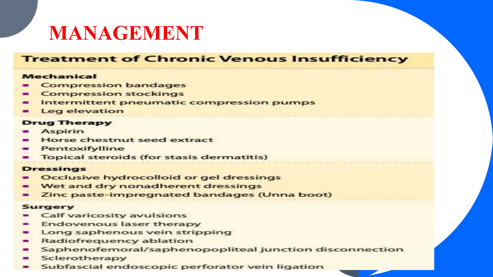
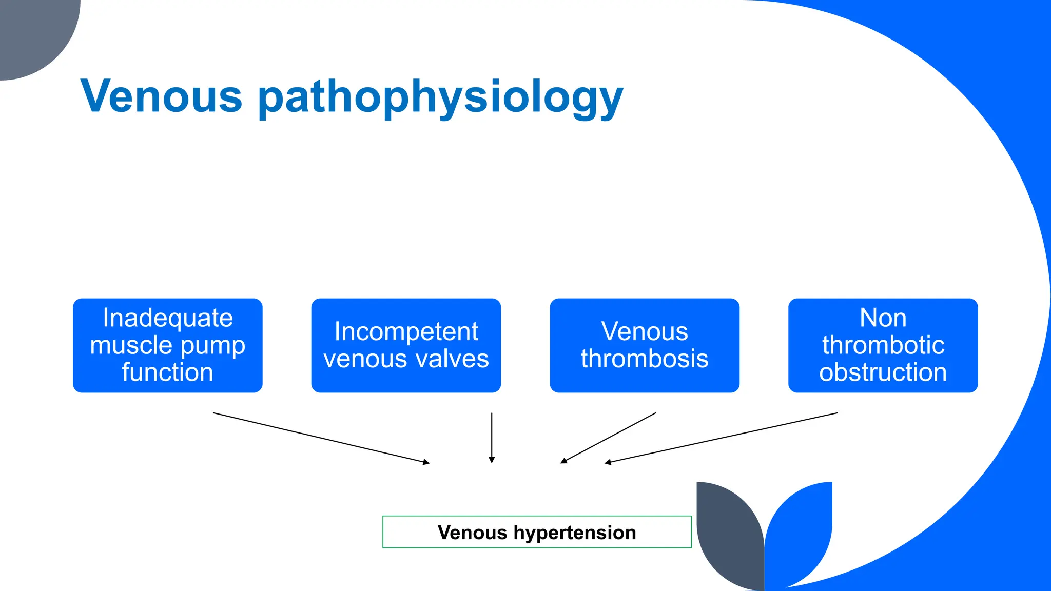
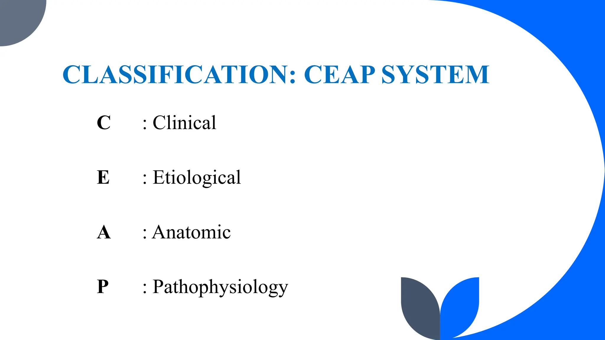
CHRONIC VENOUS INSUFFICIENCY
DEFINITION
Result of persistent ambulatory venous hypertension of the
lower-extremity venous system leading to a spectrum of clinical
findings from edema and tenderness to venous ulceration.
Common Signs and Symptoms of Peripheral
Venous Disease
 Symptoms
• Leg fullness
• Aching discomfort
• Heaviness
• Nocturnal leg cramps
• Bursting pain on standing
Common Signs and Symptoms of Peripheral
Venous Disease
• Signs
• Very early
• Tenderness to palpation
• Early
• Edema, hyperpigmentation, stasis dermatitis, varicose veins
• Late
• Venous ulcers
• atrophie blanche,
• lipodermatosclerosis,
• acroangiodermatitis of Mali,
EPIDEMIOLOGY
Extremely common; prevalence of 30% in adults
Venous ulcers cause significant disability = 1% of adults
● Risk factors:
○ Heredity
○ Age
○ Female sex
○ Obesity
○ Pregnancy
○ Prolonged standing
○ Phlebitis
○ Previous leg injury
ETIOLOGY & PATHOGENESIS
 A venous ulcer occurs after failure of the calf muscle pump.
 The heart pumps blood down to the foot; the calf muscle
pump (when upright) returns venous blood to the heart.
 Venous blood from the skin and subcutis collects in the
superficial venous system, including the greater and lesser
saphenous veins and its tributaries, moves through the fascia in a
series of “perforating” or “communicating” veins, and fills the
muscle-enveloped deep venous system.
 In all patients with venous disease there is failure of these one-way
valves, and this can result in varicose veins.
 Any obstruction to venous return (eg, thrombosis, radiation fibrosis)
or elevation of right atrial pressure (eg, pulmonary hypertension,
heart failure) further compromises venous return.
 The most common cause of venous valvular failure is thrombosis.
 The nidus for venous thrombosis is typically the valve cusp, and
when the thrombus is lysed by plasmin, valve function is often lost
as well.
 Calf muscle pump failure after deep venous thrombosis is often
referred to as the postphlebitic syndrome.
 • High-pressure blood in the deep system refluxes into the
unsupported veins of the skin.
 • Vascular leakage of fibrinogen-producing “fibrin cuffs” that may
interfere with tissue nourishment
 • Soft-tissue injury, inflammation and fibrosis
 • Subcutaneous fat is replaced by scar, resulting in
lipodermatosclerosis.
CLINICAL FINDINGS - Cutaneous Lesions
● Spectrum of clinical manifestations start with telangiectasias and reticular
veins on one end of the spectrum, and advanced chronic venous insufficiency at
the other end
● Findings of chronic venous insufficiency:
○ Dilated veins (varicose veins)
○ Edema
○ Leg pain
○ Cutaneous changes
● Earliest finding:
Peri malleolar edema that ascends the leg, followed by soft tissue
tenderness
CLINICAL FINDINGS - Cutaneous Lesions
● Varicose veins and smaller varicosities appear about the dorsum of
the foot and ankle
○ Usually asymptomatic
○ Patient’s complaints: Aching, cramping, itching, fatigue, and
swelling that worse with prolonged standing
○ Superficial thrombophlebitis can develop
CLINICAL FINDINGS - Cutaneous Lesions
● Stasis dermatitis- occurs during any stage of chronic venous
insufficiency
○ Erythema, scaling, pruritus, erosions, crusting, and occasional
vesicles
and serous drainage
○ Typical location: Medial supramalleolar region
○ Overtime, lesions may lichenified
○ Evaluate for coexisting allergic contact dermatitis
CLINICAL FINDINGS - Cutaneous Lesions
● Lipodermatosclerosis (aka. Sclerosing panniculitis, hypodermatitis
sclerodermiformis)
○ Fibrosing panniculitis characterized by a bound-down plaque
○ Begins at the medial ankle and extends circumferentially around
the entire distal lower leg
○ As the fibrosis increases → constricts and strangles the lower leg
→ impedes venous and lymph flow → Brawny edema above and
below the fibrosis (inverted champagne bottle
○ Lack of response to oral antibiotics and a relapsing nature = leads
to its diagnosis.
CLINICAL FINDINGS - Cutaneous Lesions
● Atrophie blanche
○ Skin overlying areas of fibrosis appearing in porcelain white and
atrophic
○ Fully established: Irregular, smooth, atrophic stellate plaques
surrounded with hyperpigmentation and telangiectasia
○ Related to venous stasis; may be associated with an underlying
disorder of hypercoagulation, livedoid vasculitis, or autoimmune
disease
CLINICAL FINDINGS - Cutaneous Lesions
● Acroangiodermatitis
○ Has purple macules, nodules, or verrucous plaques on the dorsal feet and
toes of patients with long-standing venous insufficiency
○ Mimics Kaposi sarcoma clinically and histologically
●
○ Ulcers occurring anywhere below the knee
○ Tender, shallow, irregular, and have a red base
○ Location: Medial ankle or along the line of the long or short saphenous
veins
DIAGNOSIS
Screening for Peripheral Arterial Disease: The Ankle–Brachial Index
● Measure systolic blood pressure in the arms and in the pedal
pulses, using Doppler ultrasound.
○ Normal ABI: greater than or equal to 1
○ Less than 1 may indicate PAD (the lower the ratio the
more severe the arterial obstruction).
DIAGNOSIS
● Skin biopsy- for diagnosis that in doubt, tissue should be sent
for both histology and tissue culture.
○ Histologic signs of venous hypertension:
■ Hemosiderin deposition
■ Lobular superficial
■ Deep dermal neovascularization
■ Fibrosis of dermis and subcutaneous tissue in later stage
DIAGNOSIS
● Functional testing of calf muscle pump function and venous
valvular function using plethysmography- occasionally useful
● Duplex Doppler ultrasonography- to document valvular
incompetence and to evaluate patients for possible sclerotherapy or
surgery.
MANAGEMENT
MANAGEMENT
Compression bandage using a zinc paste primary layer (Unna boot)
followed by Coban at full stretch
Mechanical therapy - mainstay of treatment
Daily use of elastic compression stockings:
 Reduces swelling in some patients with post thrombotic syndrome
 Prevent worsening of established post thrombotic syndrome
 Reduce recurrence of healed venous ulcers
MANAGEMENT
• Diuretics -severe edema
• Aspirin- (300 to 325 mg/day)
• Pentoxifylline- improves healing of chronic venous ulcers
• Topical steroids and emollients- aid resolution of stasis dermatitis
• Mupirocin- useful for folliculitis due to S. aureus and streptococci
• Horse chestnut seed extract (Aesculus hippocastanum L.)- 50 mg
escin,
short-term treatment for leg pain and swelling
MANAGEMENT
 Continual hemodynamic support
 Graduated stockings (minimum of 30-40 mmHg) at the ankle-
carefully fitted
 Avoid elastic stockings on edematous limbs
 Compression bandaging- used until all edema, inflammation, and
tenderness have resolved
MANAGEMENT
 Sclerotherapy or surgical techniques
 Endovenous ablation - close incompetent perforators and
correct the hemodynamic abnormalities that lead to venous ulcer
COMPLICATIONS
• Recurrent ulceration
• Cellulitis
• Contact dermatitis
• Thrombi and venous thrombosis
• Lymphatic impairment
• Elephantiasis nostras
PREVENTION
● Identify and treat patients at extra risk for thrombosis.
● Venous thrombosis: Elastic compression stockings- the only
proven method to reduce the risk of post-thrombotic syndrome.
● Supportive stockings- maternity can be recommended since
valvular failure may develop during pregnancy
● Stockings are advisable for occupation or lifestyle involving long
periods of immobility (long-distance flights).
VENOUS
ULCER
ULCER
Break in the continuity of covering epithelium(skin or
mucous membrane) following molecular death of surface
epithelium or its traumatic removal.
CLASSIFICATION
Clinically
1. Spreading Ulcer
2. Healing Ulcer
3. Callous/Chronic
ulceR
Pathologically
Nonspecific Ulcers
Traumatic
Arterial
Venous
Neurogenic (trophic)
A/w malnutrition
2. Specific ulcers
3. Malignant ulcers
Venous system of lower limb
Venous drainage of foot Deep venous system
Saphenous vein axis: Superficial system Perforator system
Venous pathophysiology
Inadequate
muscle pump
function
Incompetent
venous valves
Venous
thrombosis
Non
thrombotic
obstruction
Venous hypertension
RISK FACTORS
Advancing age
Family history of venous
disease
Ligamentous laxity
Prolonged standing
Increased BMI
Smoking
Lower extremity trauma
Prior venous thrombosis
Pregnancy
High estrogen states
Some hereditary conditions
CLASSIFICATION: CEAP SYSTEM
C : Clinical
E : Etiological
A : Anatomic
P : Pathophysiology
Clinical Etiological
C0 No visible or palpable sign of venous disease Ep Primary
C1 Telangiectasia, reticular veins Es Secondary
C2 Varicose veins Ec Congenital
C3 Edema En No cause identified
C4 Changes in skin and subcutaneous tissue
C5 Healed
C6 Active venous ulcer
S With symptoms
A Without symptoms
Anatomical Pathophysiology
As Superficial veins Pr Reflux
Ad Deep veins Po Obstruction
Ap Perforator veins Pr,o Reflux and
obstruction
An No venous anatomic
location identified
Pn No venous
pathology
identifiable
CLINICAL FEATURES: SYMPTOMS
Limb discomfort including leg heaviness or aching.
Throbbing or burning pain: Worse on standing/ sitting with feet
dependent for a prolonged period.
Itching.
Limb swelling.
Skin changes
Clinical signs by CEAP category
C-0
20% of patients with clinical symptoms have no visible
signs.
C1: Telangiectasia / reticular veins
Most frequently encountered sign.
Telangiectasia: Confluence of dilated
intradermal venules/ capillaries less than
1 mm in diameter.
Reticular veins: Dilated, bluish
subdermal veins, 1-3 mm in diameter.
C2: Varicose veins
Subcutaneous, dilated, tortuous
veins more than 3 mm in diameter.
Often a/w superficial axial
venous reflux.
C3: Edema
Dependent ankle edema may
progress to include the calf region.
In the early stage, present only at
end of the day.
C4a: Skin pigmentation/ dermatitis
Initially prominent at medial ankle but
later encroach upon foot and lower
leg.
Pigmentation due to hemosiderin
deposition.
Venous reflux predisposes to stasis
dermatitis
Presents as eczematous rash.
C4b: Lipodermatosclerosis
Fibrosing panniculitis of subcutaneous tissue.
Characterized by firm are of induration,
initially located at medial ankle.
In late stage, entire legs can be
circumferentially involved.
Fibrosis may be extensive enough to strangle
lower leg.
Prone to repeated bouts of cellulitis.
C4c: Corona phlebectatita
Crown of numerous small,
intradermal veins on medial or lateral
aspect of ankle or foot.
Indicator of venous hypertension due
to saphenous or perforator venous
insufficiency.
C5,C6: Venous ulceration
Chronic venous disease common cause of
lower extremity ulcers.
Commonly have superficial, deep or
perforator reflux; alone or in combination.
Located low on medial ankle or near the
lateral malleolus.
Never in forefoot or above the level of knee.
Venous leg ulcer: Epidemiology
Responsible for 85% of all chronic lower limb ulcers in
resource rich countries.
Prevalence: 0.1-0.3 % in adults (2-4% in elderly).
15-30% of patients have concomitant arterial occlusive
disease
Venous ulcer: Pathology
Static blood flow in veins
Hypoxia Tissue death
Not confirmed: Venous oxygen saturation found higher in ulcerated
Ambulatory
venous
hypertension
Incompetence of perforating
veins
Valve incompetence of saphenous veins
Incompetence or obstruction
of deep veins
Venous ulcer: Pathology
High venous pressure
Pericapillary infiltrate (
fibrin)
Fibrosis
Acts as impediment to
diffusion of nutrients.
Ulcer
environment
Growth factors inhibited
Increased ROS:
generate free
radicals; cause
tissue damage
Increased no. of
mast cells
monocytes and
lymphocytes
History
A complete medical history should be
obtained.
Diabetes, hypertension, CKD.
Smoking history.
Occupation history.
Past history of DVT.
Family history.
Increased JVP: Sign of RHF
Examination
General and systemic
examination.
Assessment of signs of venous
disease.
Ulcer: Size, shape, number,
position, edge, base, margin,
discharge.
Pulse examination.
Neurological assessment Types of ulcer edges
Investigations
All new patients: CBC, RBS, ESR, CRP; Sickle cell test
a. Duplex scan
Presence of reflux in the deep and superficial venous system.
Extent and distribution of reflux
Presence of obstruction in the deep venous system.
Presence of thrombus in the superficial system.
Investigations
b. Venography
Ascending: Dye injected into a superficial pedal vein.
Descending: Dye into the deep venous system at the
groin or popliteal vein.
Observation of flow defines regions of thrombus or
obstruction.
Adjunct to clinical exam and duplex scanning to identify
specific valvular incompetence.
Management
General measures
Compression therapy
Ulcer care
Pharmacotherapy
Surgical therapy
a. General measures
Avoidance of prolonged standing.
Elevation of feet to at least heart level for
30 mins 3 to 4 times a day.
Daily walking and ankle flexion exercises.
Skin care: Skin cleansing and use of
emollients.
Skincare
a. Stasis dermatitis
For chronic dryness, petroleum-based emollient.
If oozing or vesiculation: Mid potency topical steroid.
b. Contact dermatitis
Avoid use of topical moisturizers/ antibiotics.
b. Compression therapy
The keystone of management is to decrease venous HTN.
Primary way: Use of compression
Four-layer bandaging system
a. Orthopedic Wool
b. Elastic bandage
c. Cotton crepe
d. Cohesive bandage
…Bandaging
The ideal interface pressure is 35-40 mm Hg.
The interval between bandage applications is based on the amount of
exudate and speed of healing.
Compression in mixed ulcer controversial.
For ABPI >0.5, a pressure of 30 mm Hg is safe and effective.
For ABPI <0.5, must undergo revascularisation before any
compression.
c. Ulcer care
Basic wound care techniques are required.
i. Role of antibiotics
Only in those with acute cellulitis or clinically infected ulcer.
Routine use in uncomplicated ulcers doesn’t reduce bacterial
colonization or improve the healing rate; can cause emergence of
resistance.
Ulcer care: Debridement
Removal of necrotic tissue and fibrinous debris aids in the formation of granulation
tissue and enhances re-epithelialization.
Regular dressing to control exudate, maintain moisture balance, and control odor and
pain.
Options include simple nonadhesive dressing, paraffin gauze, hydrogels,
hydrocolloids, and silver-impregnated dressings.
Few trials support a role of skin grafting for very large ulcers or ulcers present >12
months
d. Pharmacotherapy
Variety of agents that affect venous tone used.
Increase tone by a mechanism related to NE pathway.
Other actions include reduction of capillary hyperpermeability,
improved lymphatic drainage, anti-inflammatory effects, and
decreased blood viscosity.
…Pharmacotherapy
a. Flavonoids
Hydroxyethylrutoside (HR)
Escin (horse chestnut seed extract: HCE)
Micronized purified flavonoid fraction
Meta-analysis of 15 RCTs showed HR has a higher response in pain and swelling
control vs placebo.
Two meta-analyses: HCE improved symptoms related to chronic venous
insufficiency vs placebo
..Pharmacotherapy
Aspirin: Insufficient evidence to detect beneficial effects or even
harm.
Stanazolol: Several RCTs have noted improvement in
lipodermatosclerosis areas and possibly faster ulcer healing rates.
Pentoxifylline: Metanalaysis of 11 trials: Significantly more effective
for complete or partial ulcer healing than placebo (800 mg TDS).
Sulodexide, calcium dobesilate: low-quality evidence.
e. Surgical therapy
Speciality referral in case of:
a. Arterial insufficiency.
b. Nonhealing ulcer.
c. Ulcer recurrence.
d. Suspected contact dermatitis.
e. Resistant or recurrent cellulitis.
Surgical therapy
Treat superficial
reflux
Ligation and
stripping
Saphenofemoral
ligation and great
saphenous stripping
Saphenopopliteal
junction ligation and
small saphenous
stripping
Endothermal
ablation
Laser ablation
Radiofrequency
ablation
Ultrasound guided
sclerotherapy
i. Ligation and stripping
Fully dissect the point of junctional
incompetence and remove the refluxing
axial vein and dilated tributaries.
Groin incision made, GSV dissected up to
SFJ, GSF tributaries ligated, then SFJ
ligated and GSF stripped to around knee.
ii. Endothermal ablation
Safer, faster recovery, cost effective.
Concept:
A device inserted into incompetent axial vein p/c.
Vein surrounded by LA solution; compresses the vein emptying
it of blood.
Device produces thermal energy that destroys structure of vein ,
resulting in permanent occlusion.
iii. Ultrasound guided sclerotherapy
Used for treatment of incompetent
perforators and large venous tributaries
causes by neovascularisation.
Injection of sclerosing agent directly into
veins.
Most commn agent: Sodium tetradecyl
sulphate.
Causes cellular death resulting in
thrombosis, fibrosis and sclerosis.
venous ulcer and venous insufficiency.ppt

venous ulcer and venous insufficiency.ppt

  • 1.
  • 2.
    DEFINITION Result of persistentambulatory venous hypertension of the lower-extremity venous system leading to a spectrum of clinical findings from edema and tenderness to venous ulceration.
  • 3.
    Common Signs andSymptoms of Peripheral Venous Disease  Symptoms • Leg fullness • Aching discomfort • Heaviness • Nocturnal leg cramps • Bursting pain on standing
  • 4.
    Common Signs andSymptoms of Peripheral Venous Disease • Signs • Very early • Tenderness to palpation • Early • Edema, hyperpigmentation, stasis dermatitis, varicose veins • Late • Venous ulcers • atrophie blanche, • lipodermatosclerosis, • acroangiodermatitis of Mali,
  • 5.
    EPIDEMIOLOGY Extremely common; prevalenceof 30% in adults Venous ulcers cause significant disability = 1% of adults
  • 6.
    ● Risk factors: ○Heredity ○ Age ○ Female sex ○ Obesity ○ Pregnancy ○ Prolonged standing ○ Phlebitis ○ Previous leg injury
  • 7.
    ETIOLOGY & PATHOGENESIS A venous ulcer occurs after failure of the calf muscle pump.  The heart pumps blood down to the foot; the calf muscle pump (when upright) returns venous blood to the heart.  Venous blood from the skin and subcutis collects in the superficial venous system, including the greater and lesser saphenous veins and its tributaries, moves through the fascia in a series of “perforating” or “communicating” veins, and fills the muscle-enveloped deep venous system.
  • 8.
     In allpatients with venous disease there is failure of these one-way valves, and this can result in varicose veins.  Any obstruction to venous return (eg, thrombosis, radiation fibrosis) or elevation of right atrial pressure (eg, pulmonary hypertension, heart failure) further compromises venous return.
  • 9.
     The mostcommon cause of venous valvular failure is thrombosis.  The nidus for venous thrombosis is typically the valve cusp, and when the thrombus is lysed by plasmin, valve function is often lost as well.  Calf muscle pump failure after deep venous thrombosis is often referred to as the postphlebitic syndrome.
  • 10.
     • High-pressureblood in the deep system refluxes into the unsupported veins of the skin.  • Vascular leakage of fibrinogen-producing “fibrin cuffs” that may interfere with tissue nourishment  • Soft-tissue injury, inflammation and fibrosis  • Subcutaneous fat is replaced by scar, resulting in lipodermatosclerosis.
  • 11.
    CLINICAL FINDINGS -Cutaneous Lesions ● Spectrum of clinical manifestations start with telangiectasias and reticular veins on one end of the spectrum, and advanced chronic venous insufficiency at the other end ● Findings of chronic venous insufficiency: ○ Dilated veins (varicose veins) ○ Edema ○ Leg pain ○ Cutaneous changes ● Earliest finding: Peri malleolar edema that ascends the leg, followed by soft tissue tenderness
  • 12.
    CLINICAL FINDINGS -Cutaneous Lesions ● Varicose veins and smaller varicosities appear about the dorsum of the foot and ankle ○ Usually asymptomatic ○ Patient’s complaints: Aching, cramping, itching, fatigue, and swelling that worse with prolonged standing ○ Superficial thrombophlebitis can develop
  • 13.
    CLINICAL FINDINGS -Cutaneous Lesions ● Stasis dermatitis- occurs during any stage of chronic venous insufficiency ○ Erythema, scaling, pruritus, erosions, crusting, and occasional vesicles and serous drainage ○ Typical location: Medial supramalleolar region ○ Overtime, lesions may lichenified ○ Evaluate for coexisting allergic contact dermatitis
  • 14.
    CLINICAL FINDINGS -Cutaneous Lesions ● Lipodermatosclerosis (aka. Sclerosing panniculitis, hypodermatitis sclerodermiformis) ○ Fibrosing panniculitis characterized by a bound-down plaque ○ Begins at the medial ankle and extends circumferentially around the entire distal lower leg ○ As the fibrosis increases → constricts and strangles the lower leg → impedes venous and lymph flow → Brawny edema above and below the fibrosis (inverted champagne bottle ○ Lack of response to oral antibiotics and a relapsing nature = leads to its diagnosis.
  • 15.
    CLINICAL FINDINGS -Cutaneous Lesions ● Atrophie blanche ○ Skin overlying areas of fibrosis appearing in porcelain white and atrophic ○ Fully established: Irregular, smooth, atrophic stellate plaques surrounded with hyperpigmentation and telangiectasia ○ Related to venous stasis; may be associated with an underlying disorder of hypercoagulation, livedoid vasculitis, or autoimmune disease
  • 16.
    CLINICAL FINDINGS -Cutaneous Lesions ● Acroangiodermatitis ○ Has purple macules, nodules, or verrucous plaques on the dorsal feet and toes of patients with long-standing venous insufficiency ○ Mimics Kaposi sarcoma clinically and histologically ● ○ Ulcers occurring anywhere below the knee ○ Tender, shallow, irregular, and have a red base ○ Location: Medial ankle or along the line of the long or short saphenous veins
  • 17.
    DIAGNOSIS Screening for PeripheralArterial Disease: The Ankle–Brachial Index ● Measure systolic blood pressure in the arms and in the pedal pulses, using Doppler ultrasound. ○ Normal ABI: greater than or equal to 1 ○ Less than 1 may indicate PAD (the lower the ratio the more severe the arterial obstruction).
  • 20.
    DIAGNOSIS ● Skin biopsy-for diagnosis that in doubt, tissue should be sent for both histology and tissue culture. ○ Histologic signs of venous hypertension: ■ Hemosiderin deposition ■ Lobular superficial ■ Deep dermal neovascularization ■ Fibrosis of dermis and subcutaneous tissue in later stage
  • 21.
    DIAGNOSIS ● Functional testingof calf muscle pump function and venous valvular function using plethysmography- occasionally useful ● Duplex Doppler ultrasonography- to document valvular incompetence and to evaluate patients for possible sclerotherapy or surgery.
  • 22.
  • 23.
    MANAGEMENT Compression bandage usinga zinc paste primary layer (Unna boot) followed by Coban at full stretch Mechanical therapy - mainstay of treatment Daily use of elastic compression stockings:  Reduces swelling in some patients with post thrombotic syndrome  Prevent worsening of established post thrombotic syndrome  Reduce recurrence of healed venous ulcers
  • 24.
    MANAGEMENT • Diuretics -severeedema • Aspirin- (300 to 325 mg/day) • Pentoxifylline- improves healing of chronic venous ulcers • Topical steroids and emollients- aid resolution of stasis dermatitis • Mupirocin- useful for folliculitis due to S. aureus and streptococci • Horse chestnut seed extract (Aesculus hippocastanum L.)- 50 mg escin, short-term treatment for leg pain and swelling
  • 25.
    MANAGEMENT  Continual hemodynamicsupport  Graduated stockings (minimum of 30-40 mmHg) at the ankle- carefully fitted  Avoid elastic stockings on edematous limbs  Compression bandaging- used until all edema, inflammation, and tenderness have resolved
  • 26.
    MANAGEMENT  Sclerotherapy orsurgical techniques  Endovenous ablation - close incompetent perforators and correct the hemodynamic abnormalities that lead to venous ulcer
  • 27.
    COMPLICATIONS • Recurrent ulceration •Cellulitis • Contact dermatitis • Thrombi and venous thrombosis • Lymphatic impairment • Elephantiasis nostras
  • 28.
    PREVENTION ● Identify andtreat patients at extra risk for thrombosis. ● Venous thrombosis: Elastic compression stockings- the only proven method to reduce the risk of post-thrombotic syndrome. ● Supportive stockings- maternity can be recommended since valvular failure may develop during pregnancy ● Stockings are advisable for occupation or lifestyle involving long periods of immobility (long-distance flights).
  • 29.
  • 30.
    ULCER Break in thecontinuity of covering epithelium(skin or mucous membrane) following molecular death of surface epithelium or its traumatic removal.
  • 31.
    CLASSIFICATION Clinically 1. Spreading Ulcer 2.Healing Ulcer 3. Callous/Chronic ulceR Pathologically Nonspecific Ulcers Traumatic Arterial Venous Neurogenic (trophic) A/w malnutrition 2. Specific ulcers 3. Malignant ulcers
  • 32.
    Venous system oflower limb Venous drainage of foot Deep venous system
  • 33.
    Saphenous vein axis:Superficial system Perforator system
  • 34.
    Venous pathophysiology Inadequate muscle pump function Incompetent venousvalves Venous thrombosis Non thrombotic obstruction Venous hypertension
  • 35.
    RISK FACTORS Advancing age Familyhistory of venous disease Ligamentous laxity Prolonged standing Increased BMI Smoking Lower extremity trauma Prior venous thrombosis Pregnancy High estrogen states Some hereditary conditions
  • 36.
    CLASSIFICATION: CEAP SYSTEM C: Clinical E : Etiological A : Anatomic P : Pathophysiology
  • 37.
    Clinical Etiological C0 Novisible or palpable sign of venous disease Ep Primary C1 Telangiectasia, reticular veins Es Secondary C2 Varicose veins Ec Congenital C3 Edema En No cause identified C4 Changes in skin and subcutaneous tissue C5 Healed C6 Active venous ulcer S With symptoms A Without symptoms
  • 38.
    Anatomical Pathophysiology As Superficialveins Pr Reflux Ad Deep veins Po Obstruction Ap Perforator veins Pr,o Reflux and obstruction An No venous anatomic location identified Pn No venous pathology identifiable
  • 39.
    CLINICAL FEATURES: SYMPTOMS Limbdiscomfort including leg heaviness or aching. Throbbing or burning pain: Worse on standing/ sitting with feet dependent for a prolonged period. Itching. Limb swelling. Skin changes
  • 40.
    Clinical signs byCEAP category C-0 20% of patients with clinical symptoms have no visible signs.
  • 41.
    C1: Telangiectasia /reticular veins Most frequently encountered sign. Telangiectasia: Confluence of dilated intradermal venules/ capillaries less than 1 mm in diameter. Reticular veins: Dilated, bluish subdermal veins, 1-3 mm in diameter.
  • 42.
    C2: Varicose veins Subcutaneous,dilated, tortuous veins more than 3 mm in diameter. Often a/w superficial axial venous reflux.
  • 43.
    C3: Edema Dependent ankleedema may progress to include the calf region. In the early stage, present only at end of the day.
  • 44.
    C4a: Skin pigmentation/dermatitis Initially prominent at medial ankle but later encroach upon foot and lower leg. Pigmentation due to hemosiderin deposition. Venous reflux predisposes to stasis dermatitis Presents as eczematous rash.
  • 45.
    C4b: Lipodermatosclerosis Fibrosing panniculitisof subcutaneous tissue. Characterized by firm are of induration, initially located at medial ankle. In late stage, entire legs can be circumferentially involved. Fibrosis may be extensive enough to strangle lower leg. Prone to repeated bouts of cellulitis.
  • 46.
    C4c: Corona phlebectatita Crownof numerous small, intradermal veins on medial or lateral aspect of ankle or foot. Indicator of venous hypertension due to saphenous or perforator venous insufficiency.
  • 47.
    C5,C6: Venous ulceration Chronicvenous disease common cause of lower extremity ulcers. Commonly have superficial, deep or perforator reflux; alone or in combination. Located low on medial ankle or near the lateral malleolus. Never in forefoot or above the level of knee.
  • 48.
    Venous leg ulcer:Epidemiology Responsible for 85% of all chronic lower limb ulcers in resource rich countries. Prevalence: 0.1-0.3 % in adults (2-4% in elderly). 15-30% of patients have concomitant arterial occlusive disease
  • 49.
    Venous ulcer: Pathology Staticblood flow in veins Hypoxia Tissue death Not confirmed: Venous oxygen saturation found higher in ulcerated Ambulatory venous hypertension Incompetence of perforating veins Valve incompetence of saphenous veins Incompetence or obstruction of deep veins
  • 50.
    Venous ulcer: Pathology Highvenous pressure Pericapillary infiltrate ( fibrin) Fibrosis Acts as impediment to diffusion of nutrients. Ulcer environment Growth factors inhibited Increased ROS: generate free radicals; cause tissue damage Increased no. of mast cells monocytes and lymphocytes
  • 51.
    History A complete medicalhistory should be obtained. Diabetes, hypertension, CKD. Smoking history. Occupation history. Past history of DVT. Family history. Increased JVP: Sign of RHF
  • 52.
    Examination General and systemic examination. Assessmentof signs of venous disease. Ulcer: Size, shape, number, position, edge, base, margin, discharge. Pulse examination. Neurological assessment Types of ulcer edges
  • 53.
    Investigations All new patients:CBC, RBS, ESR, CRP; Sickle cell test a. Duplex scan Presence of reflux in the deep and superficial venous system. Extent and distribution of reflux Presence of obstruction in the deep venous system. Presence of thrombus in the superficial system.
  • 54.
    Investigations b. Venography Ascending: Dyeinjected into a superficial pedal vein. Descending: Dye into the deep venous system at the groin or popliteal vein. Observation of flow defines regions of thrombus or obstruction. Adjunct to clinical exam and duplex scanning to identify specific valvular incompetence.
  • 55.
    Management General measures Compression therapy Ulcercare Pharmacotherapy Surgical therapy
  • 56.
    a. General measures Avoidanceof prolonged standing. Elevation of feet to at least heart level for 30 mins 3 to 4 times a day. Daily walking and ankle flexion exercises. Skin care: Skin cleansing and use of emollients.
  • 57.
    Skincare a. Stasis dermatitis Forchronic dryness, petroleum-based emollient. If oozing or vesiculation: Mid potency topical steroid. b. Contact dermatitis Avoid use of topical moisturizers/ antibiotics.
  • 58.
    b. Compression therapy Thekeystone of management is to decrease venous HTN. Primary way: Use of compression Four-layer bandaging system a. Orthopedic Wool b. Elastic bandage c. Cotton crepe d. Cohesive bandage
  • 59.
    …Bandaging The ideal interfacepressure is 35-40 mm Hg. The interval between bandage applications is based on the amount of exudate and speed of healing. Compression in mixed ulcer controversial. For ABPI >0.5, a pressure of 30 mm Hg is safe and effective. For ABPI <0.5, must undergo revascularisation before any compression.
  • 60.
    c. Ulcer care Basicwound care techniques are required. i. Role of antibiotics Only in those with acute cellulitis or clinically infected ulcer. Routine use in uncomplicated ulcers doesn’t reduce bacterial colonization or improve the healing rate; can cause emergence of resistance.
  • 61.
    Ulcer care: Debridement Removalof necrotic tissue and fibrinous debris aids in the formation of granulation tissue and enhances re-epithelialization. Regular dressing to control exudate, maintain moisture balance, and control odor and pain. Options include simple nonadhesive dressing, paraffin gauze, hydrogels, hydrocolloids, and silver-impregnated dressings. Few trials support a role of skin grafting for very large ulcers or ulcers present >12 months
  • 62.
    d. Pharmacotherapy Variety ofagents that affect venous tone used. Increase tone by a mechanism related to NE pathway. Other actions include reduction of capillary hyperpermeability, improved lymphatic drainage, anti-inflammatory effects, and decreased blood viscosity.
  • 63.
    …Pharmacotherapy a. Flavonoids Hydroxyethylrutoside (HR) Escin(horse chestnut seed extract: HCE) Micronized purified flavonoid fraction Meta-analysis of 15 RCTs showed HR has a higher response in pain and swelling control vs placebo. Two meta-analyses: HCE improved symptoms related to chronic venous insufficiency vs placebo
  • 64.
    ..Pharmacotherapy Aspirin: Insufficient evidenceto detect beneficial effects or even harm. Stanazolol: Several RCTs have noted improvement in lipodermatosclerosis areas and possibly faster ulcer healing rates. Pentoxifylline: Metanalaysis of 11 trials: Significantly more effective for complete or partial ulcer healing than placebo (800 mg TDS). Sulodexide, calcium dobesilate: low-quality evidence.
  • 65.
    e. Surgical therapy Specialityreferral in case of: a. Arterial insufficiency. b. Nonhealing ulcer. c. Ulcer recurrence. d. Suspected contact dermatitis. e. Resistant or recurrent cellulitis.
  • 66.
    Surgical therapy Treat superficial reflux Ligationand stripping Saphenofemoral ligation and great saphenous stripping Saphenopopliteal junction ligation and small saphenous stripping Endothermal ablation Laser ablation Radiofrequency ablation Ultrasound guided sclerotherapy
  • 67.
    i. Ligation andstripping Fully dissect the point of junctional incompetence and remove the refluxing axial vein and dilated tributaries. Groin incision made, GSV dissected up to SFJ, GSF tributaries ligated, then SFJ ligated and GSF stripped to around knee.
  • 68.
    ii. Endothermal ablation Safer,faster recovery, cost effective. Concept: A device inserted into incompetent axial vein p/c. Vein surrounded by LA solution; compresses the vein emptying it of blood. Device produces thermal energy that destroys structure of vein , resulting in permanent occlusion.
  • 69.
    iii. Ultrasound guidedsclerotherapy Used for treatment of incompetent perforators and large venous tributaries causes by neovascularisation. Injection of sclerosing agent directly into veins. Most commn agent: Sodium tetradecyl sulphate. Causes cellular death resulting in thrombosis, fibrosis and sclerosis.