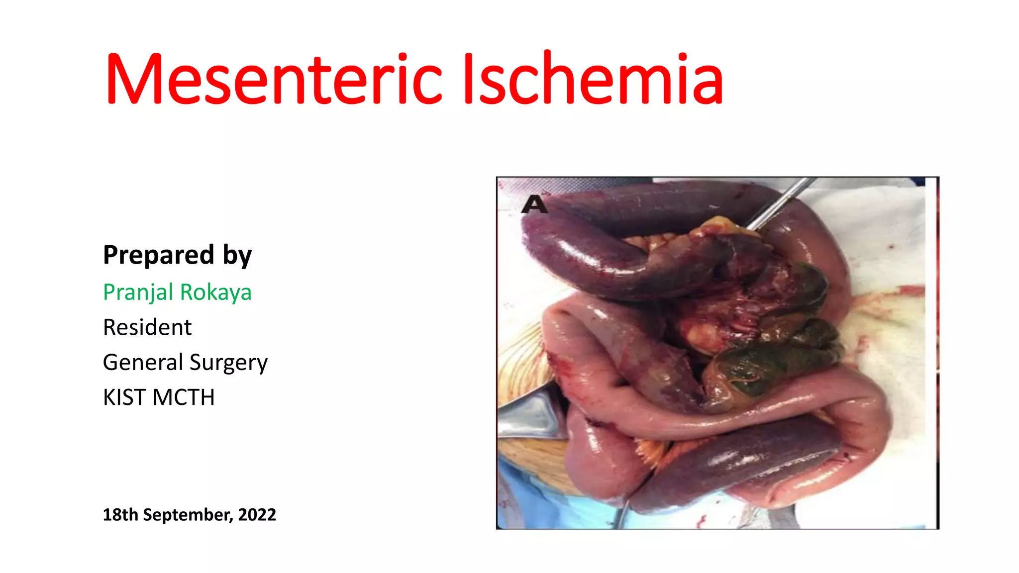
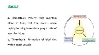
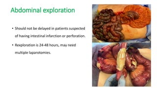
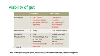
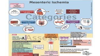
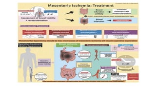
Mesenteric Ischemia
Prepared by
Pranjal Rokaya
Resident
General Surgery
KIST MCTH
18th September, 2022
Outline
โ€ข Terminologies
โ€ข Blood supply of gut
โ€ข Introduction and classification
โ€ข Clinical features
โ€ข Diagnosis
โ€ข Management
โ€ข Prognosis
Basics
a. Hemostasis: Process that maintain
blood in fluid, clot free state , while
rapidly forming hemostatic plug at site of
vascular injury.
b. Thrombosis: Formation of blod clot
within intact vessels
c. Embolism
๏ƒ˜An embolus is an intravascular solid, liquid, or gaseous mass that is carried by the
blood to a site distant from its point of origin.
โ€ข Thrombus : Thromboembolus
โ€ข Tissue : Amniotic fluid, Tumor cells
โ€ข Lipids : Fat embolism, Atheromatous embolism
Both thrombus and embolus cause tissue injury by vascular occlusion.
d. Ischemia
โ€ข Reduced blood flow to the tissue.
e. Infarction
โ€ข An infarct is an area of ischemic necrosis caused by occlusion of either arterial
supply or venous drainage.
Arterial supply of gut
Overview of arterial circulation of gut
Arterial supply of small intestine
Arterial supply of colon and rectum
Venous drainage of gut
Venous drainage of small intestine
Venous drainage of colon and rectum
Introduction
โ€ข Vasocclusive disease of mesenteric vessels.
โ€ข More common in older age (>60 years) and females (3 times more frequent).
โ€ข Relatively uncommon but devastating condition.
โ€ข Represents 2% of revascularisation operations for atheromatous lesions.
Risk factors
Embolism Thrombosis
1. Cardiac arrthmias 1. Coronary artery disease
2. Valvular heart disease 2. Peripheral arterial disease
3. Recent myocardial infarction 3. Advance age
4. Infective endocarditis 4. Low cardiac output states
5. Aortic atherosclerosis 5. Traumatic injury
6. Cardiogenic shock 6. Acquired hypercoagulable states:
COVID-19
Types of Mesenteric occlusive diseases
a. Acute mesenteric ischemia
b. Chronic mesenteric ischemia
c. Non occlusive mesenteric Ischemia (NOMI)
a. Acute Mesenteric Ischemia
๏ƒ˜Sudden onset of small intestine hypoperfusion.
๏ƒ˜Arterial occlusion in 67 to 95% of cases.
๏ƒ˜SMA is most commonly involved.
๏ƒผAcute thrombotic obstruction with underlying atherosclerosis: usually involves
origin of artery.
๏ƒผAcute embolism: usually at the branching point of an artery.
Clinical features
๏ƒ˜Severe periumbilical abdominal pain.
๏ƒ˜Out of proportion to physical findings (No signs of peritonism).
๏ƒ˜Nausea, vomiting and diarrhea.
๏ƒ˜Blood in stool in advanced ischemia.
๏ƒ˜Peritonism develops in advanced stage.
b. Chronic mesenteric Ischemia
๏ƒ˜ Intestinal angina
๏ƒ˜Episodic or continuous hypoperfusion of small intestine occuring in patient
with multivessel mesenteric artery stenosis or occlusion.
๏ƒ˜Majority caused by atherosclerotic narrowing of origins of CA or SMA.
๏ƒ˜ Rare causes include fibromuscular dysplasia, aortic or mesenteric artery
dissection, vasculitis, Takayasu disease.
Clinical features
๏ƒ˜ Most asymptomatic due to extensive collateral blood supply.
๏ƒ˜ Reccurent episodes of acute abodminal pain after eating.
๏ƒ˜ Pain usually subsides over 2 hours.
๏ƒ˜ Adaptive eating pattern
๏ƒ˜Weight loss
๏ƒ˜Acute on chronic ischemia
c. Non occlusive mesenteric ischemia
๏ƒ˜ Most commonly due to primary mesenteric arterial vasoconstriction.
๏ƒ˜The majority includes spasms of branches of SMA.
๏ƒ˜Related to the homeostatic mechanism that maintains cardiac and cerebral blood
flow at expense of mesenteric circulation.
๏ƒ˜Represents 5-15% of mesenteric ischemia.
Risk factors
๏‚ง Heart failure/Cardiogenic shock
๏‚ง Peripheral arterial disease
๏‚ง Aortic insufficiency
๏‚ง Septic shock
๏‚ง Cardiac arrhythmias
๏‚ง Administration of vasoconstrictive medication.
๏‚ง Cocaine abuse.
๏‚ง Severe burns
๏‚ง Severe acute pancreatitis
Clinical features
โ€ข Location and severity of abdominal pain more variable.
โ€ข Mild abdominal pain a/w nausea/vomittting.
โ€ข Presentation overshadowed by precipitating disorders.
โ€ข Mental status changes in 1/3rd of old patients.
Physical examination
โ€ข Abdominal examination normal initially or only mild distension.
โ€ข Signs of peritoneal inflammation absent with ischemia alone.
โ€ข If infarction develops, peritoneal signs, distended abdomen and ileus develops.
Diagnosis
โ€ข Clinical suspicion in all patients with risk factors.
โ€ข Complete blood count
โ€ข Arterial blood gas
โ€ข Serum amylase
โ€ข Renal function test and lactate level.
โ€ข Patients with peritonitis or bowel perforation, diagnosis in OT.
๏ถPlain radiograph
๏ถDuplex Ultrasonography
๏ƒผNo invasive means of assessing patency of mesenteric vessels.
๏ƒผPeak systolic velocity in SMA >275 cm/s: 92% sensitivity, 96% specificity for
detecting >70% stenosis.
๏ƒผUsed for followup after successful surgical/endovascular treatment.
๏ƒผSensitivity limited for detecting distal emboli or NOMI.
Advance abdominal imaging
๏ƒ˜ CT angiography or MR angiography 1st line for a definite diagnosis.
๏ƒ˜CT is performed without oral contrast as it can obscure mesenteric vessels and
bowel wall enhancement.
๏ƒ˜MRA is more sensitive; ideal for patients with allergies.
๏ƒ˜CT can show focal or segmental bowel wall thickening, intestinal pneumatosis,
bowel dilatation, or solid organ infarction.
๏ƒ˜Lack of enhancement of arterial vasculature with contrast.
Pneumatosis coli
Acute thrombosis with calcification
3 vessel disease in aortography
Nonocclusive ischemia
Management
a. Gastrointestinal decompression
โ€ข NG tube placement
b. Fluid resuscitation
c. Hemodynamic monitoring and support
โ€ข Vasoconstricting agents and digitalis avoided
โ€ข Dobutamine or low-dose dopamine.
d. Correction of electrolyte abnormalities
e. Pain control
โ€ข Typically with parenteral opiods.
f. Broad spectrum antibiotics
G. Anticoagulation
Revascularisation
๏ƒผEndovascular techniques
๏ƒผOpen techniques
โ€ข Depends on the acuity of presentation, etiology, and lesion
characteristics.
โ€ข Usually, open techniques for acute mesenteric ischemia and
endovascular for chronic cases.
1. Endovascular approach
a.Mesenteric stenting
๏ƒผFor patients with CMI, revascularisation of SMA main goal.
๏ƒผBallon expandable covered stents are used due to better patency, fewer chances
of restenosis, and symptom recurrence.
b. Mechanical thrombectomy and lysis
c. Retrograde open mesenteric stenting
๏ƒผIn CMA, if long segment disease or excessive calcification; a hybrid approach used
2. Open approach
โ€ข Prepared from nipples to knee to allow possible saphenous or femoral vein harvest.
โ€ข NG tube placed.
โ€ข Entire bowel length evaluated but ischemic bowel not resected prior to
revascularization.
โ€ข Following revascularisation, retroperitoneal tissues are approximated to provide
layer between the intestine and graft.
Open approaches
a. SMA embolectomy
๏ƒผAfter adequate exposure, SMA opened transversely or longitudinally.
๏ƒผThrombectomy is performed using Fogarty catheters sized to the diameter
of vessel.
๏ƒผPassed retrograde and antegrade until clean pass made.
๏ƒผTransverse arteriotomy closed with interrupted suture.
๏ƒผLongitundinal requires patch closure.
Open approaches
b. Mesenteric artery bypass
๏ƒผUse inflow from supraceliac aorta or distal aorta/ iliac arteries.
๏ƒผFor SMA revascularisation, anastomosis placed just distal to the origin of middle
colic artery.
๏ƒผSaphenous or femoral vein may be harvested for bypass conduit.
Abdominal exploration
โ€ข Should not be delayed in patients suspected
of having intestinal infarction or perforation.
โ€ข Rexploration is 24-48 hours, may need
multiple laparotomies.
Viability of gut
Other techniques: Doppler exam, flourescein, perfusion flourometers, indocyanine green
Treatment of NOMI
โ€ข Remove inciting factors
โ€ข Treat underlying causes.
โ€ข Hemodynamic support and monitoring.
โ€ข Vasodilator(eg. Papaverine, prostaglandin, nitroglycerin), only intervention
available.
โ€ข Given through arteriographic cathetar placed in SMA.
โ€ข Abdominal exploration.
Prognosis
โ€ข Mortality exceeds 60% for acute mesenteric ischemia.
โ€ข Survival is worst for patients with arterial etiology.
โ€ข May suffer from short bowel syndrome.
โ€ข Long term dependence on TPN.
References
โ€ข Bailey and love textbook of surgery, 27th edition.
โ€ข Schwartz textbook of surgery, 11th edition.
โ€ข Uptodate
Thank You.
Mesenteric Ischemia
Mesenteric Ischemia

Mesenteric Ischemia

  • 1.
    Mesenteric Ischemia Prepared by PranjalRokaya Resident General Surgery KIST MCTH 18th September, 2022
  • 2.
    Outline โ€ข Terminologies โ€ข Bloodsupply of gut โ€ข Introduction and classification โ€ข Clinical features โ€ข Diagnosis โ€ข Management โ€ข Prognosis
  • 3.
    Basics a. Hemostasis: Processthat maintain blood in fluid, clot free state , while rapidly forming hemostatic plug at site of vascular injury. b. Thrombosis: Formation of blod clot within intact vessels
  • 4.
    c. Embolism ๏ƒ˜An embolusis an intravascular solid, liquid, or gaseous mass that is carried by the blood to a site distant from its point of origin. โ€ข Thrombus : Thromboembolus โ€ข Tissue : Amniotic fluid, Tumor cells โ€ข Lipids : Fat embolism, Atheromatous embolism Both thrombus and embolus cause tissue injury by vascular occlusion.
  • 5.
    d. Ischemia โ€ข Reducedblood flow to the tissue. e. Infarction โ€ข An infarct is an area of ischemic necrosis caused by occlusion of either arterial supply or venous drainage.
  • 6.
  • 7.
    Overview of arterialcirculation of gut
  • 8.
    Arterial supply ofsmall intestine
  • 9.
    Arterial supply ofcolon and rectum
  • 10.
  • 11.
    Venous drainage ofsmall intestine
  • 12.
    Venous drainage ofcolon and rectum
  • 14.
    Introduction โ€ข Vasocclusive diseaseof mesenteric vessels. โ€ข More common in older age (>60 years) and females (3 times more frequent). โ€ข Relatively uncommon but devastating condition. โ€ข Represents 2% of revascularisation operations for atheromatous lesions.
  • 15.
    Risk factors Embolism Thrombosis 1.Cardiac arrthmias 1. Coronary artery disease 2. Valvular heart disease 2. Peripheral arterial disease 3. Recent myocardial infarction 3. Advance age 4. Infective endocarditis 4. Low cardiac output states 5. Aortic atherosclerosis 5. Traumatic injury 6. Cardiogenic shock 6. Acquired hypercoagulable states: COVID-19
  • 16.
    Types of Mesentericocclusive diseases a. Acute mesenteric ischemia b. Chronic mesenteric ischemia c. Non occlusive mesenteric Ischemia (NOMI)
  • 17.
    a. Acute MesentericIschemia ๏ƒ˜Sudden onset of small intestine hypoperfusion. ๏ƒ˜Arterial occlusion in 67 to 95% of cases. ๏ƒ˜SMA is most commonly involved. ๏ƒผAcute thrombotic obstruction with underlying atherosclerosis: usually involves origin of artery. ๏ƒผAcute embolism: usually at the branching point of an artery.
  • 18.
    Clinical features ๏ƒ˜Severe periumbilicalabdominal pain. ๏ƒ˜Out of proportion to physical findings (No signs of peritonism). ๏ƒ˜Nausea, vomiting and diarrhea. ๏ƒ˜Blood in stool in advanced ischemia. ๏ƒ˜Peritonism develops in advanced stage.
  • 19.
    b. Chronic mesentericIschemia ๏ƒ˜ Intestinal angina ๏ƒ˜Episodic or continuous hypoperfusion of small intestine occuring in patient with multivessel mesenteric artery stenosis or occlusion. ๏ƒ˜Majority caused by atherosclerotic narrowing of origins of CA or SMA. ๏ƒ˜ Rare causes include fibromuscular dysplasia, aortic or mesenteric artery dissection, vasculitis, Takayasu disease.
  • 20.
    Clinical features ๏ƒ˜ Mostasymptomatic due to extensive collateral blood supply. ๏ƒ˜ Reccurent episodes of acute abodminal pain after eating. ๏ƒ˜ Pain usually subsides over 2 hours. ๏ƒ˜ Adaptive eating pattern ๏ƒ˜Weight loss ๏ƒ˜Acute on chronic ischemia
  • 21.
    c. Non occlusivemesenteric ischemia ๏ƒ˜ Most commonly due to primary mesenteric arterial vasoconstriction. ๏ƒ˜The majority includes spasms of branches of SMA. ๏ƒ˜Related to the homeostatic mechanism that maintains cardiac and cerebral blood flow at expense of mesenteric circulation. ๏ƒ˜Represents 5-15% of mesenteric ischemia.
  • 22.
    Risk factors ๏‚ง Heartfailure/Cardiogenic shock ๏‚ง Peripheral arterial disease ๏‚ง Aortic insufficiency ๏‚ง Septic shock ๏‚ง Cardiac arrhythmias ๏‚ง Administration of vasoconstrictive medication. ๏‚ง Cocaine abuse. ๏‚ง Severe burns ๏‚ง Severe acute pancreatitis
  • 23.
    Clinical features โ€ข Locationand severity of abdominal pain more variable. โ€ข Mild abdominal pain a/w nausea/vomittting. โ€ข Presentation overshadowed by precipitating disorders. โ€ข Mental status changes in 1/3rd of old patients.
  • 24.
    Physical examination โ€ข Abdominalexamination normal initially or only mild distension. โ€ข Signs of peritoneal inflammation absent with ischemia alone. โ€ข If infarction develops, peritoneal signs, distended abdomen and ileus develops.
  • 25.
    Diagnosis โ€ข Clinical suspicionin all patients with risk factors. โ€ข Complete blood count โ€ข Arterial blood gas โ€ข Serum amylase โ€ข Renal function test and lactate level. โ€ข Patients with peritonitis or bowel perforation, diagnosis in OT.
  • 26.
  • 27.
    ๏ถDuplex Ultrasonography ๏ƒผNo invasivemeans of assessing patency of mesenteric vessels. ๏ƒผPeak systolic velocity in SMA >275 cm/s: 92% sensitivity, 96% specificity for detecting >70% stenosis. ๏ƒผUsed for followup after successful surgical/endovascular treatment. ๏ƒผSensitivity limited for detecting distal emboli or NOMI.
  • 28.
    Advance abdominal imaging ๏ƒ˜CT angiography or MR angiography 1st line for a definite diagnosis. ๏ƒ˜CT is performed without oral contrast as it can obscure mesenteric vessels and bowel wall enhancement. ๏ƒ˜MRA is more sensitive; ideal for patients with allergies. ๏ƒ˜CT can show focal or segmental bowel wall thickening, intestinal pneumatosis, bowel dilatation, or solid organ infarction. ๏ƒ˜Lack of enhancement of arterial vasculature with contrast.
  • 29.
  • 30.
    Acute thrombosis withcalcification
  • 31.
    3 vessel diseasein aortography
  • 32.
  • 33.
    Management a. Gastrointestinal decompression โ€ขNG tube placement b. Fluid resuscitation c. Hemodynamic monitoring and support โ€ข Vasoconstricting agents and digitalis avoided โ€ข Dobutamine or low-dose dopamine.
  • 34.
    d. Correction ofelectrolyte abnormalities e. Pain control โ€ข Typically with parenteral opiods. f. Broad spectrum antibiotics G. Anticoagulation
  • 35.
    Revascularisation ๏ƒผEndovascular techniques ๏ƒผOpen techniques โ€ขDepends on the acuity of presentation, etiology, and lesion characteristics. โ€ข Usually, open techniques for acute mesenteric ischemia and endovascular for chronic cases.
  • 36.
    1. Endovascular approach a.Mesentericstenting ๏ƒผFor patients with CMI, revascularisation of SMA main goal. ๏ƒผBallon expandable covered stents are used due to better patency, fewer chances of restenosis, and symptom recurrence. b. Mechanical thrombectomy and lysis c. Retrograde open mesenteric stenting ๏ƒผIn CMA, if long segment disease or excessive calcification; a hybrid approach used
  • 37.
    2. Open approach โ€ขPrepared from nipples to knee to allow possible saphenous or femoral vein harvest. โ€ข NG tube placed. โ€ข Entire bowel length evaluated but ischemic bowel not resected prior to revascularization. โ€ข Following revascularisation, retroperitoneal tissues are approximated to provide layer between the intestine and graft.
  • 38.
    Open approaches a. SMAembolectomy ๏ƒผAfter adequate exposure, SMA opened transversely or longitudinally. ๏ƒผThrombectomy is performed using Fogarty catheters sized to the diameter of vessel. ๏ƒผPassed retrograde and antegrade until clean pass made. ๏ƒผTransverse arteriotomy closed with interrupted suture. ๏ƒผLongitundinal requires patch closure.
  • 39.
    Open approaches b. Mesentericartery bypass ๏ƒผUse inflow from supraceliac aorta or distal aorta/ iliac arteries. ๏ƒผFor SMA revascularisation, anastomosis placed just distal to the origin of middle colic artery. ๏ƒผSaphenous or femoral vein may be harvested for bypass conduit.
  • 40.
    Abdominal exploration โ€ข Shouldnot be delayed in patients suspected of having intestinal infarction or perforation. โ€ข Rexploration is 24-48 hours, may need multiple laparotomies.
  • 41.
    Viability of gut Othertechniques: Doppler exam, flourescein, perfusion flourometers, indocyanine green
  • 42.
    Treatment of NOMI โ€ขRemove inciting factors โ€ข Treat underlying causes. โ€ข Hemodynamic support and monitoring. โ€ข Vasodilator(eg. Papaverine, prostaglandin, nitroglycerin), only intervention available. โ€ข Given through arteriographic cathetar placed in SMA. โ€ข Abdominal exploration.
  • 43.
    Prognosis โ€ข Mortality exceeds60% for acute mesenteric ischemia. โ€ข Survival is worst for patients with arterial etiology. โ€ข May suffer from short bowel syndrome. โ€ข Long term dependence on TPN.
  • 44.
    References โ€ข Bailey andlove textbook of surgery, 27th edition. โ€ข Schwartz textbook of surgery, 11th edition. โ€ข Uptodate
  • 45.