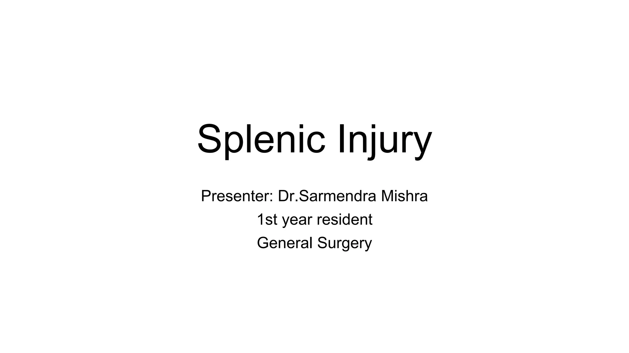
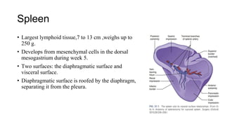
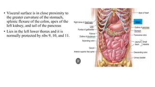
Splenic Injury
Presenter: Dr.Sarmendra Mishra
1st year resident
General Surgery
Spleen
• Largest lymphoid tissue,7 to 13 cm ,weighs up to
250 g.
• Develops from mesenchymal cells in the dorsal
mesogastrium during week 5.
• Two surfaces: the diaphragmatic surface and
visceral surface.
• Diaphragmatic surface is roofed by the diaphragm,
separating it from the pleura.
• Visceral surface is in close proximity to
the greater curvature of the stomach,
splenic flexure of the colon, apex of the
left kidney, and tail of the pancreas
• Lies in the left lower thorax and it is
normally protected by ribs 9, 10, and 11.
Vascular Anatomy
• The splenic artery, a branch of the celiac trunk ,gives off
multiple branches (16–18 branches) to the pancreas as it
travels along its posterior aspect.
• Two variants:The magistral type(30%), which branches into
terminal and polar arteries near the hilum of the spleen
• Distributed type(70%), gives off its branches early and
distant from the hilum.
• The splenic vein created by the union of several splenic
veins and the left gastroepiploic vein then travels posteriorly
to the pancreas to superior mesenteric vein to form the
portal vein.
SPLENIC FUNCTION
• Hematopoietic :During 3 to 5 weeks of fetal life, the hematopoietic functions,
which include white and red blood cell (RBC) production.
• Reservoir: one third of the platelets are pooled within the spleen.
• Immunologic Function:Contribute to humoral and cell-mediated immunity
• Filtration:
In the closed system, blood flows directly from
arteries to veins.
In the open system, the blood flows through the
arterioles and then trickles into the splenic sinuses
before draining into the venous system
Splenic Injuries.
• First or second most commonly injured ,approximately 42% of abdominal trauma.
• Direct compression of the spleen with parenchymal fracture
• Rapid deceleration tearing the splenic capsule and/or parenchyma where it is fixed to
the retroperitoneum,create a subcapsular hematoma
• Patients who respond to resuscitation managed nonoperatively
• Unstable patients have a splenic injury identified at the time of laparotomy
Types of Splenic Trauma
Blunt trauma.
• Treated with nonoperative observation.
• CT: diagnostic modality of choice.
• Close observation with continuous monitoring of vital signs and serial hematocrit
determinations.
• CT evidence of a contrast “blush” or evidence of continuing blood loss who remain
stable should undergo embolization.
• Patients who are hemodynamically unstable should undergo operative exploration.
Penetrating trauma
• Penetrating splenic injuries,diagnosed at laparotomy or imaging reports.
• Management depends on complete mobilization of the spleen.
• Initial hemostasis is possible through manual compression.
• Minor injuries contained within the splenic capsule do not require any
intervention.
• Bleeding from small capsular lacerations can be controlled with direct pressure or
topical hemostatic agents
• In the stable patient, splenorrhaphy can be employed in an attempt to preserve
immune function
• Splenorrhaphy : parenchyma saving operations of spleen
• Techniques:Electric cautery, Argon Beam Coagulator,topical packing (fibrin sealing,
omental pouch ),splenic sutures ( simple or figure-of-eight ),Capping (mesh),partial
splenectomies,splenic artery ligation
• Devitalized tissue should be debrided and the wound closed with absorbable
horizontal mattress sutures (usually 2-0 chromic).
• In unstable patients or in patients for whom splenic salvage fails, splenectomy
done
• Emergent splenectomy require postoperative immunization against Streptococcus
pneumoniae, Haemophilus influenzae, and Neisseria meningitidis.
• It is recommended that vaccines be given 14 days following trauma splenectomy;
Evaluation
Focused Assessment with Sonography for Trauma (FAST)
• identify free intraperitoneal fluid
• considered positive if the fluid is identified as an anechoic
band or a (black) rim around the spleen.
• Hemodynamic instability in the presence of free fluid requires
immediate laparotomy.
Computed Tomography (CT)
• Diagnostic modality of choice for detecting solid organ
injuries.
• Shows disruption in the normal splenic parenchyma,
surrounding hematoma, and free intra-abdominal blood.
• Abdominal CT with IV
administration(sensitivity/specificity 96%–100%)
• A splenic injury with active extravasation into a
pseudoaneurysm
AAST( American Association for the Surgery of
Trauma)Scoring Scale
World Society of Emergency Surgery Classification
(WSES)
SW stab wound, GSW gunshot wound
• Success rate for nonoperative management is approximately 90% in blunt splenic
trauma
• Advantages: reductions in hospital costs, intraabdominal complications, blood
transfusions, nontherapeutic laparotomies, and mortality.
• Stable patients who demonstrate imaging concerning for active extravasation or
pseudoaneurysm are evaluated by interventional radiology or angiography and
embolization.
• Patients without active extravasation or pseudoaneurysm but high-grade injuries (III–
V) are also evaluated by interventional radiology and proceed to angiography and
embolization within 24 hours.
• Patients with high-grade injures should undergo intensive care monitoring on
admission
• Operative management of splenic injuries may be required in the setting of
instability at the time of admission or after failed nonoperative management.
PREOPERATIVE CONSIDERATIONS
• Patients should be consulted on potential complications: Overwhelming
postsplenectomy sepsis(OPSI), splenic vein thrombosis, bleeding, arterial
thrombosis (myocardial infarction, stroke), deep vein thrombosis, and pulmonary
hypertension.
• Splenectomy for hematologic or malignant indications have the greatest risk,
splenectomy for trauma or iatrogenic injury have the lowest risk.
• OPSI can occur in 4.4% of children less than 16 years of age & 0.9% of adults
• Greatest in the first 2 years after splenectomy; however, asplenic patients remain
at lifelong risk.
• Annual influenza vaccine for asplenic patients.
Deep Vein Thrombosis Prophylaxis:
• Risk of portal vein thrombosis (PVT) may reach 50% for patients presenting with both
splenomegaly and myeloproliferative disorders.
• presents with anorexia, abdominal pain, leukocytosis, and thrombocytosis.
• early diagnosis with contrast enhanced CT, and starting anticoagulation immediately.
• DVT prophylaxis, including use of sequential compression devices and subcutaneous
administration of heparin (5000 U), should be initiated for patients undergoing
splenectomy
• elevated risk patients(obesity, history of prior venous thromboembolism, known
hypercoagulable state, older age), a postoperative antithrombotic regimen of up to two
weeks of low molecular weight heparin given
SPLENECTOMY TECHNIQUES
Patient Preparation
• Potential need for transfusion of blood products and optimization of preoperative
coagulation status are necessary.
• Thrombocytopenia corrected with platelet transfusions.
• Pooled normal human immunoglobulin is effective in elevating the platelet count in
approximately 75% of patients
• Patients maintained on corticosteroid therapy preoperatively should receive parenteral
corticosteroid therapy perioperatively.
• DVT prophylaxis
• A nasogastric (NG) tube is inserted for stomach decompression.
Open Splenectomy
• Traumatic rupture is common indication for OS. Others including massive
splenomegaly, ascites, portal hypertension, multiple prior operations, extensive
splenic irradiation, and possible splenic abscess.
• During OS,A left subcostal incision paralleling the left costal margin and lying
two fingerbreadths below is preferred for most elective splenectomies.
• A midline incision is optimal for exposure when the spleen is ruptured or
massively enlarged.
• The spleen is mobilized by dividing ligamentous attachments, usually beginning
with the splenocolic ligament
• Once lesser sac access has been achieved through either the gastrosplenic or
gastrohepatic attachments, ligating the splenic artery in continuity along the
superior border of the pancreas.
• This maneuver allows safer manipulation of the spleen and dissection of the
splenic hilum, facilitating some shrinkage of the spleen, and providing an
autotransfusion of erythrocytes and platelets.
• Medial mobilization achieved by incising its lateral peritoneal attachments,
splenophrenic ligament which follows individual ligation and sequential division
of the short gastric vessels
• Splenic hilar dissection then takes place.
• Care should be taken to dissect and individually ligate the splenic artery and vein
• During hilar dissection, great care must be taken to avoid injuring the pancreas.
• Once the spleen is excised, hemostasis is secured by irrigating, suctioning, and
scrupulously inspecting the bed of dissection.
• The splenic bed is not routinely drained.
• A thorough search for accessory spleens done when hematologic disorder has
occasioned splenectomy
Laparoscopic Splenectomy
• LS is the gold standard for elective splenectomy in patients with normal-sized spleens.
• Right lateral decubitus position(Lateral Approach)
• The lateral approach routinely involves the use of three or four trocars
• As with OS, the splenocolic ligament and the lateral peritoneal attachments are
divided with resultant medial mobilization of the spleen.
• The short gastric vessels divided usually with hemostatic energy sources such as
ultrasonic dissection, diathermy, or radiofrequency ablation.
• With the lower pole of the spleen gently retracted, the splenic hilum is accessible to
further applications of clips or an endovascular stapling device
• Visualization of the tail of the pancreas using lateral approach,and avoid injury
when placing the endovascular stapler within the sack and allows piecemeal
extraction;
• A blunt instrument should be used to disrupt and remove the spleen to avoid the risk
of sack rupture, spillage of contents, and subsequent splenosis
Preoperative Grading Score to Predict Technical
Difficulty in Laparoscopic Splenectomy
Complications of Splenectomy
• Left lower lobe atelectasis
• Pleural effusion and pneumonia
• Subphrenic hematoma
• Subphrenic abscess and wound infection
• Pancreatitis, pseudocyst, and pancreatic fistula are among the pancreatic
complications
References
• Schwartz’s Principles of Surgery
• Sabiston Textbook of Surgery
• Bailey & Love's Short Practice of Surgery, 27th Edition

Splenic Injury.pptx

  • 1.
    Splenic Injury Presenter: Dr.SarmendraMishra 1st year resident General Surgery
  • 2.
    Spleen • Largest lymphoidtissue,7 to 13 cm ,weighs up to 250 g. • Develops from mesenchymal cells in the dorsal mesogastrium during week 5. • Two surfaces: the diaphragmatic surface and visceral surface. • Diaphragmatic surface is roofed by the diaphragm, separating it from the pleura.
  • 3.
    • Visceral surfaceis in close proximity to the greater curvature of the stomach, splenic flexure of the colon, apex of the left kidney, and tail of the pancreas • Lies in the left lower thorax and it is normally protected by ribs 9, 10, and 11.
  • 4.
    Vascular Anatomy • Thesplenic artery, a branch of the celiac trunk ,gives off multiple branches (16–18 branches) to the pancreas as it travels along its posterior aspect. • Two variants:The magistral type(30%), which branches into terminal and polar arteries near the hilum of the spleen • Distributed type(70%), gives off its branches early and distant from the hilum. • The splenic vein created by the union of several splenic veins and the left gastroepiploic vein then travels posteriorly to the pancreas to superior mesenteric vein to form the portal vein.
  • 5.
    SPLENIC FUNCTION • Hematopoietic:During 3 to 5 weeks of fetal life, the hematopoietic functions, which include white and red blood cell (RBC) production. • Reservoir: one third of the platelets are pooled within the spleen. • Immunologic Function:Contribute to humoral and cell-mediated immunity
  • 6.
    • Filtration: In theclosed system, blood flows directly from arteries to veins. In the open system, the blood flows through the arterioles and then trickles into the splenic sinuses before draining into the venous system
  • 7.
    Splenic Injuries. • Firstor second most commonly injured ,approximately 42% of abdominal trauma. • Direct compression of the spleen with parenchymal fracture • Rapid deceleration tearing the splenic capsule and/or parenchyma where it is fixed to the retroperitoneum,create a subcapsular hematoma • Patients who respond to resuscitation managed nonoperatively • Unstable patients have a splenic injury identified at the time of laparotomy
  • 8.
    Types of SplenicTrauma Blunt trauma. • Treated with nonoperative observation. • CT: diagnostic modality of choice. • Close observation with continuous monitoring of vital signs and serial hematocrit determinations. • CT evidence of a contrast “blush” or evidence of continuing blood loss who remain stable should undergo embolization. • Patients who are hemodynamically unstable should undergo operative exploration.
  • 9.
    Penetrating trauma • Penetratingsplenic injuries,diagnosed at laparotomy or imaging reports. • Management depends on complete mobilization of the spleen. • Initial hemostasis is possible through manual compression. • Minor injuries contained within the splenic capsule do not require any intervention. • Bleeding from small capsular lacerations can be controlled with direct pressure or topical hemostatic agents
  • 10.
    • In thestable patient, splenorrhaphy can be employed in an attempt to preserve immune function • Splenorrhaphy : parenchyma saving operations of spleen • Techniques:Electric cautery, Argon Beam Coagulator,topical packing (fibrin sealing, omental pouch ),splenic sutures ( simple or figure-of-eight ),Capping (mesh),partial splenectomies,splenic artery ligation • Devitalized tissue should be debrided and the wound closed with absorbable horizontal mattress sutures (usually 2-0 chromic).
  • 11.
    • In unstablepatients or in patients for whom splenic salvage fails, splenectomy done • Emergent splenectomy require postoperative immunization against Streptococcus pneumoniae, Haemophilus influenzae, and Neisseria meningitidis. • It is recommended that vaccines be given 14 days following trauma splenectomy;
  • 12.
    Evaluation Focused Assessment withSonography for Trauma (FAST) • identify free intraperitoneal fluid • considered positive if the fluid is identified as an anechoic band or a (black) rim around the spleen. • Hemodynamic instability in the presence of free fluid requires immediate laparotomy.
  • 13.
    Computed Tomography (CT) •Diagnostic modality of choice for detecting solid organ injuries. • Shows disruption in the normal splenic parenchyma, surrounding hematoma, and free intra-abdominal blood. • Abdominal CT with IV administration(sensitivity/specificity 96%–100%) • A splenic injury with active extravasation into a pseudoaneurysm
  • 14.
    AAST( American Associationfor the Surgery of Trauma)Scoring Scale
  • 15.
    World Society ofEmergency Surgery Classification (WSES) SW stab wound, GSW gunshot wound
  • 16.
    • Success ratefor nonoperative management is approximately 90% in blunt splenic trauma • Advantages: reductions in hospital costs, intraabdominal complications, blood transfusions, nontherapeutic laparotomies, and mortality. • Stable patients who demonstrate imaging concerning for active extravasation or pseudoaneurysm are evaluated by interventional radiology or angiography and embolization. • Patients without active extravasation or pseudoaneurysm but high-grade injuries (III– V) are also evaluated by interventional radiology and proceed to angiography and embolization within 24 hours.
  • 17.
    • Patients withhigh-grade injures should undergo intensive care monitoring on admission • Operative management of splenic injuries may be required in the setting of instability at the time of admission or after failed nonoperative management.
  • 18.
    PREOPERATIVE CONSIDERATIONS • Patientsshould be consulted on potential complications: Overwhelming postsplenectomy sepsis(OPSI), splenic vein thrombosis, bleeding, arterial thrombosis (myocardial infarction, stroke), deep vein thrombosis, and pulmonary hypertension. • Splenectomy for hematologic or malignant indications have the greatest risk, splenectomy for trauma or iatrogenic injury have the lowest risk. • OPSI can occur in 4.4% of children less than 16 years of age & 0.9% of adults • Greatest in the first 2 years after splenectomy; however, asplenic patients remain at lifelong risk. • Annual influenza vaccine for asplenic patients.
  • 19.
    Deep Vein ThrombosisProphylaxis: • Risk of portal vein thrombosis (PVT) may reach 50% for patients presenting with both splenomegaly and myeloproliferative disorders. • presents with anorexia, abdominal pain, leukocytosis, and thrombocytosis. • early diagnosis with contrast enhanced CT, and starting anticoagulation immediately. • DVT prophylaxis, including use of sequential compression devices and subcutaneous administration of heparin (5000 U), should be initiated for patients undergoing splenectomy • elevated risk patients(obesity, history of prior venous thromboembolism, known hypercoagulable state, older age), a postoperative antithrombotic regimen of up to two weeks of low molecular weight heparin given
  • 20.
    SPLENECTOMY TECHNIQUES Patient Preparation •Potential need for transfusion of blood products and optimization of preoperative coagulation status are necessary. • Thrombocytopenia corrected with platelet transfusions. • Pooled normal human immunoglobulin is effective in elevating the platelet count in approximately 75% of patients • Patients maintained on corticosteroid therapy preoperatively should receive parenteral corticosteroid therapy perioperatively. • DVT prophylaxis • A nasogastric (NG) tube is inserted for stomach decompression.
  • 21.
    Open Splenectomy • Traumaticrupture is common indication for OS. Others including massive splenomegaly, ascites, portal hypertension, multiple prior operations, extensive splenic irradiation, and possible splenic abscess. • During OS,A left subcostal incision paralleling the left costal margin and lying two fingerbreadths below is preferred for most elective splenectomies. • A midline incision is optimal for exposure when the spleen is ruptured or massively enlarged. • The spleen is mobilized by dividing ligamentous attachments, usually beginning with the splenocolic ligament
  • 22.
    • Once lessersac access has been achieved through either the gastrosplenic or gastrohepatic attachments, ligating the splenic artery in continuity along the superior border of the pancreas. • This maneuver allows safer manipulation of the spleen and dissection of the splenic hilum, facilitating some shrinkage of the spleen, and providing an autotransfusion of erythrocytes and platelets. • Medial mobilization achieved by incising its lateral peritoneal attachments, splenophrenic ligament which follows individual ligation and sequential division of the short gastric vessels • Splenic hilar dissection then takes place.
  • 23.
    • Care shouldbe taken to dissect and individually ligate the splenic artery and vein • During hilar dissection, great care must be taken to avoid injuring the pancreas. • Once the spleen is excised, hemostasis is secured by irrigating, suctioning, and scrupulously inspecting the bed of dissection. • The splenic bed is not routinely drained. • A thorough search for accessory spleens done when hematologic disorder has occasioned splenectomy
  • 24.
    Laparoscopic Splenectomy • LSis the gold standard for elective splenectomy in patients with normal-sized spleens. • Right lateral decubitus position(Lateral Approach) • The lateral approach routinely involves the use of three or four trocars
  • 25.
    • As withOS, the splenocolic ligament and the lateral peritoneal attachments are divided with resultant medial mobilization of the spleen. • The short gastric vessels divided usually with hemostatic energy sources such as ultrasonic dissection, diathermy, or radiofrequency ablation. • With the lower pole of the spleen gently retracted, the splenic hilum is accessible to further applications of clips or an endovascular stapling device • Visualization of the tail of the pancreas using lateral approach,and avoid injury when placing the endovascular stapler within the sack and allows piecemeal extraction; • A blunt instrument should be used to disrupt and remove the spleen to avoid the risk of sack rupture, spillage of contents, and subsequent splenosis
  • 26.
    Preoperative Grading Scoreto Predict Technical Difficulty in Laparoscopic Splenectomy
  • 27.
    Complications of Splenectomy •Left lower lobe atelectasis • Pleural effusion and pneumonia • Subphrenic hematoma • Subphrenic abscess and wound infection • Pancreatitis, pseudocyst, and pancreatic fistula are among the pancreatic complications
  • 28.
    References • Schwartz’s Principlesof Surgery • Sabiston Textbook of Surgery • Bailey & Love's Short Practice of Surgery, 27th Edition