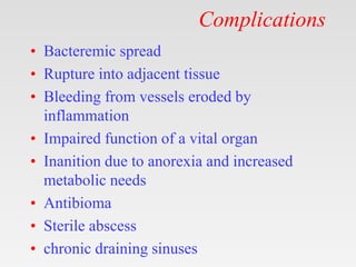
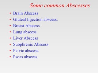
Tips on using my ppt.
1. You can freely download, edit, modify and put your
name etc.
2. Don’t be concerned about number of slides. Half the
slides are blanks except for the title.
3. First show the blank slides (eg. Aetiology ) > Ask
students what they already know about ethology of
today's topic. > Then show next slide which enumerates
aetiologies.
4. At the end rerun the show – show blank> ask questions >
show next slide.
5. This will be an ACTIVE LEARNING SESSION x
three revisions.
6. Good for self study also.
7. See notes for bibliography.
Introduction & History.
• Abscess is a localised collection of pus.
• Pus is composed of
• Tissue debris
• Dead and alive leucocytes
• Dead and alive bacteria.
• Laudable pus.- Pus was considered good
beacause it precedes cure.
• Represents near win of body”s defenses.
Etiology
Etiology
• Idiopathic
• Congenital/Genetic
• Traumatic
• Infections /Infestation
• Autoimmune
• Neoplastic (Benign/Malignant)
• Degenerative
• Iatrogenic
Etiology:Predisposing factors
• Impaired host defense mechanisms eg, HIV
• The presence of foreign bodies
• Obstruction to normal drainage (eg, in the
urinary, biliary, or respiratory tracts)
• Tissue ischemia or necrosis
• Hematoma or excessive fluid accumulation
in tissue
• Trauma
Etiology
• Numerous organisms can cause abscesses,
but the most common is
– Staphylococcus aureus.
Pathophysiology
Pathophysiology
• Abscesses may begin in an area of cellulitis
or in compromised tissue where leukocytes
accumulate.
• Progressive dissection by pus or necrosis of
surrounding cells expands the abscess.
• Highly vascularized connective tissue may
then surround the necrotic tissue,
leukocytes, and debris to wall off the
abscess and limit further spread.
Pathophysiology
Pathophysiology
• Organisms may enter the tissue by-
– Direct implantation eg im injection.
– Spread from an established, contiguous
infection
– Dissemination via lymphatic or hematogenous
routes from a distant site
– Migration from a location where there are
resident flora into an adjacent, normally sterile
area because natural barriers are disrupted (eg,
by perforation of an abdominal viscus causing
an intra-abdominal abscess)
Clinical Features
Clinical Features
• Demography
• Symptoms
• Signs
• Prognosis
• Complications
Symptoms
Symptoms
• Fever with spikes and chills.
• Local pain and tenderness
• Anorexia
• Weight loss
• Fatigue
Signs
Signs
• Signs of cutaneous and subcutaneous
abscesses are pain, heat, swelling,
tenderness, and redness.
• If superficial abscesses are ready to
spontaneously rupture, the skin over the
center of the abscess may thin, sometimes
appearing white or yellow because of the
underlying pus (termed pointing).
• The predominant manifestation of some
abscesses is abnormal organ function (eg,
hemiplegia due to a brain abscess).
Complications
Complications
• Bacteremic spread
• Rupture into adjacent tissue
• Bleeding from vessels eroded by
inflammation
• Impaired function of a vital organ
• Inanition due to anorexia and increased
metabolic needs
• Antibioma
• Sterile abscess
• chronic draining sinuses
Investigations
Investigations
• Laboratory Studies
– Routine- leucocytosis, raised CRP
– Special – Blood culture,
• Imaging Studies
• Tissue diagnosis
– Cytology
• FNAC
– Histlogy
Diagnostic Studies
Diagnostic Studies
Imaging Studies
• X-Ray
• USG
• CT
• Angiography
• MRI
• Endoscopy
• Nuclear scan- radiolabelled leucocytes.
Operative Therapy
Operative Therapy
• Drainage- Deroofing.
• Antibiotics when abscesses are large, deep,
or surrounded by significant cellulitis.
• Aspiration.
• Conseervative – Amoebic Liver abscess.
Some common Abscesses
• Brain Abscess
• Gluteal Injection abscess.
• Breast Abscess
• Lung abscess
• Liver Abscess
• Subphrenic Abscess
• Pelvic abscess.
• Psoas abscess.
Get this ppt in mobile
1. Download Microsoft
PowerPoint from play
store.
2. Open Google assistant
3. Open Google lens.
4. Scan qr code from
next slide.
Get this ppt in mobile
Get my ppt collection
• https://www.slideshare.net/drpradeeppande/
edit_my_uploads
• https://www.dropbox.com/sh/x600md3cvj8
5woy/AACVMHuQtvHvl_K8ehc3ltkEa?dl
=0
• https://www.facebook.com/doctorpradeeppa
nde/?ref=pages_you_manage

Abscess.pptx

  • 1.
    Tips on usingmy ppt. 1. You can freely download, edit, modify and put your name etc. 2. Don’t be concerned about number of slides. Half the slides are blanks except for the title. 3. First show the blank slides (eg. Aetiology ) > Ask students what they already know about ethology of today's topic. > Then show next slide which enumerates aetiologies. 4. At the end rerun the show – show blank> ask questions > show next slide. 5. This will be an ACTIVE LEARNING SESSION x three revisions. 6. Good for self study also. 7. See notes for bibliography.
  • 2.
  • 3.
    Introduction & History. •Abscess is a localised collection of pus. • Pus is composed of • Tissue debris • Dead and alive leucocytes • Dead and alive bacteria. • Laudable pus.- Pus was considered good beacause it precedes cure. • Represents near win of body”s defenses.
  • 4.
  • 5.
    Etiology • Idiopathic • Congenital/Genetic •Traumatic • Infections /Infestation • Autoimmune • Neoplastic (Benign/Malignant) • Degenerative • Iatrogenic
  • 6.
    Etiology:Predisposing factors • Impairedhost defense mechanisms eg, HIV • The presence of foreign bodies • Obstruction to normal drainage (eg, in the urinary, biliary, or respiratory tracts) • Tissue ischemia or necrosis • Hematoma or excessive fluid accumulation in tissue • Trauma
  • 7.
    Etiology • Numerous organismscan cause abscesses, but the most common is – Staphylococcus aureus.
  • 8.
  • 9.
    Pathophysiology • Abscesses maybegin in an area of cellulitis or in compromised tissue where leukocytes accumulate. • Progressive dissection by pus or necrosis of surrounding cells expands the abscess. • Highly vascularized connective tissue may then surround the necrotic tissue, leukocytes, and debris to wall off the abscess and limit further spread.
  • 10.
  • 11.
    Pathophysiology • Organisms mayenter the tissue by- – Direct implantation eg im injection. – Spread from an established, contiguous infection – Dissemination via lymphatic or hematogenous routes from a distant site – Migration from a location where there are resident flora into an adjacent, normally sterile area because natural barriers are disrupted (eg, by perforation of an abdominal viscus causing an intra-abdominal abscess)
  • 12.
  • 13.
    Clinical Features • Demography •Symptoms • Signs • Prognosis • Complications
  • 14.
  • 15.
    Symptoms • Fever withspikes and chills. • Local pain and tenderness • Anorexia • Weight loss • Fatigue
  • 16.
  • 17.
    Signs • Signs ofcutaneous and subcutaneous abscesses are pain, heat, swelling, tenderness, and redness. • If superficial abscesses are ready to spontaneously rupture, the skin over the center of the abscess may thin, sometimes appearing white or yellow because of the underlying pus (termed pointing). • The predominant manifestation of some abscesses is abnormal organ function (eg, hemiplegia due to a brain abscess).
  • 18.
  • 19.
    Complications • Bacteremic spread •Rupture into adjacent tissue • Bleeding from vessels eroded by inflammation • Impaired function of a vital organ • Inanition due to anorexia and increased metabolic needs • Antibioma • Sterile abscess • chronic draining sinuses
  • 20.
  • 21.
    Investigations • Laboratory Studies –Routine- leucocytosis, raised CRP – Special – Blood culture, • Imaging Studies • Tissue diagnosis – Cytology • FNAC – Histlogy
  • 22.
  • 23.
    Diagnostic Studies Imaging Studies •X-Ray • USG • CT • Angiography • MRI • Endoscopy • Nuclear scan- radiolabelled leucocytes.
  • 24.
  • 25.
    Operative Therapy • Drainage-Deroofing. • Antibiotics when abscesses are large, deep, or surrounded by significant cellulitis. • Aspiration. • Conseervative – Amoebic Liver abscess.
  • 26.
  • 27.
    Some common Abscesses •Brain Abscess • Gluteal Injection abscess. • Breast Abscess • Lung abscess • Liver Abscess • Subphrenic Abscess • Pelvic abscess. • Psoas abscess.
  • 28.
    Get this pptin mobile 1. Download Microsoft PowerPoint from play store. 2. Open Google assistant 3. Open Google lens. 4. Scan qr code from next slide.
  • 29.
    Get this pptin mobile
  • 30.
    Get my pptcollection • https://www.slideshare.net/drpradeeppande/ edit_my_uploads • https://www.dropbox.com/sh/x600md3cvj8 5woy/AACVMHuQtvHvl_K8ehc3ltkEa?dl =0 • https://www.facebook.com/doctorpradeeppa nde/?ref=pages_you_manage

Editor's Notes

  • #2 drpradeeppande@gmail.com 7697305442