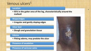
DEPARTMENT OF SURGERY
NDUTH - OKOLOBIRI
DISCUSS VASCULAR ULCERS
BY
DR BARIBOTE SAMPSON OTONYE
16TH SEPT., 2022
OUTLINE
• Introduction
• Epidemiology
• Classification
• Venous ulcer
• Arterial ulcer
• Management
• Complications
• Education and risk reduction
• Comparison between venous and arterial ulcer
• Conclusion
• Reference
Introduction
• is the loss of continuity of the skin or mucous
membrane1
An ulcer
• underlying vascular pathology
Vascular ulcers
Chronic or recurrent
• Location
• Appearance
• Bleeding and
• Associated pain and findings1
Distinct:
Epidemiology
• 500,000 to 600,000 and increases in age2
Prevalence US estimates
• estimates – 1%
• 80% thought to be venous
Ireland and Australia1
Epidemiology contd Chronic Leg Ulcer
Vascular Malignant Infective Trauma
56.7%
31.7%
Rahman GA et al2
3.3%
3.3%
Classification
Venous ulcer Arterial ulcer Mixed ulcers
Venous ulcers
A venous ulcer is an open skin lesion of the leg or foot
that occurs in an area affected by venous hypertension3
Most common cause of leg ulcers
• 60 – 80% of leg ulcers
Prevalence
• 0.18% and 1%
• 4% over age 65years4
Risk factors to developing Venous ulcers3,4
• Older age
• Female sex
• Obesity
• Trauma
• Pregnancy
• Estrogen
• Prolonged standing
• Congenital absence of veins
• Deep vein thrombosis (DVT), phlebitis,
• Varicose veins
Include
Anatomy of Leg Veins8
Pathophysiology
Arterial ulcer
• Ischemic ulcer is an open skin lesion of the leg or foot that
occurs in an area with underlying peripheral vascular disease
(PVD)
Ischemic ulcers
• 6% of vascular ulcers
Ischemic ulcers
• men older than 45 and
• women older than 55
PVD
• Atherosclerotic disease of medium and large sized arteries5
Most common cause
Causes
Atherosclerosis
Others
• diabetes,
• thromboangiitis,
• vasculitis,
• Pyoderma gangrenosum,
• thalasaemia, and
• sickle cell disease
Risk factors6
• Age
• Family history of PAD
Non modifiable
• Smoking
• Diabetes
• Hyperlipidaemia
• Hypertension
• Obesity
• Sedentary lifestyle
Modifiable
Pathophysiology of
arterial ulcer
Ankle brachial index7
• Non invasion diagnosis test
• Compares blood pressures in
upper and lower limb
• Determine presence of lower limb
PAD
Ankle brachial index
Value Interpretation
0 – 0.4 Severe PAD sufficient to cause resting pain or gangrene
0.41 – 0.9 PAD sufficient to cause claudication
0.91 –
1.30
Normal vessels
>1.30 Noncompressible, severely calcified vessel
Classification – Society for Vascular Surgery WIFI6
Wound
Grade Ulcer Gangrene
0 No ulcer No gangrene
1 Small, shallow ulcer on distal leg or foot; no exposed bone,
unless limited to distal phalax
No gangrene
2 Deeper ulcer with exposed bone, joint or tendon, generally not
involving the heel; shallow heel ulcer without calcaeneal
involvement
Gangrenous changes limited to digits
3 Extensive, deep ulcer involving forefoot and/or midfoot; deep
full thickness heel ulcer +- calcaneal involvement
Extensive gangrene involving the
forefoot/midfoot; full thickness heel
necrosis +- calcaneal involvement
Ischemia
Grade ABI Ankle systolic pressure
0 >= 0.8 >100mmHg
1 0.6 – 0.79 70 – 100mmHg
2 0.4 – 0.59 50 – 70mmHg
3 <= 0.39 <50mmHg
Infection
Grade Clinical manifestation of infection
0 No symptoms or signs of infection
1 Local infection involving only the skin and the subcutaneous tissue
2 Local infection with erythema >2 cm, or involving structures deeper than skin and
subcutaneous tissues, and no systemic inflammatory response signs
3 Local infection with the signs of SIRS, as manifested by two or more of the following:
• Temperature > 38 or < 36°C
• Heart rate > 90 beats/min
• Respiratory rate > 20 breaths/min or PaCO2 < 32 mm Hg
• White blood cell count > 12,000 or < 4,000 cu/mm or 10% immature bands
Interpretation (Risk of amputation at 1 year)
Total score
0 – 1 Very low
2 Low
3 Moderate
4+ High
Mgt
History
Physical
Exam
Investiga
tion
Treatme
nt
History
Biodata Sex and Age
Complain Chronicity
Course Associations Pain
Cause (risk
factors)
Venous Vs Arterial
Care
Complications
Effect on work
Quality of life
• General examination
• Regional lymph nodes
• Assess for obesity
• Peripheral neuropathy
• Peripheral pulses
• Documentation of the size of the ulcer and
photographs
• Characterization of the ulcers
Physical Examination
Venous ulcers5
• 95% in the gaiter area of the leg, characteristically around the
malleoli
Location
• Irregular and gently sloping edges
Shape/edges
• Slough and granulation tissue
Ulcer bed
• Pitting edema, may predate the ulcer
Edema
Presence of exudation
Presence of varicose veins
v
Hemosiderin
lipodermatosclerosis
• prolong venous insufficiency
Occurs following
• Dermal and subcutaneous
induration and fibrosis
• Atrophic skin
• Surrounding hypo or hyper
pigmentation
Characterized by
Inverted Champaign bottle
Arterial Ulcers5
Location
• Toes, heels and bony prominences of the foot
Edges
• Punched out edges
Ulcer bed
• Pale, non-granulating necrotic base
Surrounding skin
• Dusky erythema, Cold, Hairless
• Thin, brittle with Shiny texture
Pulses
• Decrease or absent dorsalis pedis and posterior tibal pulses
• Bruit in proximal arterials
Presence of gangrene
Investigation Specific
Ankle brachial
pressure index
Duplex
Doppler
scanning
CT
angiography
Magnetic
resonant
angiography
General
FBC
EUCr
Urinalysis
Wound swab
and biopsy
Others
FBS
Fasting lipid
profile etc
Treatment (Venous Ulcers)
•Compression therapy
•Limb elevation
•Calf exercises and walking
•Wound care
•Diagnostic testing for venous reflux
•Correction of superficial venous reflux
Standard treatment:
Compression Therapy
Two to four-layer bandage kits (Profore, 3M)
Unna boot
Double layer compression stocking 40-50mmHg:
• Inner stocking (10-15mmHg) stays on 24/7, remove to bathe and
change dressing
• Outer stocking (30-40mmHg) applied on waking, removed at
bedtime
Pneumatic compression pump
4 layer compression boot
2 layers compression
Wound care9,5
Dressing
• use a dressing that will maintain a moist wound-healing
environment
• Has ability to absorb exudates and protect periwound skin
Debridement
• Thorough serial debridement and irrigation
Infection control
• Empirical antibiotics and tailor to narrow spectrum in line with
sensitivity
• Impede wound
Healing
Advanced Wound Care
A large, growing array of therapies have been developed . . .
Bioengineered Alternative Tissues
Bio-Active Wound Adjuncts:
• Oasis
• Porcine intestinal submucosa
• Epifix
• Dried human amniotic membrane
Living Tissues / Growth Factors:
• Regranex (becaplermin)
• Platelet derived growth factor topical
• Apligraf
• Living human dermal fibroblasts and epidermal keratinocytes in bovine collagen matrix
Arterial ulcer
Prior to revascularization, an anatomic road map should be obtained9
Options include:
Angiogram9
Duplex Angiography9,
• Femoropopliteal segment
• Sensitivity 99%, Specificity 94%
• Tibial segment
• Sensitivity 80% Specificity 91%
Magnetic Resonance Angiography9
Contrast Tomography Angiography9
Surgical options
• Reconstructive surgery (arterial bypass)5
Diffuse disease
• Angioplasty5
Local stenosis
• Non healing ulceration
• Gangrene
• Rest pain
• Progression of disabling claudication
Indication6
Wound care9,5
Dressing
• use a dressing that will maintain a moist wound-healing environment.
• Dry gangrene or eschar is best left dry until revascularization is successful
Debridement
• Debridement of nonviable and noninfected tissue is performed ONLY AFTER the
revascularization procedure.
• Prerevascularization debridement should be indicated only in a septic foot with
and without ischemic signs
• Should be minimal
Infection control
• Empirical antibiotics and tailor to narrow spectrum in line with sensitivity
Adjuvant treatment
Hyperbaric Oxygen therapy9
• In patients with nonreconstructable anatomy
• Non healing ulcer despite revascularization
Indications
• Increase tissue oxygen
• Increase angiogenesis in hypoxic or injured tissue
• Increase signal transduction between growth factors and
receptors
Benefits
Others
Control of DM, hyperlipidaemia, hypertension etc
Cessation of smoking
Patients may find benefits from sleeping in a bed raised at the head end
Opioid for pain
complications Recurrence
Tetanus
Septicemia
Osteomyelitis
Periostitis
Gangrene
Contracture
Non healing
Guidelines for patients on protecting lower
limbs and feet5
• Examine the feet daily for broken skin, blisters, swelling, or redness
• Report worsening symptoms—for example, decreasing walking
distance, pain at rest, pain at night, changes in skin colour
• Never walk barefoot
• Ensure shoes are well fitting and free of friction and pressure points;
check them for foreign objects (such as stones) before wearing; and
avoid open toed sandals and pointed shoes
• Give up smoking
• Take regular exercise within limits of pain and tolerance
• Weight reduction
Conclusion
Management of patients with vascular ulcers has to be multidisciplinary
Should include
• Detailed history
• Physical examination
• Appropriate investigations
• Basic and newer treatment modalities
While educating patients on issues of correct skin care and the
importance of seeking early medical advice
References
1. Allen Gabriel et al: Vascular Ulcers – Practical Essentials,
Epidemiology, Etiology. https://emedicine.Medscape.com. 17th
August 2021
2. G.A. Rahman et al: Epidemiology, Etiology and Treatment of Chronic
Leg Ulcers: Experience of 60 patients. Annals of African Medicine
Vol. 9, No. 1, 2010:1-4. DOI: 10:4103/1596-3519.62615
3. Deborah A Simon et al:Management of venous leg ulcers. BMJ
VOLUME 328 5 JUNE 2004 bmj.com
4. Biju Vasudevan: Venous leg ulcers: Pathophysiology and
Classification. Indian Dermatology Online Journal - July-September
2014 - Volume 5 - Issue 3. DOI: 10.4103/2229-5178.137819
5. Joseph E Grey, Stuart Enoch, Keith G Harding: ABC of wound healing
Venous and arterial leg ulcers. BMJ 2006;332:347–50 bmj.com
Reference contd
6. Rulon L. Hardman et al: Overview of Classification Systems in Peripheral
Artery Disease. Seminars in Interventional Radiology Vol. 31 No. 4/2014.
Copyright © 2014 by Thieme Medical Publishers, Inc., 333 Seventh
Avenue, New York, NY 10001, USA. DOI http://dx.doi.org/10.1055/s-
0034-1393976. ISSN 0739-9529.
7. Chan W. Park: Ankle-Brachial Index (ABI) measurement.
https://emedicine.Medscape.com 10th Sept. 2020
8. Fronek HS, Bergan JJ et al. Fundamentals of Phlebology:venous disease
for clinicians. 2nd Edition. Pg 1, 2. American College of Phlebology
9. Harriet W. Hopf, Cristiane Ueno et al: Guidelines for the treatment of
arterial insufficiency ulcers. Wound Rep Reg (2006) 14 693–710 c 2006 by
theWound Healing Society

Vascular ulcer.pptx

  • 1.
    DEPARTMENT OF SURGERY NDUTH- OKOLOBIRI DISCUSS VASCULAR ULCERS BY DR BARIBOTE SAMPSON OTONYE 16TH SEPT., 2022
  • 2.
    OUTLINE • Introduction • Epidemiology •Classification • Venous ulcer • Arterial ulcer • Management • Complications • Education and risk reduction • Comparison between venous and arterial ulcer • Conclusion • Reference
  • 3.
    Introduction • is theloss of continuity of the skin or mucous membrane1 An ulcer • underlying vascular pathology Vascular ulcers Chronic or recurrent • Location • Appearance • Bleeding and • Associated pain and findings1 Distinct:
  • 4.
    Epidemiology • 500,000 to600,000 and increases in age2 Prevalence US estimates • estimates – 1% • 80% thought to be venous Ireland and Australia1
  • 5.
    Epidemiology contd ChronicLeg Ulcer Vascular Malignant Infective Trauma 56.7% 31.7% Rahman GA et al2 3.3% 3.3%
  • 6.
  • 7.
    Venous ulcers A venousulcer is an open skin lesion of the leg or foot that occurs in an area affected by venous hypertension3 Most common cause of leg ulcers • 60 – 80% of leg ulcers Prevalence • 0.18% and 1% • 4% over age 65years4
  • 8.
    Risk factors todeveloping Venous ulcers3,4 • Older age • Female sex • Obesity • Trauma • Pregnancy • Estrogen • Prolonged standing • Congenital absence of veins • Deep vein thrombosis (DVT), phlebitis, • Varicose veins Include
  • 9.
  • 10.
  • 12.
    Arterial ulcer • Ischemiculcer is an open skin lesion of the leg or foot that occurs in an area with underlying peripheral vascular disease (PVD) Ischemic ulcers • 6% of vascular ulcers Ischemic ulcers • men older than 45 and • women older than 55 PVD • Atherosclerotic disease of medium and large sized arteries5 Most common cause
  • 13.
    Causes Atherosclerosis Others • diabetes, • thromboangiitis, •vasculitis, • Pyoderma gangrenosum, • thalasaemia, and • sickle cell disease
  • 14.
    Risk factors6 • Age •Family history of PAD Non modifiable • Smoking • Diabetes • Hyperlipidaemia • Hypertension • Obesity • Sedentary lifestyle Modifiable
  • 15.
  • 16.
    Ankle brachial index7 •Non invasion diagnosis test • Compares blood pressures in upper and lower limb • Determine presence of lower limb PAD Ankle brachial index Value Interpretation 0 – 0.4 Severe PAD sufficient to cause resting pain or gangrene 0.41 – 0.9 PAD sufficient to cause claudication 0.91 – 1.30 Normal vessels >1.30 Noncompressible, severely calcified vessel
  • 17.
    Classification – Societyfor Vascular Surgery WIFI6 Wound Grade Ulcer Gangrene 0 No ulcer No gangrene 1 Small, shallow ulcer on distal leg or foot; no exposed bone, unless limited to distal phalax No gangrene 2 Deeper ulcer with exposed bone, joint or tendon, generally not involving the heel; shallow heel ulcer without calcaeneal involvement Gangrenous changes limited to digits 3 Extensive, deep ulcer involving forefoot and/or midfoot; deep full thickness heel ulcer +- calcaneal involvement Extensive gangrene involving the forefoot/midfoot; full thickness heel necrosis +- calcaneal involvement Ischemia Grade ABI Ankle systolic pressure 0 >= 0.8 >100mmHg 1 0.6 – 0.79 70 – 100mmHg 2 0.4 – 0.59 50 – 70mmHg 3 <= 0.39 <50mmHg
  • 18.
    Infection Grade Clinical manifestationof infection 0 No symptoms or signs of infection 1 Local infection involving only the skin and the subcutaneous tissue 2 Local infection with erythema >2 cm, or involving structures deeper than skin and subcutaneous tissues, and no systemic inflammatory response signs 3 Local infection with the signs of SIRS, as manifested by two or more of the following: • Temperature > 38 or < 36°C • Heart rate > 90 beats/min • Respiratory rate > 20 breaths/min or PaCO2 < 32 mm Hg • White blood cell count > 12,000 or < 4,000 cu/mm or 10% immature bands Interpretation (Risk of amputation at 1 year) Total score 0 – 1 Very low 2 Low 3 Moderate 4+ High
  • 19.
  • 20.
    History Biodata Sex andAge Complain Chronicity Course Associations Pain Cause (risk factors) Venous Vs Arterial Care Complications Effect on work Quality of life
  • 21.
    • General examination •Regional lymph nodes • Assess for obesity • Peripheral neuropathy • Peripheral pulses • Documentation of the size of the ulcer and photographs • Characterization of the ulcers Physical Examination
  • 22.
    Venous ulcers5 • 95%in the gaiter area of the leg, characteristically around the malleoli Location • Irregular and gently sloping edges Shape/edges • Slough and granulation tissue Ulcer bed • Pitting edema, may predate the ulcer Edema Presence of exudation Presence of varicose veins v
  • 23.
  • 24.
    lipodermatosclerosis • prolong venousinsufficiency Occurs following • Dermal and subcutaneous induration and fibrosis • Atrophic skin • Surrounding hypo or hyper pigmentation Characterized by
  • 25.
  • 26.
    Arterial Ulcers5 Location • Toes,heels and bony prominences of the foot Edges • Punched out edges Ulcer bed • Pale, non-granulating necrotic base Surrounding skin • Dusky erythema, Cold, Hairless • Thin, brittle with Shiny texture Pulses • Decrease or absent dorsalis pedis and posterior tibal pulses • Bruit in proximal arterials Presence of gangrene
  • 27.
    Investigation Specific Ankle brachial pressureindex Duplex Doppler scanning CT angiography Magnetic resonant angiography General FBC EUCr Urinalysis Wound swab and biopsy Others FBS Fasting lipid profile etc
  • 28.
    Treatment (Venous Ulcers) •Compressiontherapy •Limb elevation •Calf exercises and walking •Wound care •Diagnostic testing for venous reflux •Correction of superficial venous reflux Standard treatment:
  • 29.
    Compression Therapy Two tofour-layer bandage kits (Profore, 3M) Unna boot Double layer compression stocking 40-50mmHg: • Inner stocking (10-15mmHg) stays on 24/7, remove to bathe and change dressing • Outer stocking (30-40mmHg) applied on waking, removed at bedtime Pneumatic compression pump
  • 30.
  • 32.
  • 33.
    Wound care9,5 Dressing • usea dressing that will maintain a moist wound-healing environment • Has ability to absorb exudates and protect periwound skin Debridement • Thorough serial debridement and irrigation Infection control • Empirical antibiotics and tailor to narrow spectrum in line with sensitivity
  • 34.
  • 35.
    Advanced Wound Care Alarge, growing array of therapies have been developed . . . Bioengineered Alternative Tissues Bio-Active Wound Adjuncts: • Oasis • Porcine intestinal submucosa • Epifix • Dried human amniotic membrane Living Tissues / Growth Factors: • Regranex (becaplermin) • Platelet derived growth factor topical • Apligraf • Living human dermal fibroblasts and epidermal keratinocytes in bovine collagen matrix
  • 37.
    Arterial ulcer Prior torevascularization, an anatomic road map should be obtained9 Options include: Angiogram9 Duplex Angiography9, • Femoropopliteal segment • Sensitivity 99%, Specificity 94% • Tibial segment • Sensitivity 80% Specificity 91% Magnetic Resonance Angiography9 Contrast Tomography Angiography9
  • 38.
    Surgical options • Reconstructivesurgery (arterial bypass)5 Diffuse disease • Angioplasty5 Local stenosis • Non healing ulceration • Gangrene • Rest pain • Progression of disabling claudication Indication6
  • 39.
    Wound care9,5 Dressing • usea dressing that will maintain a moist wound-healing environment. • Dry gangrene or eschar is best left dry until revascularization is successful Debridement • Debridement of nonviable and noninfected tissue is performed ONLY AFTER the revascularization procedure. • Prerevascularization debridement should be indicated only in a septic foot with and without ischemic signs • Should be minimal Infection control • Empirical antibiotics and tailor to narrow spectrum in line with sensitivity
  • 40.
    Adjuvant treatment Hyperbaric Oxygentherapy9 • In patients with nonreconstructable anatomy • Non healing ulcer despite revascularization Indications • Increase tissue oxygen • Increase angiogenesis in hypoxic or injured tissue • Increase signal transduction between growth factors and receptors Benefits
  • 41.
    Others Control of DM,hyperlipidaemia, hypertension etc Cessation of smoking Patients may find benefits from sleeping in a bed raised at the head end Opioid for pain
  • 42.
  • 43.
    Guidelines for patientson protecting lower limbs and feet5 • Examine the feet daily for broken skin, blisters, swelling, or redness • Report worsening symptoms—for example, decreasing walking distance, pain at rest, pain at night, changes in skin colour • Never walk barefoot • Ensure shoes are well fitting and free of friction and pressure points; check them for foreign objects (such as stones) before wearing; and avoid open toed sandals and pointed shoes • Give up smoking • Take regular exercise within limits of pain and tolerance • Weight reduction
  • 45.
    Conclusion Management of patientswith vascular ulcers has to be multidisciplinary Should include • Detailed history • Physical examination • Appropriate investigations • Basic and newer treatment modalities While educating patients on issues of correct skin care and the importance of seeking early medical advice
  • 46.
    References 1. Allen Gabrielet al: Vascular Ulcers – Practical Essentials, Epidemiology, Etiology. https://emedicine.Medscape.com. 17th August 2021 2. G.A. Rahman et al: Epidemiology, Etiology and Treatment of Chronic Leg Ulcers: Experience of 60 patients. Annals of African Medicine Vol. 9, No. 1, 2010:1-4. DOI: 10:4103/1596-3519.62615 3. Deborah A Simon et al:Management of venous leg ulcers. BMJ VOLUME 328 5 JUNE 2004 bmj.com 4. Biju Vasudevan: Venous leg ulcers: Pathophysiology and Classification. Indian Dermatology Online Journal - July-September 2014 - Volume 5 - Issue 3. DOI: 10.4103/2229-5178.137819 5. Joseph E Grey, Stuart Enoch, Keith G Harding: ABC of wound healing Venous and arterial leg ulcers. BMJ 2006;332:347–50 bmj.com
  • 47.
    Reference contd 6. RulonL. Hardman et al: Overview of Classification Systems in Peripheral Artery Disease. Seminars in Interventional Radiology Vol. 31 No. 4/2014. Copyright © 2014 by Thieme Medical Publishers, Inc., 333 Seventh Avenue, New York, NY 10001, USA. DOI http://dx.doi.org/10.1055/s- 0034-1393976. ISSN 0739-9529. 7. Chan W. Park: Ankle-Brachial Index (ABI) measurement. https://emedicine.Medscape.com 10th Sept. 2020 8. Fronek HS, Bergan JJ et al. Fundamentals of Phlebology:venous disease for clinicians. 2nd Edition. Pg 1, 2. American College of Phlebology 9. Harriet W. Hopf, Cristiane Ueno et al: Guidelines for the treatment of arterial insufficiency ulcers. Wound Rep Reg (2006) 14 693–710 c 2006 by theWound Healing Society

Editor's Notes

  • #4 Considerable amount of morbidity among patients with peripheral vascular disease including work incapacity The care of chronic vascular ulcers places a significant burden on the patient and the healthcare system
  • #5 Prevalence US estimates – 500,000 to 600,000 and increases in age2 Ireland and Australia estimates – 1% 80% thought to be venous Venous Disease: 72% Arterial Disease: 6% Mixed Arterial/Venous: 22%
  • #6 Exact prevalence in Nigeria unknown but Rahman GA et al – study of CLU in 60 patients over a 3 years period in University of Ilorin Teaching Hospital
  • #7 Venous Disease: 72% Arterial Disease: 6% Mixed Arterial/Venous: 22%
  • #8 They are the most common cause of leg ulcers, accounting for 60-80% of them.[2] The prevalence of VLUs is between 0.18% and 1%.[3] Over the age of 65, the prevalence increases to 4%.[4] On an average 33-60% of these ulcers persist for more than 6 weeks and are therefore referred to as chronic VLUs.
  • #9 family history, female gender, pregnancy, estrogen, prolonged standing, sitting postures, and obesity
  • #10 The deep veins of the lower limbs are located in the deep compartment of the leg bound by the muscle fascia and accompany the main arteries of the leg and pelvis. Superficial veins of the lower limbs are those located between the deep fascia covering the muscles of the limb and the skin and primarily included the saphenous system. Perforating veins pass through the deep muscle fascia and connect the superficial to the deep venous system. Communicating veins connect veins within the same system
  • #12 CEAP classification
  • #13 Peripheral vascular disease is most common in men older The most common cause is atherosclerotic disease of the medium and large sized arteries. Other causes include diabetes, thromboangiitis, vasculitis, pyoderma gangrenosum, thalassaemia, and sickle cell disease, some of which may predispose to the formation of atheromathan 45 and women older than 55
  • #15 Modifiable risk factors for peripheral vascular disease include smoking, hyperlipidaemia, hypertension, diabetes, and obesity, with associated decreased activity
  • #17 Procedure: Supine Both brachial systolic – highest Both Dorsalis and Posterior tibial systolic – Highest ABI = ankle systolic/brachial systolic
  • #19 Infection present, as defined by presence of at least 2 of the Local swelling or induration • Erythema 0.5–2 cm around the ulcer • Local tenderness or pain • Local warmth • Purulent discharge (thick, opaque to white, or sanguineous secretion)
  • #21 Pain – Claudication pain, pain at rest, night pain. Pain Relieved on hanging leg down the bed Arterial: Smoking, Diabetes, Hyperlipidaemia, Hypertension, Obesity, Sedentary lifestyle, family hx of PVD, SCD Venous: varicose veins, DVT (previous or present), Trauma, Pregnancy, Prolonged standing, phlebitis
  • #22 Should be thorough bearing in the mind the possible causes/risk factors
  • #23 Ninety five per cent of venous ulceration is in the gaiter area of the leg, characteristically around the malleoli. Ulceration may be discrete or circumferential. The ulcer bed is often covered with a fibrinous layer mixed with granulation tissue, surrounded by an irregular, gently sloping edge. Ulcers occurring above the mid-calf or on the foot are likely to have other origins. Pitting oedema is often present and may predate the ulcer. It is often worse towards the end of the day. Extravasation of erythrocytes into the skin occurs, resulting in the deposition of haemosiderin within macrophages, which stimulates melanin production, pigmenting the skin brown.
  • #24 Extravasation of erythrocytes into the skin occurs, resulting in the deposition of haemosiderin within macrophages, which stimulates melanin production, pigmenting the skin brown
  • #25 Characterised by dermal and subcutaneous induration and fibrosis, atrophic skin with surrounding hypo or hyperpigmentation
  • #26 Severe lipodermatosclerosis
  • #27 The toenails thicken and become opaque and may be lost. Gangrene of the extremities may also occur
  • #34 Manage exudate and protect periwound skin Dressing should stay in place, minimize friction, not cause pain, atraumatic removal Cost-effectiveness, ease of use, provider time Dressings are used under compression