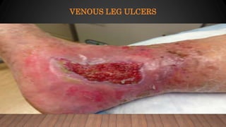
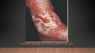
VENOUS ULCER
DR. MD. SHERAJUL ISLAM
FCPS (SURGERY), FACS(USA)
ASSISTANT PROFESSOR, SURGERY
SHEIKH SAYERA KHATUN MEDICAL COLLEGE
ULCER
An ulcer is a discontinuity of an epithelial surfaces due
to sloughing out of inflammatory necrotic tissues
CAUSES OF LOWER LIMB ULCERATION
1. Venous insufficiency (45 -60%)
2. Arterial insufficiency (10 -20%)
3. Diabetes (15 -25%)
4. Vasculitis
5. Haematological disease
CAUSES OF LOWER LIMB ULCERATION
6. Infections
7. Trauma
8. Drugs/therapy: Hydroxy urea
9. Skin conditions: Pyoderma gangrenosum
Necrobiosis lipoidica
10. Malignancies
11. Genetic : Prolidase deficiency
Klinefelter's syndrome
VENOUS LEG ULCERS
VENOUS LEG ULCERS
• Ulcers caused due to venous insufficiency
• Often at lower medial aspect of leg `gaiter region`
• With or without visible varicose veins
NORMAL PHYSIOLOGY
NORMAL PHYSIOLOGY
• Muscle contraction-- deep veins are compressed-- one way valves in
deep system allow the high pressure flow to move against gravity
• One way valves in perforators close --to prevent pressure injury to the
skin
• There is failure of these one way valves in all patients with venous
disease
• Worsened by impairment of leg muscle function or ankle joint range of
motion
INCOMPETENT PERFORATORS AND REFLUX
• Failure of venous valve (REFLUX) and poor calf muscle function
leads to ambulatory venous hypertension and sustained capillary
hypertension
• 60% of venous ulcer patients have isolated superficial vein
insufficiency
INCOMPETENT PERFORATORS AND REFLUX
PATHOGENESIS
1. Capillary stasis:
Stasis of venous blood in post-thrombotic syndrome gave rise to anoxia and
hence venous ulcers(`Homans` )
2. Fibrin cuff theory:
Venous ulcer could be result of deposition of pericapillary fibrin due to
leakage of fibrinogen through pericapillary spaces (Browse and Bermand)
fibrinogen polymerizes to form fibrin
Oxygen diffusion barrier
Increase in collagen IV in and around capillary
PATHOGENESIS
3. White cell trapping:
Leukocytes may become trapped in capillaries in static blood, obstructing
the flow(Coleridge and Smith)
4. Trapping growth factors:
Pericapillary fibrin cuff , α-2 macroglobulin interfere with growth factor
transport (`Felanga and Eaglestein)
PATHOGENESIS
5. Multicausal model/ Maastricht model:
Elevated pressures on vascular side of capillaries lead to structural
changes in capillary wall
Inter endothelial space broadens
Collagen IV layer disintegrates
Capillary wall becomes thicker
Water diffusion is affected- oedema
Larger molecules (fibrin ) escape- fibrin cuff formation
α-2 macroglobulin escapes and traps TGF-β
CAUSES OF VENOUS ULCER
SIGNS AND SYMPTOMS
• Patchy erythema or discoloration of an intense bluish red colour
(capillary congestion)
• Ischemia of the skin leads to necrosis, often following a minor episode
of trauma (scratching, small knocks, dermatitis)
• Ulcer is typically painless
• Patients develop typical ischemic pain on elevation of the ulcerated
leg, which is oedematous
SIGNS AND SYMPTOMS
• Associated with arterial disease in1/3rd of cases
• Ulcer is covered with yellowish exudate over granulation tissue
• Healing ulcers have a shallow sloping edge with healthy granulation
tissue in their base and little slough
• Pink lip of epithelium at the edge of ulcer is uniform and supplied by
relatively uncongested capillaries
VENOUS ULCER AT INNER ASPECT OF LEG
A NON HEALING ULCER WITH A THICK FIBROUS BASE WITH NECROTIC
TURGID EDGE
SIGNS AND SYMPTOMS
Signs of venous hypertension are present-
LDS
varicose veins
varicose eczema
oedema
An oedematous leg not responding to diuretics is a strong clue to the
diagnosis
LDS- LIPODERMATOSLEROSIS
COMPLICATIONS:
• 1)Infection- >105 /cm2 tissue
S. aureus
GAB Hemolytic streptococcus
Candida
• 2)Haemorrhage
• 3)Lymphoedema
• 4)Malignant change
• 5)Subcutaneous calcification
• 6)Bone changes
DIAGNOSIS
Clinical:
Gaiter area
Signs of venous hypertension
Past history of venous thrombosis
Past history of treatment for varicose veins
Family history of venous disease
Colour Doppler duplex USG: usually, after ulcer heals
Confirm venous reflux, superficial venous incompetence
Confirm deep venous compliance
DIAGNOSIS
Plethysmography:
To investigate calf muscle pump function when Colour Duplex is
normal
Skin biopsy: not indicated in venous ulcer
Skin malignancy or vasculitis suspected
Arterial disease must be excluded by
ABPI
Arterial duplex doppler scanning
arteriography
MANAGEMENT
Done by the following means:-
• 1. CONSERVATIVE TREATMENT
• 2. SYSTEMIC MEDICATIONS
• 3. SURGICAL OPTIONS
The goals of treatment are:
.To reduce edema
.Improve ulcer healing
. Prevention of recurrence
COMPRESSION THERAPY
• Contraindications to compression therapy include
Clinically significant arterial disease
Uncompensated heart failure
• Methods include
1. Inelastic
2. Elastic
3. Intermittent pneumatic compression
COMPRESSION THERAPY
• • Compression therapy is the standard of care for venous ulcers and
chronic venous insufficiency
• • Compression therapy reduces edema, improves venous reflux,
enhances healing of ulcers, and reduces pain
• • After an ulcer has healed, lifelong maintenance of compression
therapy reduce the risk of recurrence
1. INELASTIC COMPRESSION :
• Inelastic compression therapy provides high working pressure during
ambulation and muscle contraction, but no resting pressure
• The most common method of inelastic compression therapy is the
Unna boot, a zinc oxide–impregnated, moist bandage that hardens
after application
1. INELASTIC COMPRESSION :
• Disadvantages:
Because of its inelasticity, the Unna boot does not conform to changes
in leg size and may be uncomfortable to wear
The Unna boot may lead to a foul smell from the accumulation of
exudate from the ulcer, requiring frequent reapplications
UNNA BOOT
2. ELASTIC COMPRESSION :
• Elastic compression therapy methods conform to changes in leg size
and sustain compression during both rest and activity
• Stockings or bandages can be used; however, elastic wraps are not
recommended because they do not provide enough pressure
• Compression stockings are removed at night and should be replaced
every six months because they lose pressure with regular washing
2. ELASTIC COMPRESSION :
• Elastic bandages are alternatives to compression stockings (multilayer
bandages are more effective than single layer)
• Disadvantages:
Multilayer compression bandages require skilled application in the
physician’s office one or two times per week
Depending on drainage
ELASTIC COMPRESSION
3. INTERMITTENT PNEUMATIC COMPRESSION:
• Comprises a pump that delivers air to inflatable and deflatable
sleeves that embrace extremities, providing intermittent compression
• Generally reserved for bedridden patients who cannot tolerate
continuous compression therapy
• Disadvantages : Expensive
Requires immobilization of the patient
INTERMITTENT PNEUMATIC COMPRESSION
LEG ELEVATION
• Leg elevation requires raising lower extremities above the level of the
heart
• Aim of leg elevation:
Reducing edema
Improving microcirculation and oxygen delivery
Hastening ulcer healing
DRESSINGS
• Dressings are often used under compression bandages to promote
faster healing and prevent adherence of the bandage to the ulcer
• Treament of underlying cause of eczema:
Varicose
Contact allergy
Contact irritant
Emollients , Steroids if inflammed
• Cleansing and debridement:
1. Irrigation of ulcer with warm tap water, sterile saline
2. Debridement improves wound healing
SYSTEMIC THERAPY
• Antibiotics:- only used if there is clear evidence of infection
• Pentoxyphylline:-
Fibrinolytic
Reduction in leukocyte adhesion
Dose of 400 to 800mgs TDS
• Oral enteric coated aspirin:- 300mg
SYSTEMIC THERAPY
• Flavonoid drugs (e.g. oxerutins)
• Daflon 500
• Stanazolol:
androgenic steroid with fibrinolytic property.
Improves LDS
• Iloprost infusion (vasodilator that inhibits platelet aggregation)
• Sodium dobesilate:- 500 mgs BD
TYPES OF DEBRIDEMENT
• I. SHARP DEBRIDEMENT
• II. MECHANICAL DEBRIDEMENT
• III.AUTOLYTIC DEBRIDEMENT
• IV.ENZYMATIC DEBRIEMENT
• V. BIOLOGICAL DEBRIDEMENT
DRESSING AND TOPICAL THERAPIES
Should keep ulcer moist not wet
Simple ,low adherent
Left undisturbed as long as possible
`strike through` of exudate to outside of the bandage is indication
for change
DRESSING AND TOPICAL THERAPIES
1.Knitted viscose primary dressings + superimposed absorbent pad
(secondary dressing)
2. Hydrocolloid dressing- dry sloughy wounds to reduce pain
3. Absorptive dressing (alginate, foam, hydrofibre) – highly exuding
wound
4. Zinc paste bandage (unna boot)
SURGICAL TREATMENT
• Between 50-70 % of ulcers heal at 3 months
80-90% by 12 months
• 50% overall recurrence rate by 5-7 yrs. mostly in post thrombotic limbs
Surgical ligation of saphenous vein and incompetent communicating veins is `no
better than` stanozolol and stockings in preventing ulcer
Various procedures used:
• I. Ligation and stripping of saphenous veins
• II. Compressive
• III. Complete extirpation of the communicating veins `feeding` the ulcer
SURGICAL TREATMENT
• IV. Deep vein bypass
• V. Valvuloplasty
• VI. Brachial valve transplant
• Shave therapy:
Excision of ulcer and surrounding LDS followed by meshed split
skin graft
Heals 88% of ulcers
SURGICAL TREATMENT
• Skin grafting
Punch grafting
Split skin grafts
Mesh grafts
• Subfascial endoscopic perforator surgery (SEPS)
• •Improves healing rates, and recurrence
LIFELONG: COMPRESSION THERAPY
• After healing of ulcer, fit for custom stockings
• Remove and bathe each evening, apply moisturizer
• Each morning put on to prevent edema
• Pt should purchase in pairs of two, replace every 6 months
ASSOCIATIONS & COMPLICATION OF VENOUS ULCER
1. General disease
Obesity
Hypertension
Cardiovascular diseases
2. Anaemia, hypoproteinemia
3. Depression
4. Inverted foot, equinus ,calf muscle atrophy
5. Zinc depletion
ASSOCIATIONS & COMPLICATION OF VENOUS ULCER
6. Infections:
staph. Aureus
groupA β- haemolytic streptococci
pseudomonas
candida albicans
7. Contact dermatitis
8. haemorrhage
ASSOCIATIONS & COMPLICATION OF VENOUS ULCER
• 9. Lymphoedema
• 10.Malignant change
• 11.Sub cutaneous calfication
• 12.Bone changes
Venous ulcer for MBBS

Venous ulcer for MBBS

  • 1.
    VENOUS ULCER DR. MD.SHERAJUL ISLAM FCPS (SURGERY), FACS(USA) ASSISTANT PROFESSOR, SURGERY SHEIKH SAYERA KHATUN MEDICAL COLLEGE
  • 2.
    ULCER An ulcer isa discontinuity of an epithelial surfaces due to sloughing out of inflammatory necrotic tissues
  • 3.
    CAUSES OF LOWERLIMB ULCERATION 1. Venous insufficiency (45 -60%) 2. Arterial insufficiency (10 -20%) 3. Diabetes (15 -25%) 4. Vasculitis 5. Haematological disease
  • 4.
    CAUSES OF LOWERLIMB ULCERATION 6. Infections 7. Trauma 8. Drugs/therapy: Hydroxy urea 9. Skin conditions: Pyoderma gangrenosum Necrobiosis lipoidica 10. Malignancies 11. Genetic : Prolidase deficiency Klinefelter's syndrome
  • 5.
  • 6.
    VENOUS LEG ULCERS •Ulcers caused due to venous insufficiency • Often at lower medial aspect of leg `gaiter region` • With or without visible varicose veins
  • 7.
  • 8.
    NORMAL PHYSIOLOGY • Musclecontraction-- deep veins are compressed-- one way valves in deep system allow the high pressure flow to move against gravity • One way valves in perforators close --to prevent pressure injury to the skin • There is failure of these one way valves in all patients with venous disease • Worsened by impairment of leg muscle function or ankle joint range of motion
  • 9.
    INCOMPETENT PERFORATORS ANDREFLUX • Failure of venous valve (REFLUX) and poor calf muscle function leads to ambulatory venous hypertension and sustained capillary hypertension • 60% of venous ulcer patients have isolated superficial vein insufficiency
  • 10.
  • 11.
    PATHOGENESIS 1. Capillary stasis: Stasisof venous blood in post-thrombotic syndrome gave rise to anoxia and hence venous ulcers(`Homans` ) 2. Fibrin cuff theory: Venous ulcer could be result of deposition of pericapillary fibrin due to leakage of fibrinogen through pericapillary spaces (Browse and Bermand) fibrinogen polymerizes to form fibrin Oxygen diffusion barrier Increase in collagen IV in and around capillary
  • 12.
    PATHOGENESIS 3. White celltrapping: Leukocytes may become trapped in capillaries in static blood, obstructing the flow(Coleridge and Smith) 4. Trapping growth factors: Pericapillary fibrin cuff , α-2 macroglobulin interfere with growth factor transport (`Felanga and Eaglestein)
  • 13.
    PATHOGENESIS 5. Multicausal model/Maastricht model: Elevated pressures on vascular side of capillaries lead to structural changes in capillary wall Inter endothelial space broadens Collagen IV layer disintegrates Capillary wall becomes thicker Water diffusion is affected- oedema Larger molecules (fibrin ) escape- fibrin cuff formation α-2 macroglobulin escapes and traps TGF-β
  • 14.
  • 16.
    SIGNS AND SYMPTOMS •Patchy erythema or discoloration of an intense bluish red colour (capillary congestion) • Ischemia of the skin leads to necrosis, often following a minor episode of trauma (scratching, small knocks, dermatitis) • Ulcer is typically painless • Patients develop typical ischemic pain on elevation of the ulcerated leg, which is oedematous
  • 17.
    SIGNS AND SYMPTOMS •Associated with arterial disease in1/3rd of cases • Ulcer is covered with yellowish exudate over granulation tissue • Healing ulcers have a shallow sloping edge with healthy granulation tissue in their base and little slough • Pink lip of epithelium at the edge of ulcer is uniform and supplied by relatively uncongested capillaries
  • 18.
    VENOUS ULCER ATINNER ASPECT OF LEG
  • 19.
    A NON HEALINGULCER WITH A THICK FIBROUS BASE WITH NECROTIC TURGID EDGE
  • 20.
    SIGNS AND SYMPTOMS Signsof venous hypertension are present- LDS varicose veins varicose eczema oedema An oedematous leg not responding to diuretics is a strong clue to the diagnosis
  • 21.
  • 22.
    COMPLICATIONS: • 1)Infection- >105/cm2 tissue S. aureus GAB Hemolytic streptococcus Candida • 2)Haemorrhage • 3)Lymphoedema • 4)Malignant change • 5)Subcutaneous calcification • 6)Bone changes
  • 23.
    DIAGNOSIS Clinical: Gaiter area Signs ofvenous hypertension Past history of venous thrombosis Past history of treatment for varicose veins Family history of venous disease Colour Doppler duplex USG: usually, after ulcer heals Confirm venous reflux, superficial venous incompetence Confirm deep venous compliance
  • 24.
    DIAGNOSIS Plethysmography: To investigate calfmuscle pump function when Colour Duplex is normal Skin biopsy: not indicated in venous ulcer Skin malignancy or vasculitis suspected Arterial disease must be excluded by ABPI Arterial duplex doppler scanning arteriography
  • 25.
    MANAGEMENT Done by thefollowing means:- • 1. CONSERVATIVE TREATMENT • 2. SYSTEMIC MEDICATIONS • 3. SURGICAL OPTIONS The goals of treatment are: .To reduce edema .Improve ulcer healing . Prevention of recurrence
  • 26.
    COMPRESSION THERAPY • Contraindicationsto compression therapy include Clinically significant arterial disease Uncompensated heart failure • Methods include 1. Inelastic 2. Elastic 3. Intermittent pneumatic compression
  • 27.
    COMPRESSION THERAPY • •Compression therapy is the standard of care for venous ulcers and chronic venous insufficiency • • Compression therapy reduces edema, improves venous reflux, enhances healing of ulcers, and reduces pain • • After an ulcer has healed, lifelong maintenance of compression therapy reduce the risk of recurrence
  • 28.
    1. INELASTIC COMPRESSION: • Inelastic compression therapy provides high working pressure during ambulation and muscle contraction, but no resting pressure • The most common method of inelastic compression therapy is the Unna boot, a zinc oxide–impregnated, moist bandage that hardens after application
  • 29.
    1. INELASTIC COMPRESSION: • Disadvantages: Because of its inelasticity, the Unna boot does not conform to changes in leg size and may be uncomfortable to wear The Unna boot may lead to a foul smell from the accumulation of exudate from the ulcer, requiring frequent reapplications
  • 30.
  • 31.
    2. ELASTIC COMPRESSION: • Elastic compression therapy methods conform to changes in leg size and sustain compression during both rest and activity • Stockings or bandages can be used; however, elastic wraps are not recommended because they do not provide enough pressure • Compression stockings are removed at night and should be replaced every six months because they lose pressure with regular washing
  • 32.
    2. ELASTIC COMPRESSION: • Elastic bandages are alternatives to compression stockings (multilayer bandages are more effective than single layer) • Disadvantages: Multilayer compression bandages require skilled application in the physician’s office one or two times per week Depending on drainage
  • 33.
  • 35.
    3. INTERMITTENT PNEUMATICCOMPRESSION: • Comprises a pump that delivers air to inflatable and deflatable sleeves that embrace extremities, providing intermittent compression • Generally reserved for bedridden patients who cannot tolerate continuous compression therapy • Disadvantages : Expensive Requires immobilization of the patient
  • 36.
  • 37.
    LEG ELEVATION • Legelevation requires raising lower extremities above the level of the heart • Aim of leg elevation: Reducing edema Improving microcirculation and oxygen delivery Hastening ulcer healing
  • 38.
    DRESSINGS • Dressings areoften used under compression bandages to promote faster healing and prevent adherence of the bandage to the ulcer
  • 39.
    • Treament ofunderlying cause of eczema: Varicose Contact allergy Contact irritant Emollients , Steroids if inflammed • Cleansing and debridement: 1. Irrigation of ulcer with warm tap water, sterile saline 2. Debridement improves wound healing
  • 40.
    SYSTEMIC THERAPY • Antibiotics:-only used if there is clear evidence of infection • Pentoxyphylline:- Fibrinolytic Reduction in leukocyte adhesion Dose of 400 to 800mgs TDS • Oral enteric coated aspirin:- 300mg
  • 41.
    SYSTEMIC THERAPY • Flavonoiddrugs (e.g. oxerutins) • Daflon 500 • Stanazolol: androgenic steroid with fibrinolytic property. Improves LDS • Iloprost infusion (vasodilator that inhibits platelet aggregation) • Sodium dobesilate:- 500 mgs BD
  • 42.
    TYPES OF DEBRIDEMENT •I. SHARP DEBRIDEMENT • II. MECHANICAL DEBRIDEMENT • III.AUTOLYTIC DEBRIDEMENT • IV.ENZYMATIC DEBRIEMENT • V. BIOLOGICAL DEBRIDEMENT
  • 43.
    DRESSING AND TOPICALTHERAPIES Should keep ulcer moist not wet Simple ,low adherent Left undisturbed as long as possible `strike through` of exudate to outside of the bandage is indication for change
  • 44.
    DRESSING AND TOPICALTHERAPIES 1.Knitted viscose primary dressings + superimposed absorbent pad (secondary dressing) 2. Hydrocolloid dressing- dry sloughy wounds to reduce pain 3. Absorptive dressing (alginate, foam, hydrofibre) – highly exuding wound 4. Zinc paste bandage (unna boot)
  • 45.
    SURGICAL TREATMENT • Between50-70 % of ulcers heal at 3 months 80-90% by 12 months • 50% overall recurrence rate by 5-7 yrs. mostly in post thrombotic limbs Surgical ligation of saphenous vein and incompetent communicating veins is `no better than` stanozolol and stockings in preventing ulcer Various procedures used: • I. Ligation and stripping of saphenous veins • II. Compressive • III. Complete extirpation of the communicating veins `feeding` the ulcer
  • 46.
    SURGICAL TREATMENT • IV.Deep vein bypass • V. Valvuloplasty • VI. Brachial valve transplant • Shave therapy: Excision of ulcer and surrounding LDS followed by meshed split skin graft Heals 88% of ulcers
  • 47.
    SURGICAL TREATMENT • Skingrafting Punch grafting Split skin grafts Mesh grafts • Subfascial endoscopic perforator surgery (SEPS) • •Improves healing rates, and recurrence
  • 48.
    LIFELONG: COMPRESSION THERAPY •After healing of ulcer, fit for custom stockings • Remove and bathe each evening, apply moisturizer • Each morning put on to prevent edema • Pt should purchase in pairs of two, replace every 6 months
  • 49.
    ASSOCIATIONS & COMPLICATIONOF VENOUS ULCER 1. General disease Obesity Hypertension Cardiovascular diseases 2. Anaemia, hypoproteinemia 3. Depression 4. Inverted foot, equinus ,calf muscle atrophy 5. Zinc depletion
  • 50.
    ASSOCIATIONS & COMPLICATIONOF VENOUS ULCER 6. Infections: staph. Aureus groupA β- haemolytic streptococci pseudomonas candida albicans 7. Contact dermatitis 8. haemorrhage
  • 51.
    ASSOCIATIONS & COMPLICATIONOF VENOUS ULCER • 9. Lymphoedema • 10.Malignant change • 11.Sub cutaneous calfication • 12.Bone changes