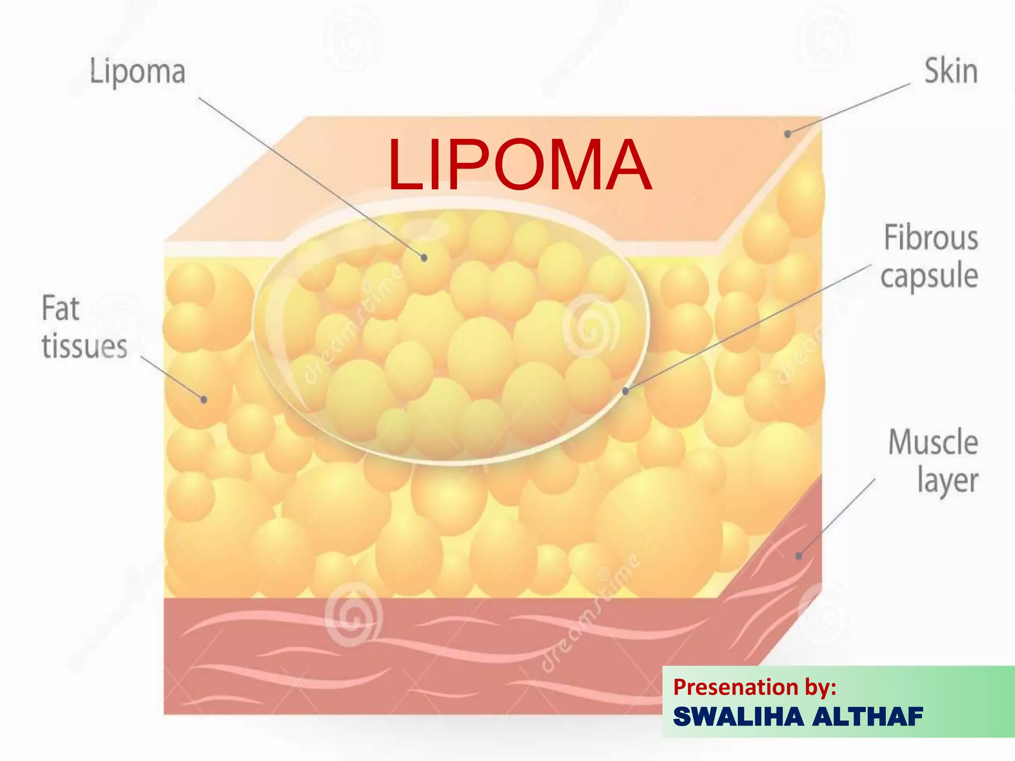
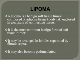
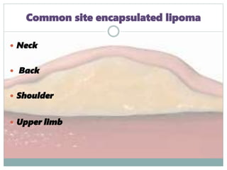
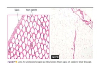
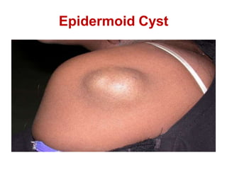
LIPOMA
Presenation by:
SWALIHA ALTHAF
OUTLINE
INTRODUCTION
EPIDEMIOLOGY
AETIOLOGY
CLASSIFICATION
CLINICAL FEATURES
DIFFERENTIALS
COMPLICATIONS
TREATMENT MODALITIES
INTRODUCTION
• Tumor (neoplasia)
• A tumor is an abnormal mass of tissues
the growth of which exceed and
uncoordinated with that of normal
tissues and persist in the same excessive
manner after cessation f stimuli which
evoked the changes , which is virtually
autonomous, preys on host.
A lipoma is a benign soft tissue tumor
composed of adipose tissue (body fat) enclosed
in a capsule of connective tissue.
It is the most common benign form of soft
tissue tumor.
It may be arranged in lobules separated by
fibrous septa.
It may also become pedunculated.
TYPES
According to presence of capsule
According to anatomical location
According to histology-
•Lipoma
•Fibrolipoma
• Neurolipoma
•Naeveolipoma
Encapsulated lipoma
 Diffuse lipoma
ACCORDING TO ANATOMICAL LOCATION
Subcutaneous
 Subfascial
Subserosal
 Intramuscular
Subsynovial
Intraarticular
 Pleural form – Lipomatosis
 Also called as universal tumor – because it can
occur in all part of body.
It occur in all part except :
 Tip of nose
 Cartilaginous part of ear
 Shaft of penis
 Eyelid
Common site encapsulated lipoma
 Neck
 Back
 Shoulder
 Upper limb
 Lipomas are commonly found in adults from 40 to 60
years of age but can also be found in younger adults and
children.
Causes of Lipomas
The tendency to develop a lipoma is not necessarily
hereditary although hereditary conditions, such as
familial multiple lipomatosis, may include lipoma
development.
Gardenerssyndrome
 Dercums’ syndrome
Cases have been reported where minor injuries are
alleged to have triggered the growth of a lipoma,
called “post traumatic lipomas”.However, the link
between trauma and the development of lipomas is
controversial.
Classification
• Based on location
Superficial subcutaneous lipomas:
The most common type of lipoma, They lie just below the
surface of theskin. Most occur on thetrunk, thigh, and
forearm, although they may be found anywhere in the body
where fat islocated.
 Adenolipomas:
Are lipomas associated with endocrine sweat
glands.
 Angiolipoleiomyomas :
Are acquired, solitary, asymptomatic acral nodules,
characterized histologically by well-circumscribed
subcutaneous tumors composed of smooth muscle
cells, blood vessels, connective tissue, and fat.
 Angiolipomas :
Painful subcutaneous nodules having all other
features of a typical lipoma.
Cerebellar pontine angle and internal auditory canal
lipomas.
Chondroid lipomas:
Are deep-seated,firm,yellow tumors that characteristically
occur on the legs of women.
Corpus callosum lipoma:
Is a rare congenital brain condition that may or may not
present with symptoms. This occurs in the corpus
callosum, also known as the colossal commissure, which is
a wide, flat bundle of neural fibers beneath the cortex in
the human brain.
Hibernomas :
Are lipoma of brownfat.
Types
Encapsulated s/c lipoma
Diffuse variety
Multiple lipomas- Dercum’s
Histologicaltypes
• Fibrolipoma
• Neurolipoma
• Naevolipoma
HISTORY
History-taking is guided by the anatomical location
of the lesion.
Questions should explore factors such as:
When the lump was first noticed
What brought the lump to the attention of the
patient
The symptoms that are related to thelump
Changes that have occurred to the lump since
it first appeared
Whether the lump ever disappears and what
causes it to reappear
Whether the patient ever had any other
lumps and what they were like
Whether there has been any loss of body
weight
Whether the lump has been treated
before and has recurred.
CLINICAL FEATURES
Most lipomas are small (under one centimeter
diameter) but can enlarge to sizes greater than six
centimeters.
Localized
Lobular
Fluctuant
Mobile
 Exhibit “Slip sign”.
(They move easily when pressure is placed on them)
Skin free
Soft
Onexamination they do not exhibit differential
warmth.
Lipomas areusually painless soft and non tender.
Diagnosis
This is usually done clinically. Any doubt about the
diagnosis calls for immediate refferall to a
dermatologist.
Ancillary investigations include:-
 Pre-operative radiography
DIAGNOSIS
 Both ultrasound and magnetic resonance imaging
have been used with some success to differentiate
lipomasand liposarcomas but are not entirely
reliable.
CT scan are occasionally required.
Alternatively, fine-needle aspiration may be used
to evaluate suspicious lesions
Differential Diagnosis
• These include but are not limited to:-
Fibrosarcomas
Abcesses (Localized)
Cold abcesses
Neurofibromas
Hernias
Pappiloma
Sebaceous cysts (contain sebum affect the
s.glands)
Epidermoid cysts (contain keratin and fat)
Nodular fascitis
Erythema nodosum
Nodular subcutaneous fat necrosis
Haematoma
Sebaceous cysts
Draining Sebum
Epidermoid Cyst
Myxomatous degeneration
Saponification
Calcification
Infection
Ulceration
Intussusception & intestinal obstruction
Some sources claim that malignant
transformation can occur while others say this
has yet to be convincingly documented.
Treatment of Lipomas
 Indications for Treatment
Usually, treatment of a lipoma is not necessary, unless the
tumor becomes painful or restrictsmovement.
 They are usually removed for cosmeticreasons
However reasons to remove lipomas include when they
grow very large, or for histopathology to check that they
are not a more dangerous type of tumor such as a lipo-
sarcoma. This last point can be important asthe actual
characteristics of a “lump" is not known until after it is
removed and medicallyexamined.
This malignancy is rare but can be found in a lesion
with the clinical appearance of a lipoma.
Liposarcoma presents in a fashion similar to that of
a lipoma and appears to be more common in the
retro peritoneum, on the shoulders and lower
extremities.
Hence some recommend an immediate and
complete excision of a lipoma with subsequent
histologic studies to exclude a possible
Liposarcoma,
Liposarcoma
Suspicious Signs that warrant immediate
removal
If the lump suddenly starts to grow verylarge
Greater than 5cm in diameter
Located in the extremities, retroperitoneally, in the
groin, in the scrotum or in the abdominalwall
Deep (beneath or fixed to superficial fascia)
Exhibiting malignant behaviour (invasion into nerve or
bone)
Surgical excision of Lipomas
 They can be left alone. They may need to be removed
for cosmetic reasons, because of compression of
surrounding structures or if the diagnosis is uncertain
 Lipomas are normally removed by simple excision. The removal
can often be done under local anaesthetic, and takes fewer than
30 minutes. This cures the great majority of cases, with about 1–
2% of lipomas recurring after excision.
 Because lipomas generally do not infiltrate into surrounding
tissue, they can usually be shelled out easily duringexcision.
 Minimal scarring can be achieved with a technique called
segmental extraction - a small stab incision followed by blind
dissection of the lipoma and extraction in a segmental fashion
 Liposuction is another option if the lipoma is softand
has a small connective tissue component.
 Liposuction typically results in less scarring; however,
with large lipomas it may fail to remove the entire
tumor, which can lead to re-growth.
 New methods under development are supposed to
remove the lipomas without scarring. One isremoval by
injecting compounds that trigger lipolysis, suchas
steroids orphosphatidylcholine.
Lipoma

Lipoma

  • 1.
  • 2.
  • 3.
    INTRODUCTION • Tumor (neoplasia) •A tumor is an abnormal mass of tissues the growth of which exceed and uncoordinated with that of normal tissues and persist in the same excessive manner after cessation f stimuli which evoked the changes , which is virtually autonomous, preys on host.
  • 4.
    A lipoma isa benign soft tissue tumor composed of adipose tissue (body fat) enclosed in a capsule of connective tissue. It is the most common benign form of soft tissue tumor. It may be arranged in lobules separated by fibrous septa. It may also become pedunculated.
  • 5.
    TYPES According to presenceof capsule According to anatomical location According to histology- •Lipoma •Fibrolipoma • Neurolipoma •Naeveolipoma
  • 6.
    Encapsulated lipoma  Diffuselipoma ACCORDING TO ANATOMICAL LOCATION Subcutaneous  Subfascial Subserosal  Intramuscular Subsynovial Intraarticular
  • 7.
     Pleural form– Lipomatosis  Also called as universal tumor – because it can occur in all part of body. It occur in all part except :  Tip of nose  Cartilaginous part of ear  Shaft of penis  Eyelid
  • 8.
    Common site encapsulatedlipoma  Neck  Back  Shoulder  Upper limb
  • 9.
     Lipomas arecommonly found in adults from 40 to 60 years of age but can also be found in younger adults and children. Causes of Lipomas The tendency to develop a lipoma is not necessarily hereditary although hereditary conditions, such as familial multiple lipomatosis, may include lipoma development. Gardenerssyndrome
  • 10.
     Dercums’ syndrome Caseshave been reported where minor injuries are alleged to have triggered the growth of a lipoma, called “post traumatic lipomas”.However, the link between trauma and the development of lipomas is controversial.
  • 11.
    Classification • Based onlocation Superficial subcutaneous lipomas: The most common type of lipoma, They lie just below the surface of theskin. Most occur on thetrunk, thigh, and forearm, although they may be found anywhere in the body where fat islocated.  Adenolipomas: Are lipomas associated with endocrine sweat glands.
  • 12.
     Angiolipoleiomyomas : Areacquired, solitary, asymptomatic acral nodules, characterized histologically by well-circumscribed subcutaneous tumors composed of smooth muscle cells, blood vessels, connective tissue, and fat.  Angiolipomas : Painful subcutaneous nodules having all other features of a typical lipoma.
  • 13.
    Cerebellar pontine angleand internal auditory canal lipomas. Chondroid lipomas: Are deep-seated,firm,yellow tumors that characteristically occur on the legs of women. Corpus callosum lipoma: Is a rare congenital brain condition that may or may not present with symptoms. This occurs in the corpus callosum, also known as the colossal commissure, which is a wide, flat bundle of neural fibers beneath the cortex in the human brain. Hibernomas : Are lipoma of brownfat.
  • 15.
    Types Encapsulated s/c lipoma Diffusevariety Multiple lipomas- Dercum’s
  • 16.
  • 18.
    HISTORY History-taking is guidedby the anatomical location of the lesion. Questions should explore factors such as: When the lump was first noticed What brought the lump to the attention of the patient The symptoms that are related to thelump Changes that have occurred to the lump since it first appeared
  • 19.
    Whether the lumpever disappears and what causes it to reappear Whether the patient ever had any other lumps and what they were like Whether there has been any loss of body weight Whether the lump has been treated before and has recurred.
  • 20.
    CLINICAL FEATURES Most lipomasare small (under one centimeter diameter) but can enlarge to sizes greater than six centimeters. Localized Lobular Fluctuant Mobile
  • 21.
     Exhibit “Slipsign”. (They move easily when pressure is placed on them) Skin free Soft Onexamination they do not exhibit differential warmth. Lipomas areusually painless soft and non tender.
  • 22.
    Diagnosis This is usuallydone clinically. Any doubt about the diagnosis calls for immediate refferall to a dermatologist. Ancillary investigations include:-  Pre-operative radiography
  • 23.
    DIAGNOSIS  Both ultrasoundand magnetic resonance imaging have been used with some success to differentiate lipomasand liposarcomas but are not entirely reliable. CT scan are occasionally required. Alternatively, fine-needle aspiration may be used to evaluate suspicious lesions
  • 24.
    Differential Diagnosis • Theseinclude but are not limited to:- Fibrosarcomas Abcesses (Localized) Cold abcesses Neurofibromas Hernias Pappiloma
  • 25.
    Sebaceous cysts (containsebum affect the s.glands) Epidermoid cysts (contain keratin and fat) Nodular fascitis Erythema nodosum Nodular subcutaneous fat necrosis Haematoma
  • 26.
  • 27.
  • 28.
  • 29.
    Myxomatous degeneration Saponification Calcification Infection Ulceration Intussusception &intestinal obstruction Some sources claim that malignant transformation can occur while others say this has yet to be convincingly documented.
  • 30.
    Treatment of Lipomas Indications for Treatment Usually, treatment of a lipoma is not necessary, unless the tumor becomes painful or restrictsmovement.  They are usually removed for cosmeticreasons However reasons to remove lipomas include when they grow very large, or for histopathology to check that they are not a more dangerous type of tumor such as a lipo- sarcoma. This last point can be important asthe actual characteristics of a “lump" is not known until after it is removed and medicallyexamined.
  • 31.
    This malignancy israre but can be found in a lesion with the clinical appearance of a lipoma. Liposarcoma presents in a fashion similar to that of a lipoma and appears to be more common in the retro peritoneum, on the shoulders and lower extremities. Hence some recommend an immediate and complete excision of a lipoma with subsequent histologic studies to exclude a possible Liposarcoma, Liposarcoma
  • 34.
    Suspicious Signs thatwarrant immediate removal If the lump suddenly starts to grow verylarge Greater than 5cm in diameter Located in the extremities, retroperitoneally, in the groin, in the scrotum or in the abdominalwall Deep (beneath or fixed to superficial fascia) Exhibiting malignant behaviour (invasion into nerve or bone)
  • 35.
    Surgical excision ofLipomas  They can be left alone. They may need to be removed for cosmetic reasons, because of compression of surrounding structures or if the diagnosis is uncertain  Lipomas are normally removed by simple excision. The removal can often be done under local anaesthetic, and takes fewer than 30 minutes. This cures the great majority of cases, with about 1– 2% of lipomas recurring after excision.  Because lipomas generally do not infiltrate into surrounding tissue, they can usually be shelled out easily duringexcision.  Minimal scarring can be achieved with a technique called segmental extraction - a small stab incision followed by blind dissection of the lipoma and extraction in a segmental fashion
  • 37.
     Liposuction isanother option if the lipoma is softand has a small connective tissue component.  Liposuction typically results in less scarring; however, with large lipomas it may fail to remove the entire tumor, which can lead to re-growth.  New methods under development are supposed to remove the lipomas without scarring. One isremoval by injecting compounds that trigger lipolysis, suchas steroids orphosphatidylcholine.