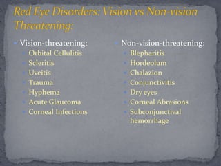
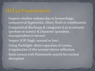
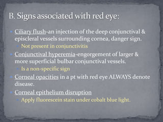
Evaluation of a red eye
@Mojee_Tuapati
 Commonest presentation of various ocular diseases.
 Need to know whether it is a minor eye irritation or a
serious eye disease.
I. Conjunctivitis (Bacterial, Viral, & Allergic)
II. Corneal Ulcer
III. Acute Glaucoma
IV. Acute Iridocyclitis
V. Sub-conjunctival hemorrhage
VI. Episcleritis, Scleritis
VII. Dacryoadenitis & Dacryocystitis
VIII.Eye lid pathology such as style, blepharitis
 Vision-threatening:
 Orbital Cellulitis
 Scleritis
 Uveitis
 Trauma
 Hyphema
 Acute Glaucoma
 Corneal Infections
 Non-vision-threatening:
 Blepharitis
 Hordeolum
 Chalazion
 Conjunctivitis
 Dry eyes
 Corneal Abrasions
 Subconjunctival
hemorrhage
 “Red eye” refers to hyperemia of superficial visible
vessels of the conjunctiva, episclera & sclera.
 Can be caused by disorders of any of the adjoining
structures:
 Conjunctiva-common, often not serious.
 Cornea-common, potentially serious
 Episclera-not common, not serious, usually allergic
 Sclera-not common, may indicate serious systemic
diseases such as Collagen Vascular Disease
 Iris & Ciliary body-serious
 Acute Glaucoma-serious
 Adnexal disease-not serious
 Inspect whether redness due to hemorrhage,
conjunctival hyperemia, ciliary flush or combination.
 Conjunctival discharge & categorize it as to amount
(profuse or scanty) & character (purulent,
mucopurulent or serous)
 Inspect IOP (high, normal or low)
 Using flashlight: detect opacities of cornea,
irregularities of the corneal mirror reflection.
 Stain cornea with fluorescein-search for corneal
disruption
 Estimate depth of anterior chamber (normal or
shallow)
 And detect blood or pus in anterior chamber, if any.
 Detect irregularities of pupil (compare bilaterally)
 Detect limitation of eye movement.
 Blurred vision-that doesn’t subside s upon blinking
 Suggests a serious ocular disease such as an inflamed
cornea, iridicyclitis or glaucoma.
 Doesn’t occur in simple conjunctivitis
 Pain-may indicate keratitis, iridicyclitis, or acute
glaucoma.
 Pts with conjunctivitis complain of scratchiness but not
of severe pain.
 Photophobia-accompanies iritis
 Pts. with conjunctivitis have normal light sensitivity
A. Symptoms associated with a Red eye:
 Halos-usually a sign of corneal edema, often resulting
from an rise in IOP.
 Exudation-a symptom of conjunctival or eyelid
inflammations
 Not in iridocyclitis or glaucoma.
 Corneal ulcer-serious, may be manifested by discharge.
 Itching-suggests allergic conjunctivitis
 Upper respiratory infection & fever may be associated
with viral conjunctivitis. (Adenovirus)
 Ciliary flush-an injection of the deep conjunctival &
episcleral vessels surrounding cornea, danger sign.
 Not present in conjunctivitis
 Conjunctival hyperemia-engorgement of larger &
more superficial bulbar conjunctival vessels.
 Is a non-specific sign
 Corneal opacities in a pt with red eye ALWAYS denote
disease.
 Corneal epithelium disruption
 Apply fluorescein stain under cobalt blue light.
 Pupil size & shape abnormalities.
 In iridicyclitis-smaller than fellow eye ‘cause of reflex
spasm of iris sphincter muscle; distorted also by
posterior synachiae
 In acute glaucoma-partially dilated & may not be quite
round.
 Conjunctivitis does NOT affect pupil.
 Shallow Ant Chamber:
 Suggests possibility of acute glaucoma(closure).
 IOP-to rule out glaucoma in any red eye without
obvious infection
 Tonometer be cleaned afterward to prevent office
transmission.
 Sudden proptosis(forward displacement):
 Looking down at pts from above.
 Common cause is thyroid disease
 Discharge:
 Purulent(creamy white) or mucopurulent (yellowish)
suggests bacterial etiology.
 Serous (watery, clear or yellow-tinged) suggests a viral
etiology
 Scanty, white, stringy exudate sometimes occurs in
allergic conjunctivitis.
 Smears of exudate or conjunctival scrapings:
 PMNs & bacteria in bacterial conjunctivitis
 Eosinophils in allergic conjunctivitis.
 Cultures for bacteria & sensitivity determination (to
Antibiotics)
 Most cases of conjunctivitis are managed without
laboratory assistance.
 Cases of presumed bacterial conjunctivitis, which do
not improve in two days with antibiotic treatment,
should be referred to an ophthalmologist for
confirmation of diagnosis & appropriate studies.
 A. Prolonged use of topical anesthetic:
 Inhibit growth & healing of corneal epithelium
 May cause severe allergic reactions.
 It eliminates the protective blink reflex, thus exposing
cornea to dehydration & injury
 B. Topical corticosteroids serious side effects:
 Herpes Simplex keratitis & fungal keratitis, both
potentiated by corticosteroids.
 Causes formation of cataracts.
 Use for 2-6weeks may cause an elevation of IOP.
 PC:
 Ask abt main complains & list in chronological order.
 HPC:
 If symptom were sudden/gradual
 Severity of symptom
 Check for common symptom:
 Visual loss/impairment
 Visual field defects
 Dazzling
 Pain/Foreign body sensation
 Discomfort/itchy/Dry eyes
 Discharge/Epiphora
 Diplopia
 Photophobia/Haloes/Floaters
 POH:
 Past & present eye problems as well as any past eye
operations
 List drugs of treatment used then.
 PMH:
 Relevant systemic medical diseases e.g. DM, HTN,
leprosy, hyperthyroidism, arthritis, cancers, etc.
 DH:
 List current drugs used for treatment of:
 Existing medical conditions
 Eye conditions
 Allergies:
 Food(s)? Drug(s)?
 Have asthma?
 Caution with treating asthmatics with anti-glaucoma beta-
blockers such as Timoptol
 FH:
 Some eye diseases run in families:
 Myopia
 Glaucoma
 Retinitis pigmentosa ,etc.
 Occupation:
 Help decide what level of vision the pt needs for work.
 Visual Acuity
 Examination of external eye & ocular adnexia
 Ocular alignment & motility exam
 Pupillary examination
 Examination of anterior segment-penlight
 Visual field examination-via
 Examination of posterior segment via fundoscopy
 Macular function test
 Tonometry to determine IOP
 Visual pathway examinations
 Snellen chart at 6meters
 If top can’t be discerned:
 Test done closer to the chart.
 If chart can’t be read at 1m:
 Pts may be asked to count fingers
 If can’t:
 Asked to detect hand movements
 If can’t:
 Asked to perceive only light (LP)
 To achieve optimal visual acuity, the pt should be
asked to look through pinhole.
 www.uptodate.com
 Monograph, by Dr. Ernest Than Tun Oo
 ABC of Eyes, 4th Ed., PT Khaw

Red eye evaluation

  • 1.
    Evaluation of ared eye @Mojee_Tuapati
  • 2.
     Commonest presentationof various ocular diseases.  Need to know whether it is a minor eye irritation or a serious eye disease.
  • 3.
    I. Conjunctivitis (Bacterial,Viral, & Allergic) II. Corneal Ulcer III. Acute Glaucoma IV. Acute Iridocyclitis V. Sub-conjunctival hemorrhage VI. Episcleritis, Scleritis VII. Dacryoadenitis & Dacryocystitis VIII.Eye lid pathology such as style, blepharitis
  • 4.
     Vision-threatening:  OrbitalCellulitis  Scleritis  Uveitis  Trauma  Hyphema  Acute Glaucoma  Corneal Infections  Non-vision-threatening:  Blepharitis  Hordeolum  Chalazion  Conjunctivitis  Dry eyes  Corneal Abrasions  Subconjunctival hemorrhage
  • 5.
     “Red eye”refers to hyperemia of superficial visible vessels of the conjunctiva, episclera & sclera.  Can be caused by disorders of any of the adjoining structures:  Conjunctiva-common, often not serious.  Cornea-common, potentially serious  Episclera-not common, not serious, usually allergic  Sclera-not common, may indicate serious systemic diseases such as Collagen Vascular Disease  Iris & Ciliary body-serious  Acute Glaucoma-serious  Adnexal disease-not serious
  • 6.
     Inspect whetherredness due to hemorrhage, conjunctival hyperemia, ciliary flush or combination.  Conjunctival discharge & categorize it as to amount (profuse or scanty) & character (purulent, mucopurulent or serous)  Inspect IOP (high, normal or low)  Using flashlight: detect opacities of cornea, irregularities of the corneal mirror reflection.  Stain cornea with fluorescein-search for corneal disruption
  • 7.
     Estimate depthof anterior chamber (normal or shallow)  And detect blood or pus in anterior chamber, if any.  Detect irregularities of pupil (compare bilaterally)  Detect limitation of eye movement.
  • 8.
     Blurred vision-thatdoesn’t subside s upon blinking  Suggests a serious ocular disease such as an inflamed cornea, iridicyclitis or glaucoma.  Doesn’t occur in simple conjunctivitis  Pain-may indicate keratitis, iridicyclitis, or acute glaucoma.  Pts with conjunctivitis complain of scratchiness but not of severe pain.  Photophobia-accompanies iritis  Pts. with conjunctivitis have normal light sensitivity A. Symptoms associated with a Red eye:
  • 9.
     Halos-usually asign of corneal edema, often resulting from an rise in IOP.  Exudation-a symptom of conjunctival or eyelid inflammations  Not in iridocyclitis or glaucoma.  Corneal ulcer-serious, may be manifested by discharge.  Itching-suggests allergic conjunctivitis  Upper respiratory infection & fever may be associated with viral conjunctivitis. (Adenovirus)
  • 10.
     Ciliary flush-aninjection of the deep conjunctival & episcleral vessels surrounding cornea, danger sign.  Not present in conjunctivitis  Conjunctival hyperemia-engorgement of larger & more superficial bulbar conjunctival vessels.  Is a non-specific sign  Corneal opacities in a pt with red eye ALWAYS denote disease.  Corneal epithelium disruption  Apply fluorescein stain under cobalt blue light.
  • 11.
     Pupil size& shape abnormalities.  In iridicyclitis-smaller than fellow eye ‘cause of reflex spasm of iris sphincter muscle; distorted also by posterior synachiae  In acute glaucoma-partially dilated & may not be quite round.  Conjunctivitis does NOT affect pupil.  Shallow Ant Chamber:  Suggests possibility of acute glaucoma(closure).  IOP-to rule out glaucoma in any red eye without obvious infection  Tonometer be cleaned afterward to prevent office transmission.
  • 12.
     Sudden proptosis(forwarddisplacement):  Looking down at pts from above.  Common cause is thyroid disease  Discharge:  Purulent(creamy white) or mucopurulent (yellowish) suggests bacterial etiology.  Serous (watery, clear or yellow-tinged) suggests a viral etiology  Scanty, white, stringy exudate sometimes occurs in allergic conjunctivitis.
  • 13.
     Smears ofexudate or conjunctival scrapings:  PMNs & bacteria in bacterial conjunctivitis  Eosinophils in allergic conjunctivitis.  Cultures for bacteria & sensitivity determination (to Antibiotics)  Most cases of conjunctivitis are managed without laboratory assistance.  Cases of presumed bacterial conjunctivitis, which do not improve in two days with antibiotic treatment, should be referred to an ophthalmologist for confirmation of diagnosis & appropriate studies.
  • 14.
     A. Prolongeduse of topical anesthetic:  Inhibit growth & healing of corneal epithelium  May cause severe allergic reactions.  It eliminates the protective blink reflex, thus exposing cornea to dehydration & injury  B. Topical corticosteroids serious side effects:  Herpes Simplex keratitis & fungal keratitis, both potentiated by corticosteroids.  Causes formation of cataracts.  Use for 2-6weeks may cause an elevation of IOP.
  • 15.
     PC:  Askabt main complains & list in chronological order.  HPC:  If symptom were sudden/gradual  Severity of symptom  Check for common symptom:  Visual loss/impairment  Visual field defects  Dazzling  Pain/Foreign body sensation  Discomfort/itchy/Dry eyes  Discharge/Epiphora  Diplopia  Photophobia/Haloes/Floaters
  • 16.
     POH:  Past& present eye problems as well as any past eye operations  List drugs of treatment used then.  PMH:  Relevant systemic medical diseases e.g. DM, HTN, leprosy, hyperthyroidism, arthritis, cancers, etc.  DH:  List current drugs used for treatment of:  Existing medical conditions  Eye conditions
  • 17.
     Allergies:  Food(s)?Drug(s)?  Have asthma?  Caution with treating asthmatics with anti-glaucoma beta- blockers such as Timoptol  FH:  Some eye diseases run in families:  Myopia  Glaucoma  Retinitis pigmentosa ,etc.  Occupation:  Help decide what level of vision the pt needs for work.
  • 18.
     Visual Acuity Examination of external eye & ocular adnexia  Ocular alignment & motility exam  Pupillary examination  Examination of anterior segment-penlight  Visual field examination-via  Examination of posterior segment via fundoscopy  Macular function test  Tonometry to determine IOP  Visual pathway examinations
  • 19.
     Snellen chartat 6meters  If top can’t be discerned:  Test done closer to the chart.  If chart can’t be read at 1m:  Pts may be asked to count fingers  If can’t:  Asked to detect hand movements  If can’t:  Asked to perceive only light (LP)  To achieve optimal visual acuity, the pt should be asked to look through pinhole.
  • 20.
     www.uptodate.com  Monograph,by Dr. Ernest Than Tun Oo  ABC of Eyes, 4th Ed., PT Khaw