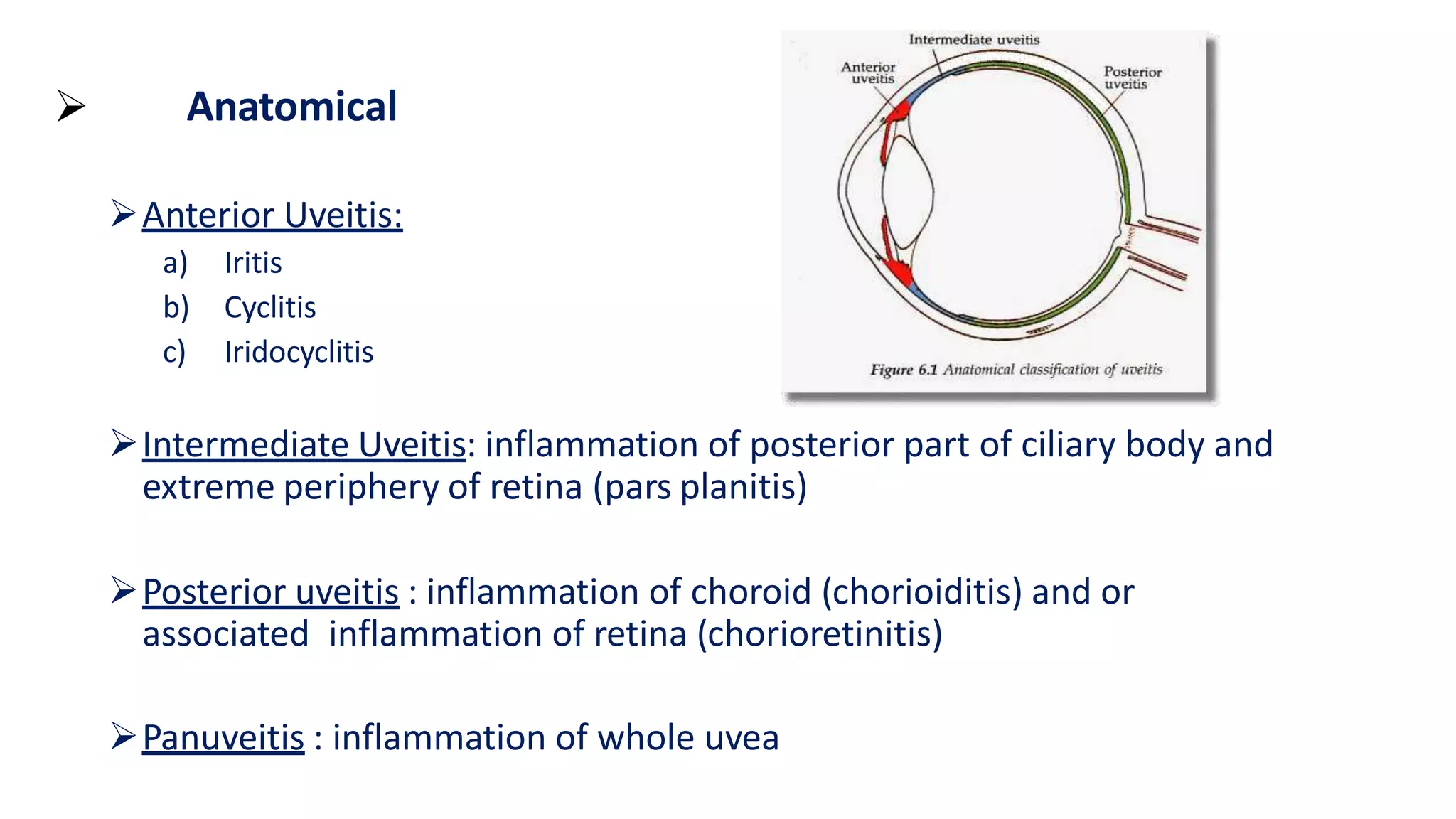
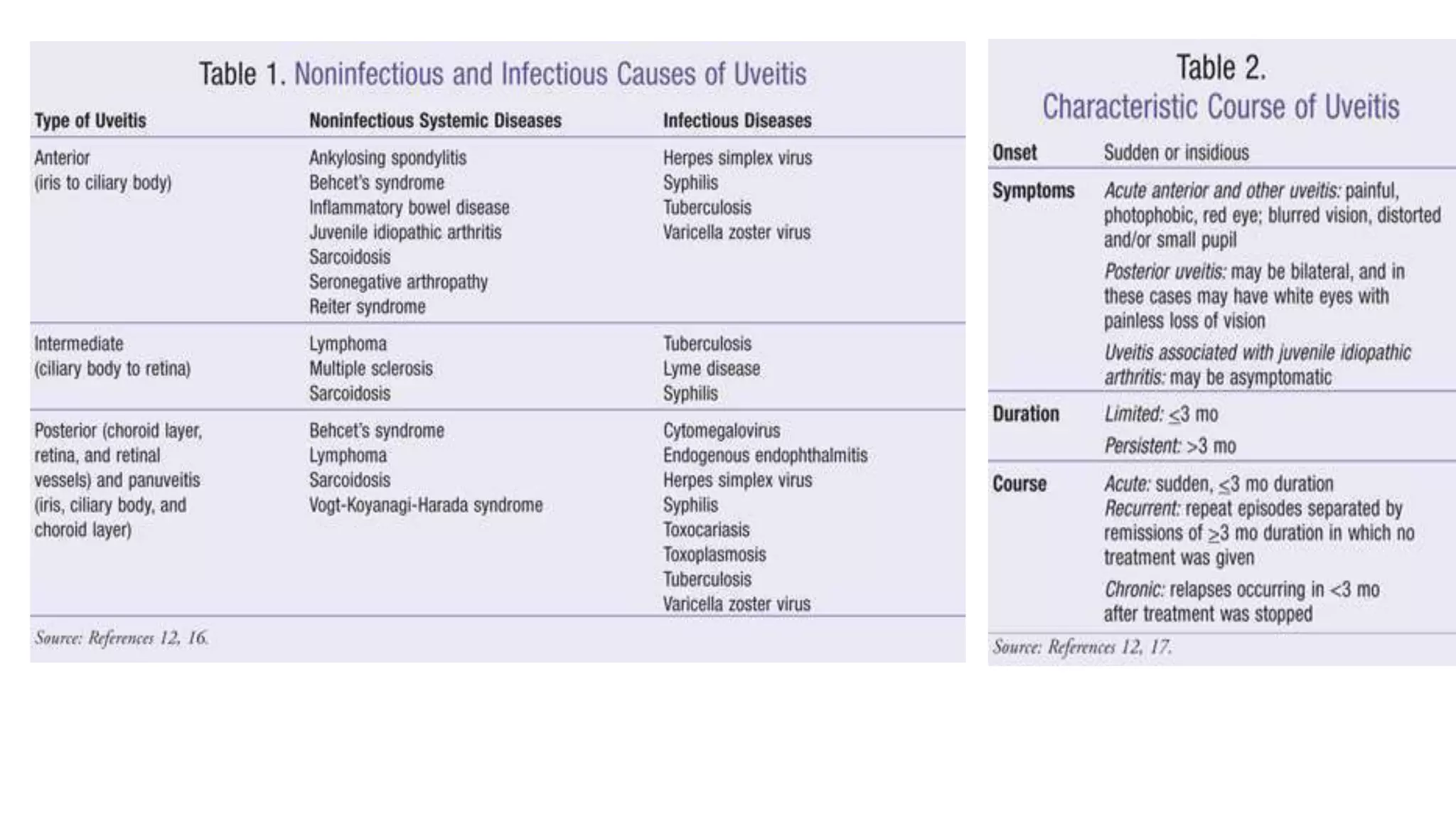
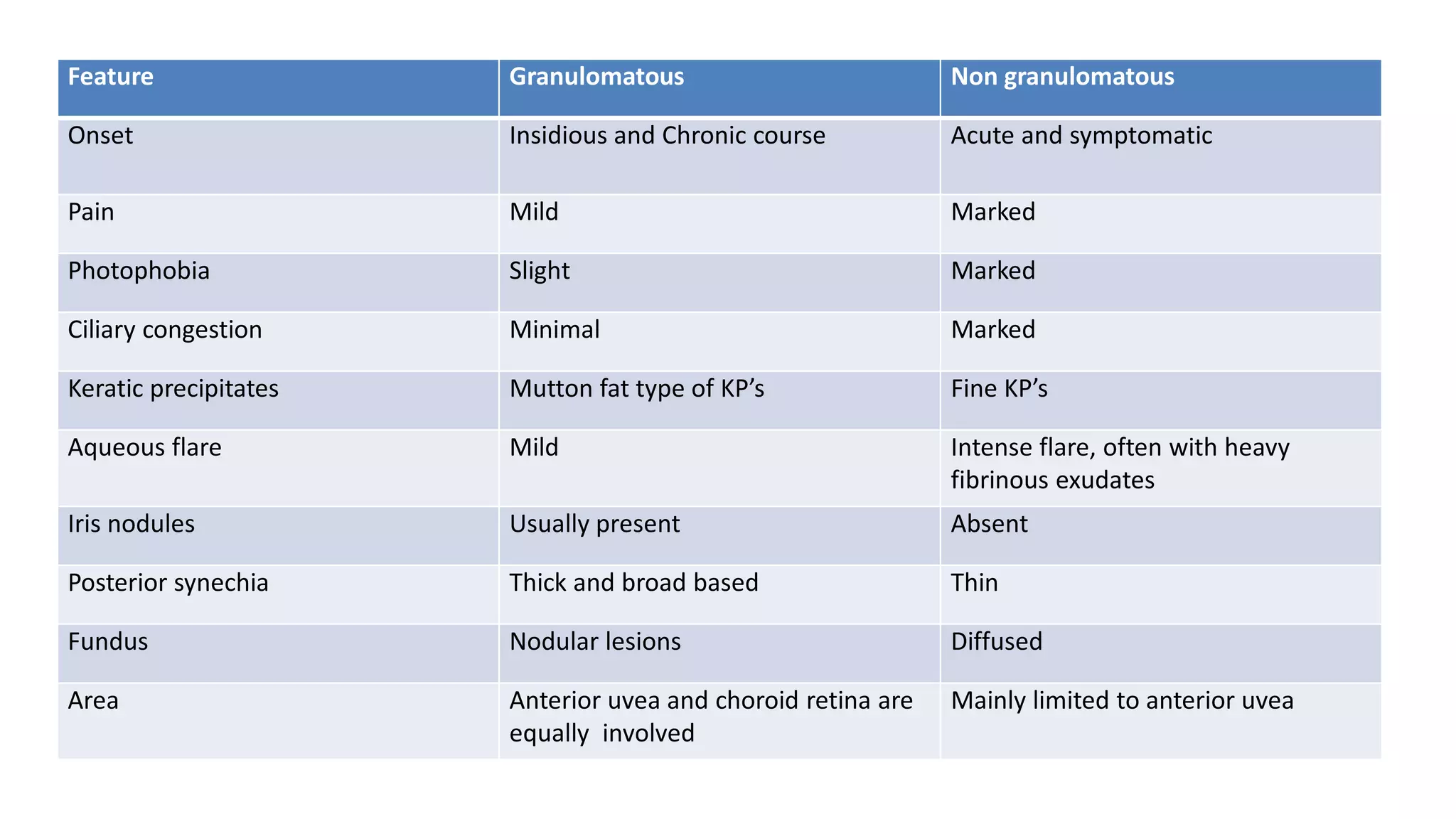
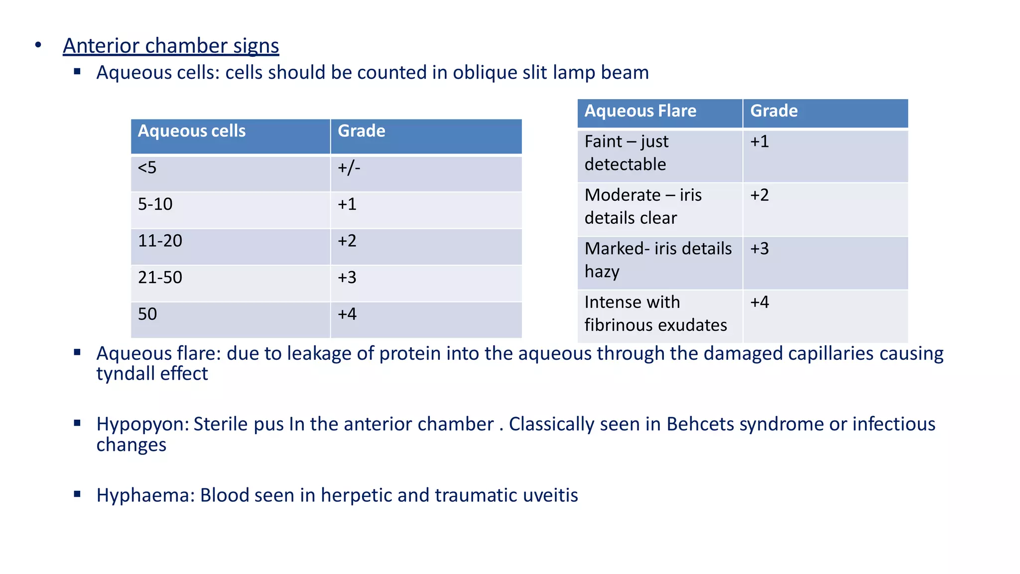
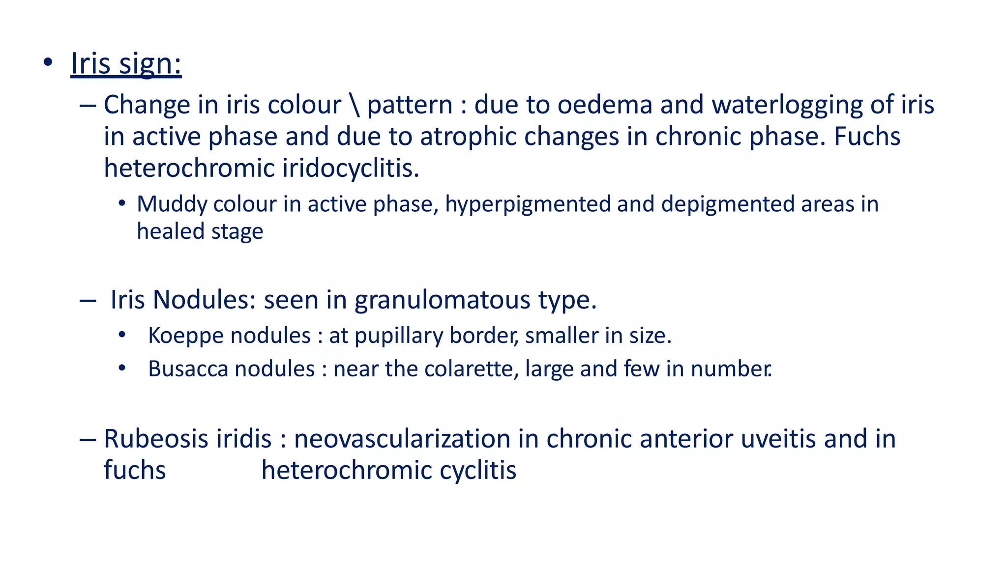
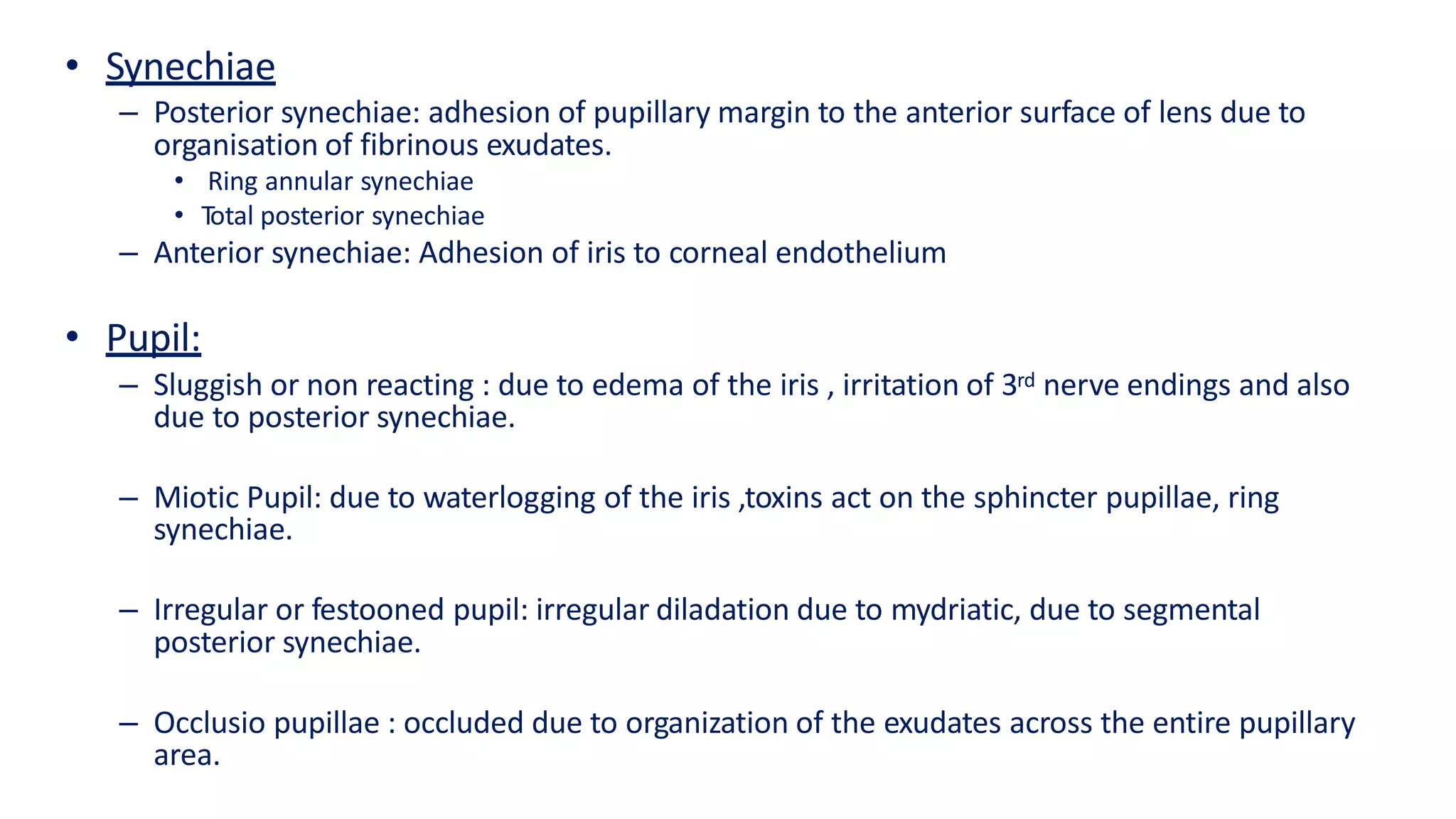
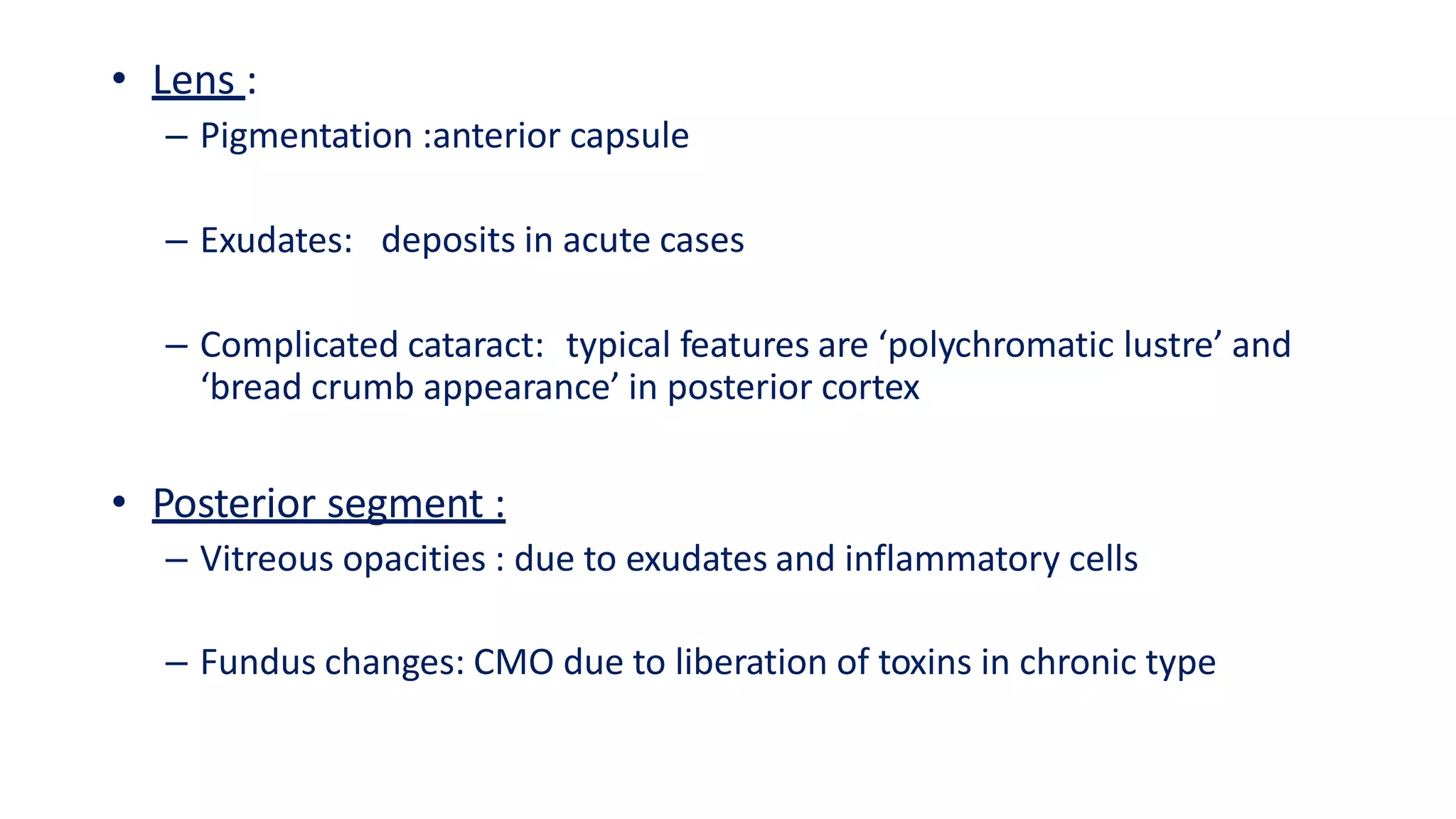
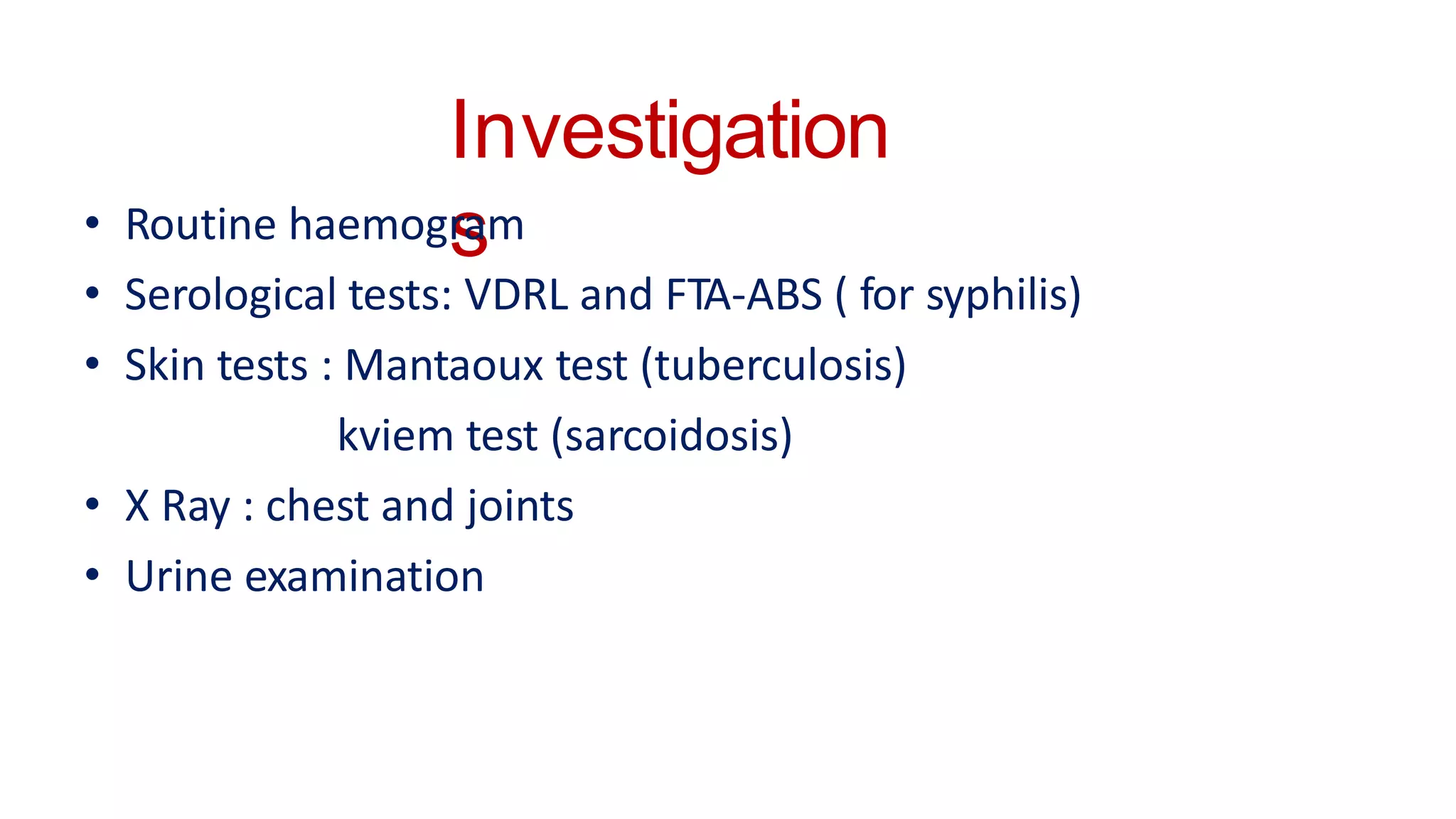
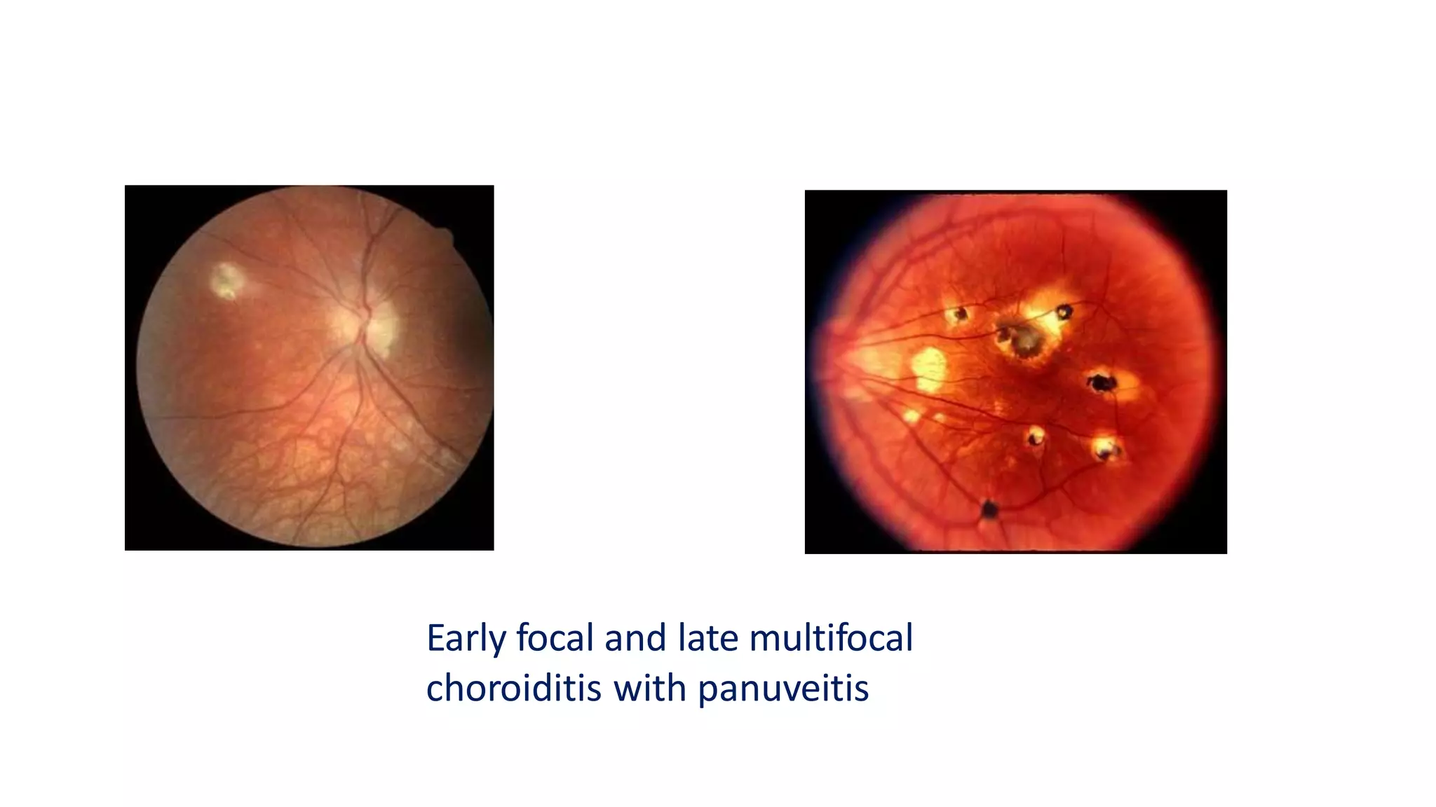
Uveiti
s
By
Dr. Moin Munir
Ul Haq
Introduction
• Inflammation of the uveal tract with associated inflammation
of the adjacent structures such as cornea, sclera, vitreous and
retina
Classification
 Anatomical
 Clinical
 Pathological
 Aetiological
 Anatomical
Anterior Uveitis:
a) Iritis
b) Cyclitis
c) Iridocyclitis
Intermediate Uveitis: inflammation of posterior part of ciliary body and
extreme periphery of retina (pars planitis)
Posterior uveitis : inflammation of choroid (chorioiditis) and or
associated inflammation of retina (chorioretinitis)
Panuveitis : inflammation of whole uvea
 Clinical
– Acute uveitis: sudden symptomatic onset , last for six weeks or more
– Chronic uveitis : insidious and asymptomatic onset ,last for more than three
months or even years
 Pathological
– Granulomatous
– Non Granulomatous
 Etiological
– Infective uveitis
– Allergic uveitis
– Toxic Uveitis
– Idiopathic Uveitis
– Uveitis associated with systemic diseases
a) Non granulomatous
– Uveitis due to tissue invasion by leptospirae represents the
manifestation of non granulomatous uveitis
– It is acute ,occurring due to physical and toxic insult to the tissue
– The alterations consists of dilatation and increase permeability of
vessels, breakdown of blood aqueous barrier and infiltration of
lymphocytes, plasma cells and large macrophages of the uveal tissue
– As a consequence mobility is reduced, pupil becomes small due to
sphincter irritation and engorgement of iris vessels
Etiopathogenesi
s
b) Granulomatous:
– Chronical inflammation of proliferative nature due to irritant foreign body, a
haemorrhage or a necrotic tissue in the eye
– Characterized by infiltration with lymphocytes and proliferation of large
mononuclear cells which aggregate into nodules
– Necrosis of adjacent structures leads to reparative process resulting in fibrosis
and gliosis of the involved area
Feature Granulomatous Non granulomatous
Onset Insidious and Chronic course Acute and symptomatic
Pain Mild Marked
Photophobia Slight Marked
Ciliary congestion Minimal Marked
Keratic precipitates Mutton fat type of KP’s Fine KP’s
Aqueous flare Mild Intense flare, often with heavy
fibrinous exudates
Iris nodules Usually present Absent
Posterior synechia Thick and broad based Thin
Fundus Nodular lesions Diffused
Area Anterior uvea and choroid retina are
equally involved
Mainly limited to anterior uvea
Anterior Uveitis
• Clinical picture:
 Symptoms:-
i. Pain
ii. Redness
iii. Photophobia
iv. Lacrimation
v. Dimness of vison
• Ocular signs :
i. Circumcorneal congestion
ii. Corneal signs:
 The KP’s (cellular deposits on the endothelium) are small, medium, large mutton
fat KP’s arranged base down at the lower part of the cornea due to gravitation
 Fresh KP’s; round white and hydrated
 Old KP’s; shrunken, pigmented, haloes surrounding them
• Anterior chamber signs
 Aqueous cells: cells should be counted in oblique slit lamp beam
 Aqueous flare: due to leakage of protein into the aqueous through the damaged capillaries causing
tyndall effect
 Hypopyon: Sterile pus In the anterior chamber . Classically seen in Behcets syndrome or infectious
changes
 Hyphaema: Blood seen in herpetic and traumatic uveitis
Aqueous Flare Grade
Faint – just
detectable
+1
Moderate – iris
details clear
+2
Marked- iris details
hazy
+3
Intense with
fibrinous exudates
+4
Aqueous cells Grade
<5 +/-
5-10 +1
11-20 +2
21-50 +3
50 +4
Depth and shape: deep and regular in posterior synachiae. Funnel shape
in iris bombe
While gonioscopy we see cellular deposits in active stage and peripheral
anterior synachiae in chronic stage
• Iris sign:
– Change in iris colour  pattern : due to oedema and waterlogging of iris
in active phase and due to atrophic changes in chronic phase. Fuchs
heterochromic iridocyclitis.
• Muddy colour in active phase, hyperpigmented and depigmented areas in
healed stage
– Iris Nodules: seen in granulomatous type.
• Koeppe nodules : at pupillary border, smaller in size.
• Busacca nodules : near the colarette, large and few in number.
– Rubeosis iridis : neovascularization in chronic anterior uveitis and in
fuchs heterochromic cyclitis
• Synechiae
– Posterior synechiae: adhesion of pupillary margin to the anterior surface of lens due to
organisation of fibrinous exudates.
• Ring annular synechiae
• Total posterior synechiae
– Anterior synechiae: Adhesion of iris to corneal endothelium
• Pupil:
– Sluggish or non reacting : due to edema of the iris , irritation of 3rd nerve endings and also
due to posterior synechiae.
– Miotic Pupil: due to waterlogging of the iris ,toxins act on the sphincter pupillae, ring
synechiae.
– Irregular or festooned pupil: irregular diladation due to mydriatic, due to segmental
posterior synechiae.
– Occlusio pupillae : occluded due to organization of the exudates across the entire pupillary
area.
• Lens :
– Pigmentation :anterior capsule
– Exudates: deposits in acute cases
– Complicated cataract: typical features are ‘polychromatic lustre’ and
‘bread crumb appearance’ in posterior cortex
• Posterior segment :
– Vitreous opacities : due to exudates and inflammatory cells
– Fundus changes: CMO due to liberation of toxins in chronic type
Complications
• Complicated cataract
• Secondary glaucoma
• Posterior synechiae
• Occlusio pupillae
• Cystoid macular oedema
• Band shaped keratopathy
• Phthisis bulbi
• Retinal complications
Investigation
s
• Routine haemogram
• Serological tests: VDRL and FTA-ABS ( for syphilis)
• Skin tests : Mantaoux test (tuberculosis)
kviem test (sarcoidosis)
• X Ray : chest and joints
• Urine examination
Treatment
 The Aims of treating uveitis are :
1) To prevent vision-threatening complications
2) To relieve the patients discomfort and pain
3) To treat the underlying cause of uveitis
• Non-specific treatment :
1. Short Acting :
i. Tropicamide (0.5 and 1 %)
ii. Cyclopentolate ( 0.5 and 1%)
iii. Phenylephrine (2.5 to 10%)
2. Long Acting :
i. Atropine (lasts for 2 weeks)
3. Sub conjunctival injection(0.25 ml) (mydriacaine,atropine, adrenaline,
procaine)
4. Steroids/ Corticosteroids
i. Topically as drops or ointments(dexamethasone, betamethasone,
prednisolone)
ii. Periocular injection( tissue plasminogen activater) (triamcinolone
acetonide(kenalog), methylprednisolone acetate(depomedrone))
iii. Intravitral injection
iv. Systemic Therapy
4. NSAIDS (aspirin) (phenylbutazone , oxyphenylbutazone) in uvietis of
rheumatoid type
Treatment for
complications
• Inflammatory glaucoma :
i. Drugs to lower intraocular pressure e.g. Tab.Diamox, timolol maleate eyedrops.
etc.
ii. Pilocarpine and latanaprost are contra-indicated in such cases
• Complicated cataract :
i. Requires lens extraction
ii. Presence of fresh KP’s is considered a contraindication for intraocular surgery
• Retinal Detachment :
i. If an exudative type the RD will settle down on itself if uveitis is treated properly
ii. If an tractional type vitrectomy should be done
• Pthisis bulbi : especially painful , requires removal by enucleation operation
Posterior uveitis
• Posterior uveitis refers to the inflammation of the choroid
(chorioditis)
• Since the outer layers of the retina is in contact with the choroid
, the inflamed choroid almost always involves the adjoining
retina and the resultant lesion is called chorioretinitis
Clinical types
• Suppurative chorioditis (purulent inflammation of the choroid):
It usually does not occur alone mainly seen with endopthalmitis
• Non - suppurative chorioditis :
It is characterised by exudation and cellular infiltration resulting
to greyish white lesion on the red choroidal vessels
depending upon the number of lesion it can be classified into:
i. Diffuse
ii. Desseminated
iii. Localized/focal - central, papillary, periphery and equator
Clinical picture
• Symptoms :
i. Defective vision
ii. Photopsia
iii. Black floaters
iv. Metamorphopsia
v. Micropsia
vi. Macropsia
vii. Scotoma
• Signs
i. Vitreous opacities
 Fine opacities
 Coarse opacities
 Stringy opacities
 Snowball opacities
ii. Features of patches on choroid
 Active stage : pale yellow or dirty white patches with irregular
edges
 Healed stage : black pigmented clumps at periphery of the lesion
involved area shows sclera below the healed choroid
Early focal and late multifocal
choroiditis with panuveitis
Acute (A) and healed (B)
Pneumocystis carinii choroiditis in
a patient with AIDS.
Complications
i. Extension of inflammation towards the anterior section
ii. Complicated cataract
iii. Vitreous degeneration
iv. Macular oedema
v. Retinal Detachment
Investigation
• Investigation for tuberculosis
• Investigation for sarcoidosis
• VDRL and FTA-ABS for syphilis
• ELISA for toxocariasis
Treatment
• Non- specific treatment:
i. Topical and Systemic corticosteroids
ii. Posterior sub-tenon injections
iii. Rarely immunosuppressive agents may be needed
• Specific treatment:
 is required for causative diseases such as toxoplasmosis ,toxocariasis,
tuberculosis, syphilis, etc.
Thank You

uveitis.pptx

  • 1.
  • 2.
    Introduction • Inflammation ofthe uveal tract with associated inflammation of the adjacent structures such as cornea, sclera, vitreous and retina Classification  Anatomical  Clinical  Pathological  Aetiological
  • 3.
     Anatomical Anterior Uveitis: a)Iritis b) Cyclitis c) Iridocyclitis Intermediate Uveitis: inflammation of posterior part of ciliary body and extreme periphery of retina (pars planitis) Posterior uveitis : inflammation of choroid (chorioiditis) and or associated inflammation of retina (chorioretinitis) Panuveitis : inflammation of whole uvea
  • 4.
     Clinical – Acuteuveitis: sudden symptomatic onset , last for six weeks or more – Chronic uveitis : insidious and asymptomatic onset ,last for more than three months or even years  Pathological – Granulomatous – Non Granulomatous  Etiological – Infective uveitis – Allergic uveitis – Toxic Uveitis – Idiopathic Uveitis – Uveitis associated with systemic diseases
  • 6.
    a) Non granulomatous –Uveitis due to tissue invasion by leptospirae represents the manifestation of non granulomatous uveitis – It is acute ,occurring due to physical and toxic insult to the tissue – The alterations consists of dilatation and increase permeability of vessels, breakdown of blood aqueous barrier and infiltration of lymphocytes, plasma cells and large macrophages of the uveal tissue – As a consequence mobility is reduced, pupil becomes small due to sphincter irritation and engorgement of iris vessels Etiopathogenesi s
  • 7.
    b) Granulomatous: – Chronicalinflammation of proliferative nature due to irritant foreign body, a haemorrhage or a necrotic tissue in the eye – Characterized by infiltration with lymphocytes and proliferation of large mononuclear cells which aggregate into nodules – Necrosis of adjacent structures leads to reparative process resulting in fibrosis and gliosis of the involved area
  • 8.
    Feature Granulomatous Nongranulomatous Onset Insidious and Chronic course Acute and symptomatic Pain Mild Marked Photophobia Slight Marked Ciliary congestion Minimal Marked Keratic precipitates Mutton fat type of KP’s Fine KP’s Aqueous flare Mild Intense flare, often with heavy fibrinous exudates Iris nodules Usually present Absent Posterior synechia Thick and broad based Thin Fundus Nodular lesions Diffused Area Anterior uvea and choroid retina are equally involved Mainly limited to anterior uvea
  • 9.
    Anterior Uveitis • Clinicalpicture:  Symptoms:- i. Pain ii. Redness iii. Photophobia iv. Lacrimation v. Dimness of vison
  • 10.
    • Ocular signs: i. Circumcorneal congestion ii. Corneal signs:  The KP’s (cellular deposits on the endothelium) are small, medium, large mutton fat KP’s arranged base down at the lower part of the cornea due to gravitation  Fresh KP’s; round white and hydrated  Old KP’s; shrunken, pigmented, haloes surrounding them
  • 11.
    • Anterior chambersigns  Aqueous cells: cells should be counted in oblique slit lamp beam  Aqueous flare: due to leakage of protein into the aqueous through the damaged capillaries causing tyndall effect  Hypopyon: Sterile pus In the anterior chamber . Classically seen in Behcets syndrome or infectious changes  Hyphaema: Blood seen in herpetic and traumatic uveitis Aqueous Flare Grade Faint – just detectable +1 Moderate – iris details clear +2 Marked- iris details hazy +3 Intense with fibrinous exudates +4 Aqueous cells Grade <5 +/- 5-10 +1 11-20 +2 21-50 +3 50 +4
  • 12.
    Depth and shape:deep and regular in posterior synachiae. Funnel shape in iris bombe While gonioscopy we see cellular deposits in active stage and peripheral anterior synachiae in chronic stage
  • 13.
    • Iris sign: –Change in iris colour pattern : due to oedema and waterlogging of iris in active phase and due to atrophic changes in chronic phase. Fuchs heterochromic iridocyclitis. • Muddy colour in active phase, hyperpigmented and depigmented areas in healed stage – Iris Nodules: seen in granulomatous type. • Koeppe nodules : at pupillary border, smaller in size. • Busacca nodules : near the colarette, large and few in number. – Rubeosis iridis : neovascularization in chronic anterior uveitis and in fuchs heterochromic cyclitis
  • 14.
    • Synechiae – Posteriorsynechiae: adhesion of pupillary margin to the anterior surface of lens due to organisation of fibrinous exudates. • Ring annular synechiae • Total posterior synechiae – Anterior synechiae: Adhesion of iris to corneal endothelium • Pupil: – Sluggish or non reacting : due to edema of the iris , irritation of 3rd nerve endings and also due to posterior synechiae. – Miotic Pupil: due to waterlogging of the iris ,toxins act on the sphincter pupillae, ring synechiae. – Irregular or festooned pupil: irregular diladation due to mydriatic, due to segmental posterior synechiae. – Occlusio pupillae : occluded due to organization of the exudates across the entire pupillary area.
  • 15.
    • Lens : –Pigmentation :anterior capsule – Exudates: deposits in acute cases – Complicated cataract: typical features are ‘polychromatic lustre’ and ‘bread crumb appearance’ in posterior cortex • Posterior segment : – Vitreous opacities : due to exudates and inflammatory cells – Fundus changes: CMO due to liberation of toxins in chronic type
  • 16.
    Complications • Complicated cataract •Secondary glaucoma • Posterior synechiae • Occlusio pupillae • Cystoid macular oedema • Band shaped keratopathy • Phthisis bulbi • Retinal complications
  • 17.
    Investigation s • Routine haemogram •Serological tests: VDRL and FTA-ABS ( for syphilis) • Skin tests : Mantaoux test (tuberculosis) kviem test (sarcoidosis) • X Ray : chest and joints • Urine examination
  • 18.
    Treatment  The Aimsof treating uveitis are : 1) To prevent vision-threatening complications 2) To relieve the patients discomfort and pain 3) To treat the underlying cause of uveitis
  • 19.
    • Non-specific treatment: 1. Short Acting : i. Tropicamide (0.5 and 1 %) ii. Cyclopentolate ( 0.5 and 1%) iii. Phenylephrine (2.5 to 10%) 2. Long Acting : i. Atropine (lasts for 2 weeks) 3. Sub conjunctival injection(0.25 ml) (mydriacaine,atropine, adrenaline, procaine)
  • 20.
    4. Steroids/ Corticosteroids i.Topically as drops or ointments(dexamethasone, betamethasone, prednisolone) ii. Periocular injection( tissue plasminogen activater) (triamcinolone acetonide(kenalog), methylprednisolone acetate(depomedrone)) iii. Intravitral injection iv. Systemic Therapy 4. NSAIDS (aspirin) (phenylbutazone , oxyphenylbutazone) in uvietis of rheumatoid type
  • 21.
    Treatment for complications • Inflammatoryglaucoma : i. Drugs to lower intraocular pressure e.g. Tab.Diamox, timolol maleate eyedrops. etc. ii. Pilocarpine and latanaprost are contra-indicated in such cases • Complicated cataract : i. Requires lens extraction ii. Presence of fresh KP’s is considered a contraindication for intraocular surgery • Retinal Detachment : i. If an exudative type the RD will settle down on itself if uveitis is treated properly ii. If an tractional type vitrectomy should be done • Pthisis bulbi : especially painful , requires removal by enucleation operation
  • 22.
    Posterior uveitis • Posterioruveitis refers to the inflammation of the choroid (chorioditis) • Since the outer layers of the retina is in contact with the choroid , the inflamed choroid almost always involves the adjoining retina and the resultant lesion is called chorioretinitis
  • 23.
    Clinical types • Suppurativechorioditis (purulent inflammation of the choroid): It usually does not occur alone mainly seen with endopthalmitis • Non - suppurative chorioditis : It is characterised by exudation and cellular infiltration resulting to greyish white lesion on the red choroidal vessels depending upon the number of lesion it can be classified into: i. Diffuse ii. Desseminated iii. Localized/focal - central, papillary, periphery and equator
  • 24.
    Clinical picture • Symptoms: i. Defective vision ii. Photopsia iii. Black floaters iv. Metamorphopsia v. Micropsia vi. Macropsia vii. Scotoma
  • 25.
    • Signs i. Vitreousopacities  Fine opacities  Coarse opacities  Stringy opacities  Snowball opacities ii. Features of patches on choroid  Active stage : pale yellow or dirty white patches with irregular edges  Healed stage : black pigmented clumps at periphery of the lesion involved area shows sclera below the healed choroid
  • 26.
    Early focal andlate multifocal choroiditis with panuveitis
  • 27.
    Acute (A) andhealed (B) Pneumocystis carinii choroiditis in a patient with AIDS.
  • 28.
    Complications i. Extension ofinflammation towards the anterior section ii. Complicated cataract iii. Vitreous degeneration iv. Macular oedema v. Retinal Detachment
  • 29.
    Investigation • Investigation fortuberculosis • Investigation for sarcoidosis • VDRL and FTA-ABS for syphilis • ELISA for toxocariasis
  • 30.
    Treatment • Non- specifictreatment: i. Topical and Systemic corticosteroids ii. Posterior sub-tenon injections iii. Rarely immunosuppressive agents may be needed • Specific treatment:  is required for causative diseases such as toxoplasmosis ,toxocariasis, tuberculosis, syphilis, etc.
  • 31.