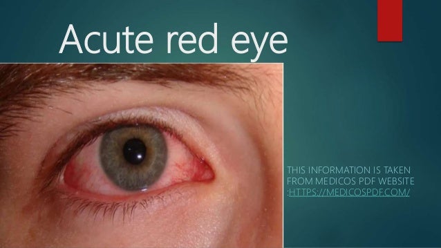
Acute red eye
THIS INFORMATION IS TAKEN
FROM MEDICOS PDF WEBSITE
:HTTPS://MEDICOSPDF.COM/
Differential diagnosis
Common causes:
 Conjunctivitis
 Episcleritis
 Subconjunctival haemorrhage: spontaneous (e.g. coughing) or
traumatic. Self-resolves, but consider bleeding disorder if recurrent.
 Trauma: corneal abrasion (+ve fluorescein test), foreign body.
Site-threatening causes (often painful):
 Keratitis (corneal infection).
 Scleritis
 Anterior uveitis (iritis or iridocyclitis).
 Acute angle closure glaucoma.
Conjunctivitis
Inflammation of the conjunctiva, the clear moist
membrane covering the exposed sclera and inner
eyelids. Usually bilateral, but may start in one eye
before spreading.
Viral conjunctivitis
Most commonly due to adenovirus, which can come in
epidemic outbreaks.
Presentation:
 Acute red eye.
 Watery discharge. May dry to form yellow
crust, so don't just assume pus if you see
yellow!
 Mild foreign body sensation.
 Associated viral URTI.
Management:
 Supportive treatment with topical lubricant.
 Strict hand hygiene as highly contagious.
Bacterial conjunctivitis
Cause:
 Staph or strep.
 Gonorrhoea or chlamydia can be the cause in concurrent STI,
from hand transfer.
Presentation:
 Acute red eye.
 Mucopurulent discharge leading to crusting
and difficulty opening eyes in morning.
 Gonorrhoea may cause a severe
manifestation requiring referral.
Management:
Chloramphenicol eyedrops, though
usually self-resolves anyway. Apply to
both eyes, even if unilateral, to prevent
amblyopia.
Do not wear contact lenses.
Allergic conjunctivitis
Presentation:
 Acute red eye ± chemosis.
 Itchy, watery eyes.
 Usually seasonal (e.g. hay fever) or perennial (e.g.
dust mites).
Management:
Antihistamines for rapid relief and long-
term control, oral (e.g. cetirizine) or
topical (e.g. azelastine).
Topical mast cell stabilizer (e.g. sodium
cromoglicate) is an alternative for long-
term control.
Episcleritis
Pathophysiology
 Inflammation of the episclera, the thin vascular
sheet between the conjunctiva and sclera.
 Usually idiopathic, or secondary to RA, IBD,
polyarteritis nodosa, or sarcoidosis.
Presentation
 Acute red eye.
 Mild symptoms, perhaps with foreign body sensation.
 Resolves in 1-2 weeks.
Management
 Topical lubricants.
 Oral NSAIDs.
Scleritis
Pathophysiology
 Inflammation of the sclera.
 50% linked to connective tissue disease: RA, GPA
(Wegner's). May be the presenting complaint.
Presentation
 Severe, dull eye pain developing over days, tender to
touch.
 Bluish-red eye due to deep vascular engorgement.
 Blurred vision, photophobia.
Management
Rapid immunosuppression may be needed to preserve
sight.
Keratitis
Pathophysiology
 Inflammation of the cornea. Can progress to
ulceration.
 Cause can be bacterial, occurring in a contact-lens
wearer with inadequate lens care, or viral, usually
herpes simplex.
Presentation
 Acute red eye.
 Photophobia, severe pain, foreign body sensation.
 Purulent discharge and hypopyon – pus in anterior
chamber – may occur with Pseudomonas aeruginosa.
 Fluorescein staining may show abrasion (possible
precipitant), corneal ulcer, or dendritic lesion (herpes
simplex keratitis).
Management
 Urgent referral for corneal scrape and treatment, due to the
risk of sight loss from perforation.
 In contact lens wearers, culture lens, case, and cleaning
solution.
Non-infectious keratitis
 Can be caused by UV light, including in welders or sunbed
users with inadequate eye protection.
 Treat with cool compress and oral analgesia.
Anterior uveitis
Pathophysiology
Includes iritis – inflammation of the anterior chamber
and iris – and iridocyclitis – also affecting the ciliary
body.
Causes:
 Often idiopathic.
 HLA-B27 disease is the commonest identified cause: ankylosing
spondylitis, psoriatic arthritis, reactive arthritis, IBD.
 Less commonly: HSV, toxoplasma, sarcoidosis, Behcet's, juvenile
idiopathic arthritis, rheumatoid arthritis.
Intermediate uveitis (anterior
vitreous and ciliary body) and
posterior uveitis (choroid):
Less common than anterior uveitis.
Causes are similar to anterior uveitis.
Usually painless.
Presentation
Symptoms:
 Acute red eye.
 Photophobia
 Blurred vision.
 Deep aching pain.
 Floaters
Signs:
 Non-reactive, small pupil.
 Slit-lamp exam: cloudy aqueous humour ('flare') due
to WBCs and protein.
Management
 Topical steroids e.g. dexamethasone.
 Antimuscarinic cytoplegics e.g. cyclopentolate.
Endophthalmitis
Pathophysiology
Inflammation of the aqueous or vitreous humour, usually
infective.
Causes:
 Exogenous: pathogen inoculation during ophthalmic surgery
(commonly coagulase-negative staph) or trauma.
 Endogenous: infectious spread from elsewhere.
Presentation
Most commonly presents 3-5 days post-surgery
with:
 Red eye, hypopyon, and hazy cornea.
 Blurred vision.
 Pain (though can be painless).
 Lid swelling.
Investigations
 Slit-lamp exam: cells and cloudy aqueous
humour. Ultrasound if retina can't be
visualised.
 Vitreous aspiration or biopsy (vitrectomy) for
microbiology.
Management
Intravitreal antibiotics.
Acute angle closure glaucoma
Pathophysiology
 Closure of the iridocorneal angle leading to
↑intraocular pressure (IOP). A sight-threatening
emergency.
 Can also be chronic.
 Risk factors: hyperopia, female, age >60.
Presentation
Symptoms:
 Painful red eye, frontal headache.
 Haloes around lights, blurring, and visual loss.
Signs:
 Corneal edema or haziness.
 Non-reactive, mid-dilated pupil.
 Cupping of optic disc.
Investigations
Tonometry: IOP typically >30 mmHg.
Gonioscopy to examine anterior chamber
angle is the gold standard.
Management
 Immediate ophthalmology referral if suspected.
 Reduce IOP: topical agents (muscarinic agonist e.g.
pilocarpine +/- β-blocker e.g. timolol +/- α2-
agonist e.g. clonidine) plus acetazolamide PO/IV.
 Ophthalmology may continue medical
management, or use laser peripheral iridotomy if
refractory.
Thank you
Keep supporting Medicos PDF website. To get more
slides, news articles and books of medical field. You
can download the website for free from
https://medicospdf.com/

Acute red eye

  • 1.
    Acute red eye THISINFORMATION IS TAKEN FROM MEDICOS PDF WEBSITE :HTTPS://MEDICOSPDF.COM/
  • 2.
    Differential diagnosis Common causes: Conjunctivitis  Episcleritis  Subconjunctival haemorrhage: spontaneous (e.g. coughing) or traumatic. Self-resolves, but consider bleeding disorder if recurrent.  Trauma: corneal abrasion (+ve fluorescein test), foreign body.
  • 3.
    Site-threatening causes (oftenpainful):  Keratitis (corneal infection).  Scleritis  Anterior uveitis (iritis or iridocyclitis).  Acute angle closure glaucoma.
  • 4.
    Conjunctivitis Inflammation of theconjunctiva, the clear moist membrane covering the exposed sclera and inner eyelids. Usually bilateral, but may start in one eye before spreading. Viral conjunctivitis Most commonly due to adenovirus, which can come in epidemic outbreaks.
  • 5.
    Presentation:  Acute redeye.  Watery discharge. May dry to form yellow crust, so don't just assume pus if you see yellow!  Mild foreign body sensation.  Associated viral URTI.
  • 6.
    Management:  Supportive treatmentwith topical lubricant.  Strict hand hygiene as highly contagious. Bacterial conjunctivitis Cause:  Staph or strep.  Gonorrhoea or chlamydia can be the cause in concurrent STI, from hand transfer.
  • 7.
    Presentation:  Acute redeye.  Mucopurulent discharge leading to crusting and difficulty opening eyes in morning.  Gonorrhoea may cause a severe manifestation requiring referral.
  • 8.
    Management: Chloramphenicol eyedrops, though usuallyself-resolves anyway. Apply to both eyes, even if unilateral, to prevent amblyopia. Do not wear contact lenses.
  • 9.
    Allergic conjunctivitis Presentation:  Acutered eye ± chemosis.  Itchy, watery eyes.  Usually seasonal (e.g. hay fever) or perennial (e.g. dust mites).
  • 10.
    Management: Antihistamines for rapidrelief and long- term control, oral (e.g. cetirizine) or topical (e.g. azelastine). Topical mast cell stabilizer (e.g. sodium cromoglicate) is an alternative for long- term control.
  • 11.
    Episcleritis Pathophysiology  Inflammation ofthe episclera, the thin vascular sheet between the conjunctiva and sclera.  Usually idiopathic, or secondary to RA, IBD, polyarteritis nodosa, or sarcoidosis.
  • 12.
    Presentation  Acute redeye.  Mild symptoms, perhaps with foreign body sensation.  Resolves in 1-2 weeks. Management  Topical lubricants.  Oral NSAIDs.
  • 13.
    Scleritis Pathophysiology  Inflammation ofthe sclera.  50% linked to connective tissue disease: RA, GPA (Wegner's). May be the presenting complaint. Presentation  Severe, dull eye pain developing over days, tender to touch.  Bluish-red eye due to deep vascular engorgement.  Blurred vision, photophobia.
  • 14.
    Management Rapid immunosuppression maybe needed to preserve sight. Keratitis Pathophysiology  Inflammation of the cornea. Can progress to ulceration.  Cause can be bacterial, occurring in a contact-lens wearer with inadequate lens care, or viral, usually herpes simplex.
  • 15.
    Presentation  Acute redeye.  Photophobia, severe pain, foreign body sensation.  Purulent discharge and hypopyon – pus in anterior chamber – may occur with Pseudomonas aeruginosa.  Fluorescein staining may show abrasion (possible precipitant), corneal ulcer, or dendritic lesion (herpes simplex keratitis).
  • 16.
    Management  Urgent referralfor corneal scrape and treatment, due to the risk of sight loss from perforation.  In contact lens wearers, culture lens, case, and cleaning solution. Non-infectious keratitis  Can be caused by UV light, including in welders or sunbed users with inadequate eye protection.  Treat with cool compress and oral analgesia.
  • 17.
  • 18.
    Pathophysiology Includes iritis –inflammation of the anterior chamber and iris – and iridocyclitis – also affecting the ciliary body. Causes:  Often idiopathic.  HLA-B27 disease is the commonest identified cause: ankylosing spondylitis, psoriatic arthritis, reactive arthritis, IBD.  Less commonly: HSV, toxoplasma, sarcoidosis, Behcet's, juvenile idiopathic arthritis, rheumatoid arthritis.
  • 19.
    Intermediate uveitis (anterior vitreousand ciliary body) and posterior uveitis (choroid): Less common than anterior uveitis. Causes are similar to anterior uveitis. Usually painless.
  • 20.
    Presentation Symptoms:  Acute redeye.  Photophobia  Blurred vision.  Deep aching pain.  Floaters
  • 21.
    Signs:  Non-reactive, smallpupil.  Slit-lamp exam: cloudy aqueous humour ('flare') due to WBCs and protein. Management  Topical steroids e.g. dexamethasone.  Antimuscarinic cytoplegics e.g. cyclopentolate.
  • 22.
    Endophthalmitis Pathophysiology Inflammation of theaqueous or vitreous humour, usually infective. Causes:  Exogenous: pathogen inoculation during ophthalmic surgery (commonly coagulase-negative staph) or trauma.  Endogenous: infectious spread from elsewhere.
  • 23.
    Presentation Most commonly presents3-5 days post-surgery with:  Red eye, hypopyon, and hazy cornea.  Blurred vision.  Pain (though can be painless).  Lid swelling.
  • 24.
    Investigations  Slit-lamp exam:cells and cloudy aqueous humour. Ultrasound if retina can't be visualised.  Vitreous aspiration or biopsy (vitrectomy) for microbiology. Management Intravitreal antibiotics.
  • 25.
  • 26.
    Pathophysiology  Closure ofthe iridocorneal angle leading to ↑intraocular pressure (IOP). A sight-threatening emergency.  Can also be chronic.  Risk factors: hyperopia, female, age >60.
  • 27.
    Presentation Symptoms:  Painful redeye, frontal headache.  Haloes around lights, blurring, and visual loss. Signs:  Corneal edema or haziness.  Non-reactive, mid-dilated pupil.  Cupping of optic disc.
  • 28.
    Investigations Tonometry: IOP typically>30 mmHg. Gonioscopy to examine anterior chamber angle is the gold standard.
  • 29.
    Management  Immediate ophthalmologyreferral if suspected.  Reduce IOP: topical agents (muscarinic agonist e.g. pilocarpine +/- β-blocker e.g. timolol +/- α2- agonist e.g. clonidine) plus acetazolamide PO/IV.  Ophthalmology may continue medical management, or use laser peripheral iridotomy if refractory.
  • 30.
    Thank you Keep supportingMedicos PDF website. To get more slides, news articles and books of medical field. You can download the website for free from https://medicospdf.com/