Download to read offline

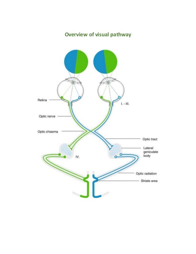
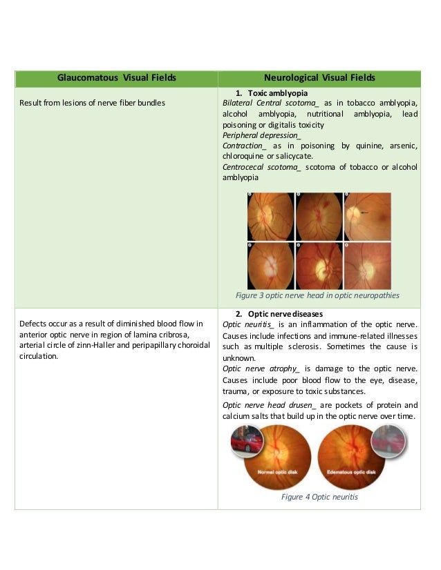
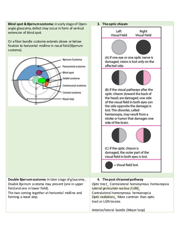
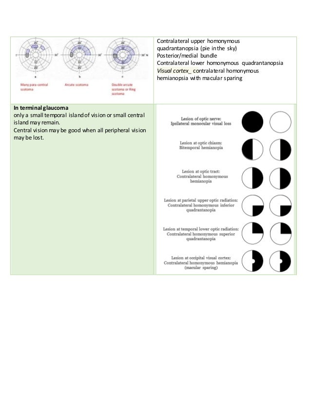

The document discusses visual fields, defining them as the area visible in peripheral vision. It classifies visual field defects into categories related to glaucomatous and neurological conditions, detailing their causes and characteristics, such as scotomas and their association with optic nerve diseases. The document outlines the visual pathway and how lesions affect visual perception, highlighting specific visual field loss patterns associated with various conditions.