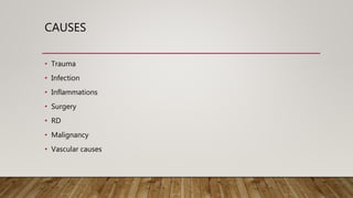
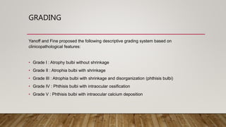
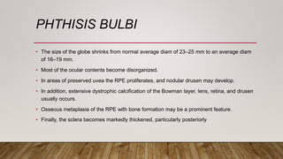
This document defines and describes phthisis bulbi, a condition where the eye shrinks and its contents become disorganized due to severe injury or disease damage. It outlines the causes of phthisis bulbi, including trauma, infection, inflammation, surgery, retinal detachment, and malignancy. A grading system is proposed based on clinical features, from atrophy without shrinkage to phthisis bulbi with intraocular ossification or calcium deposition. Signs, symptoms, pathophysiology, diagnosis, and management are discussed, with management focusing on alleviating pain and cosmetic rehabilitation rather than restoring vision.
Title: Sense of Taste
Presenter: Dr. Faiza, Assistant Professor of Physiology
Qualifications:
MBBS (Best Graduate, AIMC Lahore)
FCPS Physiology
ICMT, CHPE, DHPE (STMU)
MPH (GC University, Faisalabad)
MBA (Virtual University of Pakistan)
Learning Objectives:
Describe the structure and function of taste buds.
Describe the relationship between the taste threshold and taste index of common substances.
Explain the chemical basis and signal transduction of taste perception for each type of primary taste sensation.
Recognize different abnormalities of taste perception and their causes.
Key Topics:
Significance of Taste Sensation:
Differentiation between pleasant and harmful food
Influence on behavior
Selection of food based on metabolic needs
Receptors of Taste:
Taste buds on the tongue
Influence of sense of smell, texture of food, and pain stimulation (e.g., by pepper)
Primary and Secondary Taste Sensations:
Primary taste sensations: Sweet, Sour, Salty, Bitter, Umami
Chemical basis and signal transduction mechanisms for each taste
Taste Threshold and Index:
Taste threshold values for Sweet (sucrose), Salty (NaCl), Sour (HCl), and Bitter (Quinine)
Taste index relationship: Inversely proportional to taste threshold
Taste Blindness:
Inability to taste certain substances, particularly thiourea compounds
Example: Phenylthiocarbamide
Structure and Function of Taste Buds:
Composition: Epithelial cells, Sustentacular/Supporting cells, Taste cells, Basal cells
Features: Taste pores, Taste hairs/microvilli, and Taste nerve fibers
Location of Taste Buds:
Found in papillae of the tongue (Fungiform, Circumvallate, Foliate)
Also present on the palate, tonsillar pillars, epiglottis, and proximal esophagus
Mechanism of Taste Stimulation:
Interaction of taste substances with receptors on microvilli
Signal transduction pathways for Umami, Sweet, Bitter, Sour, and Salty tastes
Taste Sensitivity and Adaptation:
Decrease in sensitivity with age
Rapid adaptation of taste sensation
Role of Saliva in Taste:
Dissolution of tastants to reach receptors
Washing away the stimulus
Taste Preferences and Aversions:
Mechanisms behind taste preference and aversion
Influence of receptors and neural pathways
Impact of Sensory Nerve Damage:
Degeneration of taste buds if the sensory nerve fiber is cut
Abnormalities of Taste Detection:
Conditions: Ageusia, Hypogeusia, Dysgeusia (parageusia)
Causes: Nerve damage, neurological disorders, infections, poor oral hygiene, adverse drug effects, deficiencies, aging, tobacco use, altered neurotransmitter levels
Neurotransmitters and Taste Threshold:
Effects of serotonin (5-HT) and norepinephrine (NE) on taste sensitivity
Supertasters:
25% of the population with heightened sensitivity to taste, especially bitterness
Increased number of fungiform papillae
NVBDCP.pptx Nation vector borne disease control programSapna Thakur
NVBDCP was launched in 2003-2004 . Vector-Borne Disease: Disease that results from an infection transmitted to humans and other animals by blood-feeding arthropods, such as mosquitoes, ticks, and fleas. Examples of vector-borne diseases include Dengue fever, West Nile Virus, Lyme disease, and malaria.
Title: Sense of Smell
Presenter: Dr. Faiza, Assistant Professor of Physiology
Qualifications:
MBBS (Best Graduate, AIMC Lahore)
FCPS Physiology
ICMT, CHPE, DHPE (STMU)
MPH (GC University, Faisalabad)
MBA (Virtual University of Pakistan)
Learning Objectives:
Describe the primary categories of smells and the concept of odor blindness.
Explain the structure and location of the olfactory membrane and mucosa, including the types and roles of cells involved in olfaction.
Describe the pathway and mechanisms of olfactory signal transmission from the olfactory receptors to the brain.
Illustrate the biochemical cascade triggered by odorant binding to olfactory receptors, including the role of G-proteins and second messengers in generating an action potential.
Identify different types of olfactory disorders such as anosmia, hyposmia, hyperosmia, and dysosmia, including their potential causes.
Key Topics:
Olfactory Genes:
3% of the human genome accounts for olfactory genes.
400 genes for odorant receptors.
Olfactory Membrane:
Located in the superior part of the nasal cavity.
Medially: Folds downward along the superior septum.
Laterally: Folds over the superior turbinate and upper surface of the middle turbinate.
Total surface area: 5-10 square centimeters.
Olfactory Mucosa:
Olfactory Cells: Bipolar nerve cells derived from the CNS (100 million), with 4-25 olfactory cilia per cell.
Sustentacular Cells: Produce mucus and maintain ionic and molecular environment.
Basal Cells: Replace worn-out olfactory cells with an average lifespan of 1-2 months.
Bowman’s Gland: Secretes mucus.
Stimulation of Olfactory Cells:
Odorant dissolves in mucus and attaches to receptors on olfactory cilia.
Involves a cascade effect through G-proteins and second messengers, leading to depolarization and action potential generation in the olfactory nerve.
Quality of a Good Odorant:
Small (3-20 Carbon atoms), volatile, water-soluble, and lipid-soluble.
Facilitated by odorant-binding proteins in mucus.
Membrane Potential and Action Potential:
Resting membrane potential: -55mV.
Action potential frequency in the olfactory nerve increases with odorant strength.
Adaptation Towards the Sense of Smell:
Rapid adaptation within the first second, with further slow adaptation.
Psychological adaptation greater than receptor adaptation, involving feedback inhibition from the central nervous system.
Primary Sensations of Smell:
Camphoraceous, Musky, Floral, Pepperminty, Ethereal, Pungent, Putrid.
Odor Detection Threshold:
Examples: Hydrogen sulfide (0.0005 ppm), Methyl-mercaptan (0.002 ppm).
Some toxic substances are odorless at lethal concentrations.
Characteristics of Smell:
Odor blindness for single substances due to lack of appropriate receptor protein.
Behavioral and emotional influences of smell.
Transmission of Olfactory Signals:
From olfactory cells to glomeruli in the olfactory bulb, involving lateral inhibition.
Primitive, less old, and new olfactory systems with different path
Lung Cancer: Artificial Intelligence, Synergetics, Complex System Analysis, S...Oleg Kshivets
RESULTS: Overall life span (LS) was 2252.1±1742.5 days and cumulative 5-year survival (5YS) reached 73.2%, 10 years – 64.8%, 20 years – 42.5%. 513 LCP lived more than 5 years (LS=3124.6±1525.6 days), 148 LCP – more than 10 years (LS=5054.4±1504.1 days).199 LCP died because of LC (LS=562.7±374.5 days). 5YS of LCP after bi/lobectomies was significantly superior in comparison with LCP after pneumonectomies (78.1% vs.63.7%, P=0.00001 by log-rank test). AT significantly improved 5YS (66.3% vs. 34.8%) (P=0.00000 by log-rank test) only for LCP with N1-2. Cox modeling displayed that 5YS of LCP significantly depended on: phase transition (PT) early-invasive LC in terms of synergetics, PT N0—N12, cell ratio factors (ratio between cancer cells- CC and blood cells subpopulations), G1-3, histology, glucose, AT, blood cell circuit, prothrombin index, heparin tolerance, recalcification time (P=0.000-0.038). Neural networks, genetic algorithm selection and bootstrap simulation revealed relationships between 5YS and PT early-invasive LC (rank=1), PT N0—N12 (rank=2), thrombocytes/CC (3), erythrocytes/CC (4), eosinophils/CC (5), healthy cells/CC (6), lymphocytes/CC (7), segmented neutrophils/CC (8), stick neutrophils/CC (9), monocytes/CC (10); leucocytes/CC (11). Correct prediction of 5YS was 100% by neural networks computing (area under ROC curve=1.0; error=0.0).
CONCLUSIONS: 5YS of LCP after radical procedures significantly depended on: 1) PT early-invasive cancer; 2) PT N0--N12; 3) cell ratio factors; 4) blood cell circuit; 5) biochemical factors; 6) hemostasis system; 7) AT; 8) LC characteristics; 9) LC cell dynamics; 10) surgery type: lobectomy/pneumonectomy; 11) anthropometric data. Optimal diagnosis and treatment strategies for LC are: 1) screening and early detection of LC; 2) availability of experienced thoracic surgeons because of complexity of radical procedures; 3) aggressive en block surgery and adequate lymph node dissection for completeness; 4) precise prediction; 5) adjuvant chemoimmunoradiotherapy for LCP with unfavorable prognosis.
New Directions in Targeted Therapeutic Approaches for Older Adults With Mantl...i3 Health
i3 Health is pleased to make the speaker slides from this activity available for use as a non-accredited self-study or teaching resource.
This slide deck presented by Dr. Kami Maddocks, Professor-Clinical in the Division of Hematology and
Associate Division Director for Ambulatory Operations
The Ohio State University Comprehensive Cancer Center, will provide insight into new directions in targeted therapeutic approaches for older adults with mantle cell lymphoma.
STATEMENT OF NEED
Mantle cell lymphoma (MCL) is a rare, aggressive B-cell non-Hodgkin lymphoma (NHL) accounting for 5% to 7% of all lymphomas. Its prognosis ranges from indolent disease that does not require treatment for years to very aggressive disease, which is associated with poor survival (Silkenstedt et al, 2021). Typically, MCL is diagnosed at advanced stage and in older patients who cannot tolerate intensive therapy (NCCN, 2022). Although recent advances have slightly increased remission rates, recurrence and relapse remain very common, leading to a median overall survival between 3 and 6 years (LLS, 2021). Though there are several effective options, progress is still needed towards establishing an accepted frontline approach for MCL (Castellino et al, 2022). Treatment selection and management of MCL are complicated by the heterogeneity of prognosis, advanced age and comorbidities of patients, and lack of an established standard approach for treatment, making it vital that clinicians be familiar with the latest research and advances in this area. In this activity chaired by Michael Wang, MD, Professor in the Department of Lymphoma & Myeloma at MD Anderson Cancer Center, expert faculty will discuss prognostic factors informing treatment, the promising results of recent trials in new therapeutic approaches, and the implications of treatment resistance in therapeutic selection for MCL.
Target Audience
Hematology/oncology fellows, attending faculty, and other health care professionals involved in the treatment of patients with mantle cell lymphoma (MCL).
Learning Objectives
1.) Identify clinical and biological prognostic factors that can guide treatment decision making for older adults with MCL
2.) Evaluate emerging data on targeted therapeutic approaches for treatment-naive and relapsed/refractory MCL and their applicability to older adults
3.) Assess mechanisms of resistance to targeted therapies for MCL and their implications for treatment selection
- Video recording of this lecture in English language: https://youtu.be/lK81BzxMqdo
- Video recording of this lecture in Arabic language: https://youtu.be/Ve4P0COk9OI
- Link to download the book free: https://nephrotube.blogspot.com/p/nephrotube-nephrology-books.html
- Link to NephroTube website: www.NephroTube.com
- Link to NephroTube social media accounts: https://nephrotube.blogspot.com/p/join-nephrotube-on-social-media.html
These simplified slides by Dr. Sidra Arshad present an overview of the non-respiratory functions of the respiratory tract.
Learning objectives:
1. Enlist the non-respiratory functions of the respiratory tract
2. Briefly explain how these functions are carried out
3. Discuss the significance of dead space
4. Differentiate between minute ventilation and alveolar ventilation
5. Describe the cough and sneeze reflexes
Study Resources:
1. Chapter 39, Guyton and Hall Textbook of Medical Physiology, 14th edition
2. Chapter 34, Ganong’s Review of Medical Physiology, 26th edition
3. Chapter 17, Human Physiology by Lauralee Sherwood, 9th edition
4. Non-respiratory functions of the lungs https://academic.oup.com/bjaed/article/13/3/98/278874
Knee anatomy and clinical tests 2024.pdfvimalpl1234
This includes all relevant anatomy and clinical tests compiled from standard textbooks, Campbell,netter etc..It is comprehensive and best suited for orthopaedicians and orthopaedic residents.
Recomendações da OMS sobre cuidados maternos e neonatais para uma experiência pós-natal positiva.
Em consonância com os ODS – Objetivos do Desenvolvimento Sustentável e a Estratégia Global para a Saúde das Mulheres, Crianças e Adolescentes, e aplicando uma abordagem baseada nos direitos humanos, os esforços de cuidados pós-natais devem expandir-se para além da cobertura e da simples sobrevivência, de modo a incluir cuidados de qualidade.
Estas diretrizes visam melhorar a qualidade dos cuidados pós-natais essenciais e de rotina prestados às mulheres e aos recém-nascidos, com o objetivo final de melhorar a saúde e o bem-estar materno e neonatal.
Uma “experiência pós-natal positiva” é um resultado importante para todas as mulheres que dão à luz e para os seus recém-nascidos, estabelecendo as bases para a melhoria da saúde e do bem-estar a curto e longo prazo. Uma experiência pós-natal positiva é definida como aquela em que as mulheres, pessoas que gestam, os recém-nascidos, os casais, os pais, os cuidadores e as famílias recebem informação consistente, garantia e apoio de profissionais de saúde motivados; e onde um sistema de saúde flexível e com recursos reconheça as necessidades das mulheres e dos bebês e respeite o seu contexto cultural.
Estas diretrizes consolidadas apresentam algumas recomendações novas e já bem fundamentadas sobre cuidados pós-natais de rotina para mulheres e neonatos que recebem cuidados no pós-parto em unidades de saúde ou na comunidade, independentemente dos recursos disponíveis.
É fornecido um conjunto abrangente de recomendações para cuidados durante o período puerperal, com ênfase nos cuidados essenciais que todas as mulheres e recém-nascidos devem receber, e com a devida atenção à qualidade dos cuidados; isto é, a entrega e a experiência do cuidado recebido. Estas diretrizes atualizam e ampliam as recomendações da OMS de 2014 sobre cuidados pós-natais da mãe e do recém-nascido e complementam as atuais diretrizes da OMS sobre a gestão de complicações pós-natais.
O estabelecimento da amamentação e o manejo das principais intercorrências é contemplada.
Recomendamos muito.
Vamos discutir essas recomendações no nosso curso de pós-graduação em Aleitamento no Instituto Ciclos.
Esta publicação só está disponível em inglês até o momento.
Prof. Marcus Renato de Carvalho
www.agostodourado.com
Prix Galien International 2024 Forum ProgramLevi Shapiro
June 20, 2024, Prix Galien International and Jerusalem Ethics Forum in ROME. Detailed agenda including panels:
- ADVANCES IN CARDIOLOGY: A NEW PARADIGM IS COMING
- WOMEN’S HEALTH: FERTILITY PRESERVATION
- WHAT’S NEW IN THE TREATMENT OF INFECTIOUS,
ONCOLOGICAL AND INFLAMMATORY SKIN DISEASES?
- ARTIFICIAL INTELLIGENCE AND ETHICS
- GENE THERAPY
- BEYOND BORDERS: GLOBAL INITIATIVES FOR DEMOCRATIZING LIFE SCIENCE TECHNOLOGIES AND PROMOTING ACCESS TO HEALTHCARE
- ETHICAL CHALLENGES IN LIFE SCIENCES
- Prix Galien International Awards Ceremony
Couples presenting to the infertility clinic- Do they really have infertility...Sujoy Dasgupta
Dr Sujoy Dasgupta presented the study on "Couples presenting to the infertility clinic- Do they really have infertility? – The unexplored stories of non-consummation" in the 13th Congress of the Asia Pacific Initiative on Reproduction (ASPIRE 2024) at Manila on 24 May, 2024.
Ozempic: Preoperative Management of Patients on GLP-1 Receptor Agonists Saeid Safari
Preoperative Management of Patients on GLP-1 Receptor Agonists like Ozempic and Semiglutide
ASA GUIDELINE
NYSORA Guideline
2 Case Reports of Gastric Ultrasound
New Drug Discovery and Development .....NEHA GUPTA
The "New Drug Discovery and Development" process involves the identification, design, testing, and manufacturing of novel pharmaceutical compounds with the aim of introducing new and improved treatments for various medical conditions. This comprehensive endeavor encompasses various stages, including target identification, preclinical studies, clinical trials, regulatory approval, and post-market surveillance. It involves multidisciplinary collaboration among scientists, researchers, clinicians, regulatory experts, and pharmaceutical companies to bring innovative therapies to market and address unmet medical needs.
3. DEFINITION AND INTRODUCTION
• Phthisis bulbi is defined as atrophy, shrinkage, and disorganization of the eye and
intraocular contents
• Clinical condition representing end-stage ocular response to severe eye injury or disease
damage
• Not all eyes rendered sightless by trauma become phthisical
• A mnemonic rule used to describe phthisis bulbi is ''7S" referring to :
Soft - Shrunken - Shapeless - Sightless - Structureless - Squared - Sore.
5. GRADING
Yanoff and Fine proposed the following descriptive grading system based on
clinicopathological features:
• Grade I : Atrophy bulbi without shrinkage
• Grade II : Atrophia bulbi with shrinkage
• Grade III : Atrophia bulbi with shrinkage and disorganization (phthisis bulbi)
• Grade IV : Phthisis bulbi with intraocular ossification
• Grade V : Phthisis bulbi with intraocular calcium deposition
6. ATROPHIA BULBI WITHOUT
SHRINKAGE.
• The size and shape of the eye are maintained despite the atrophy of intraocular tissues.
• The following structures are most sensitive to loss of nutrition
• The lens, which becomes cataractous;
• The retina,which atrophies and becomes separated from RPE by serous fluid
accumulation;
• The aqueous outflow tract, where anterior and posterior synechiae develop
7. ATROPHIA BULBI WITH
SHRINKAGE
• The eye becomes soft because of ciliary body dysfunction and progressive reduction of
IOP.
• The globe becomes smaller and assumes a squared-off configuration as a result of the
influence of the 4 EOM
• The anterior chamber collapses.
• Associated corneal endothelial cell damage initially results in corneal edema, followed by
opacification with degenerative pannus, stromal scarring, and vascularization.
• Most of the remaining internal structures of the eye will be atrophic but recognizable
histologically
8. PHTHISIS BULBI
• The size of the globe shrinks from normal average diam of 23–25 mm to an average diam
of 16–19 mm.
• Most of the ocular contents become disorganized.
• In areas of preserved uvea the RPE proliferates, and nodular drusen may develop.
• In addition, extensive dystrophic calcification of the Bowman layer, lens, retina, and drusen
usually occurs.
• Osseous metaplasia of the RPE with bone formation may be a prominent feature.
• Finally, the sclera becomes markedly thickened, particularly posteriorly
9.
10. PATHOPHYSIOLOGY
• The major factors associated with the pathogenesis of phthisis are ocular hypotony,
deranged blood-ocular barriers, and intraocular inflammation.
• Hypotony causes alteration of oxygenation, nutrition and metabolism,
• Breakdown of the ocular blood barrier of the plasma proteins and release of cytokines,
chemotactic & angiogenetic factors
11. DIAGNOSIS
Physical examination
• Phthisical eyes can usually be easily detected by inspection of the patient’s face
• Diagnosis is simplified due to the unilaterality of the disease
• With asymmetry of the eyeballs and interpalpebral fissures.
12. SIGNS
• Microphthalmos
• Enophthalmos
• Lagophthalmos
• Reduced eyelid fissure
• Strabismus
• Conjunctival chemosis and hyperemia
• Corneoscleral scarring, thickening, vascularization, dystrophic
calcification and shrinkage
• Flattening of the anterior chamber with a narrow to closed
chamber angle
• Hypotony
• Neovascularization of the iris and chamber angle (rubeosis
iridis)
• Intraocular inflammation (uveitis/endophthalmitis)
• Synechia (peripheral/posterior)
• Cyclitic/epiretinal membranes (fibrous/fibrovascular)
• Cataract formation
• Phacodonesis (anterior displacement of the ciliary body)
• Choroidal or/and retinal detachment
• Epiretinal membranes
• Choroidal/ciliary body, retinal, and optic nerve
degeneration/atrophy
• Intraocular hemorrhages
• Dystrophic calcification and heterotopic ossification
14. MANAGEMENT
• Treatment approach for a phthisic eye is often futile,
• Mainly aims at alleviating ocular pain and at cosmetic rehabilitation of the affected eye
• Not to restore vision.
• It can be removed, a procedure called enucleation of the eye.
• Sometimes, though, it is possible to transplant only parts of the eye, and some vision can
be restored.
Phthisis bulbi is defined as atrophy,
shrinkage, and disorganization
of the eye and intraocular contents. Not all eyes rendered
sightless by trauma become phthisical. If the nutritional status of the eye and near-normal
intraocular pressure (IOP) are maintained during the repair process,
the globe will
remain
clinically stable. However, blind eyes are at high risk for repeated trauma, with cumulative
destructive effects. Slow, progressive functional decompensation may also prevail
Etiology
Phthisis bulbi is an end stage ocular disease which can result from a variety of ocular insults including. Common causes include trauma, surgery, infection, inflammation, malignancy, retinal detachment, and vascular lesion[2]
Traumatic
Open globe injury
Closed globe injury
Chemical injury —alkali burn
Radiation
Infections
Keratitis
Endophthalmitis
Panophthalmitis
Chronic retinal detachment
Postoperative
Anterior segment surgeries (cataract surgery via anterior route and lensectomy, glaucoma surgery, corneal surgeries)
Posterior segment surgeries (vitrectomy for endophthalmitis, retained intraocular foreign bodies, suprachoroidal hemorrhage, silicone oil removal, vitreoretinal surgery for retinal detachment with complex proliferative vitreoretinopathy, after penetrating trauma, combined rhegmatogenous, & tractional retinal detachment in diabetics, retinectomy for intractable glaucoma pneumatic retinopexy, suprachoroidal hemorrhage drainage)
Inflammation
Intermediate uveitis is the most common inflammatory cause of end-stage eye disease. In the advanced stages of intermediate uveitis, cyclitic membranes may form leading to hypotony and phthisis.
JIA & Childhood-onset Behçet disease
Sympathetic ophthalmia
Vogt-Koyanagi-Harada syndrome
Autosomal dominant neovascular inflammatory vitreoretinopathy.
Vascular disorders
Ocular ischemic syndrome
Coat's disease
Retinopathy of prematurity (ROP)
Proliferative diabetic retinopathy (PDR)
Retinal capillary hemangioblastoma
Congenital Causes
Persistent hyperplastic primary vitreous
Peters’ anomaly type 2
Norrie disease
Familial exudative vitreoretinopathy
Incontinentia pigmenti
Osteoporosis-pseudoglioma syndrome (OPPG)
Tumors
Retinoblastoma
Choroidal melanoma
Rare tumors including adenoma of the nonpigmented ciliary epithelium, double pseudo-neoplastic proliferation of the RPE, primary retinal glial tumor with features of myxopapillary ependymoma, osteogenic sarcoma, concurrent benign teratoid medulloepithelioma and pineoblastoma, and conjunctival spindle cell carcinoma.
Medications
Cidofovir
Miscellaneous
Parry–Romberg syndrome
Uveal effusion syndrome
Sickle cell disease
Neurofibromatosis
STAGES OF OCULAR DEGENERATION Many blind eyes pass through several stages of atrophy
and disorganization
before progressing to the end stage of phthisis bulbi:
• Atrophia bulbi without shrinkage. In this initial stage, the size and shape of the eye
are maintained despite the atrophy of intraocular tissues. The following structures
are most sensitive to loss of nutrition: the lens, which becomes cataractous; the retina,
which atrophies and becomes separated from the retinal
pigment epithelium
(RPE) by serous fluid accumulation; and the aqueous outflow tract, where anterior
and posterior synechiae develop.
A B
C
Figure 1-10 Types of multinucleated giant
cells. A, Langhans (circle). Note the peripheral
arrangement
of nuclei. B, Touton giant
cell (circle). Note the central eosinophilic cytoplasm and
annulus of nuclei surrounded by a foamy, lipid-filled
pale outer ring. C, Foreign body giant
cell
(circle). Note the haphazardly arranged nuclei.
CHAPTER 1: Introduction to Part I ● 15
C
A B
*
*
Figure 1-11 Phthisis bulbi. A, Gross photograph
of a whole
globe. Note the squared-off
shape
of the globe (arrow), resulting from hypotony and the force of the 4 rectus muscles on the
sclera. B, Gross photograph
of a phthisical globe that has been opened. Note the irregular
contour, cataractous lens with calcification (asterisk), cyclitic membrane with adherent retina
(arrowheads),
and bone formation (between green arrows). C, Photomicrograph demonstrating
the histopathologic correlation with the gross photograph
shown in part B. In addition,
organized
ciliochoroidal effusions are apparent histologically (blue arrows). (Part A courtesy of
Ralph C. Eagle,
MD; parts B and C courtesy of Robert H. Rosa Jr, MD.)
• Atrophia bulbi with shrinkage. In this stage, the eye becomes soft because
of ciliary
body dysfunction and progressive reduction of IOP. The globe becomes smaller
and assumes a squared-off
configuration as a result of the influence of the 4 rectus
muscles. The anterior chamber collapses. Associated corneal endothelial cell
damage initially results in corneal edema, followed by opacification with degenerative
pannus, stromal scarring, and vascularization. Most of the remaining internal
structures of the eye will
be atrophic but recognizable histologically.
• Phthisis bulbi (Fig 1-11). In this end stage, the size of the globe shrinks from a
normal average diameter
of 23–25 mm to an average diameter
of 16–19 mm. Most
of the ocular contents become disorganized.
In areas of preserved uvea the RPE
proliferates, and nodular drusen may develop. In addition, extensive dystrophic
calcification of the Bowman layer, lens, retina,
and drusen usually occurs. Osseous
metaplasia of the RPE with bone formation may be a prominent feature. Finally,
the
sclera becomes markedly thickened, particularly posteriorly
Figure 1-11 Phthisis bulbi. A, Gross photograph
of a whole
globe. Note the squared-off
shape
of the globe (arrow), resulting from hypotony and the force of the 4 rectus muscles on the
sclera. B, Gross photograph
of a phthisical globe that has been opened. Note the irregular
contour, cataractous lens with calcification (asterisk), cyclitic membrane with adherent retina
(arrowheads),
and bone formation (between green arrows). C, Photomicrograph demonstrating
the histopathologic correlation with the gross photograph
shown in part B. In addition,
organized
ciliochoroidal effusions are apparent histologically (blue arrows).
History
The term phthisis bulbi derives from the Greek word phthiein or phthinein , meaning shrinkage or consuming, and was first used by Galen. Over the last 200 years, the clinical interpretation of phthisis bulbi has often been modified according to the underlying disease and structural changes; a clear distinction from ocular atrophy was often difficult and controversial. Hogan and Zimmerman were the first ones who stated that both terms – atrophy and phthisis bulbi – refer to consecutive stages in the degeneration process of a severely damaged eye. Their descriptive classification system including three different stages – (1) ocular atrophy without shrinkage; (2) with shrinkage; and (3) with shrinkage and disorganization.
General Pathology
Microscopic features include internal disorganization, inflammatory reaction, a reactive proliferation of various cells, calcification, and ossification.The globe is reduced in size (usually <20 mm) with a thickened/folded posterior sclera. Dystrophic calcification is common, and osseous metaplasia sometimes occurs, forming what is called "intraocular bone".[3]
Pathophysiology
The major factors associated with the pathogenesis of phthisis are ocular hypotony, deranged blood-ocular barriers, and intraocular inflammation.[2] Hypotony causes alteration of oxygenation, nutrition and metabolism, while the breakdown of the ocular blood barrier of the plasma proteins and release of cytokines, chemotactic & angiogenetic factors
Differential diagnosis
Although the underlying diseases and the clinical course of phthisis bulbi are quite variable, the end-stage disease is rarely missed because of characteristic clinical features (i.e., small, soft, atrophic eyes), which are often associated with decreased or lost vision. However, clinicians should be aware of any potential disease entity which, if not treated properly, may result in a blind, often painful phthisical eye. Intraocular malignancies (i.e., retinoblastoma, malignant uveal melanoma) should be taken into consideration if the ocular history is limited and an obvious cause for phthisis is missing. In addition, congenital abnormalities like microphthalmos and microcornea should be kept in the differential diagnosis of phthisis bulbi.
Management
General treatment
Treatment approach for a phthisic eye is often futile, and mainly aims at alleviating ocular pain and at cosmetic rehabilitation of the affected eye, not to restore vision. It can be removed, a procedure called enucleation of the eye. Sometimes, though, it is possible to transplant only parts of the eye, and some vision can be restored.