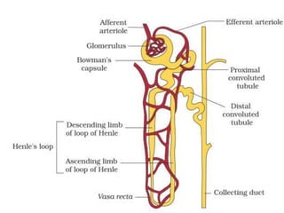
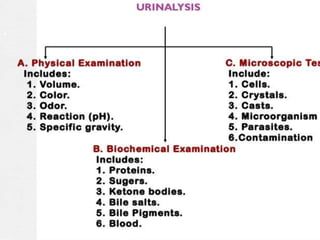
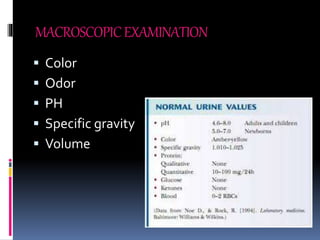
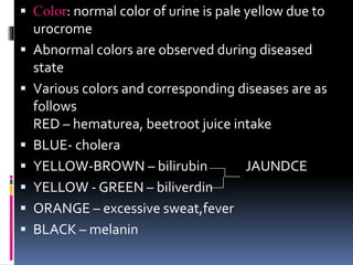
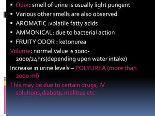
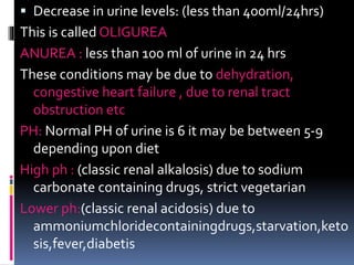
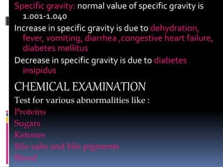
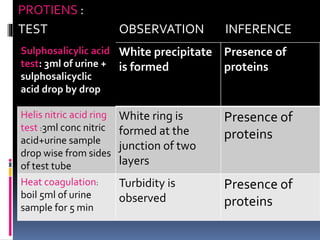
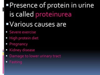
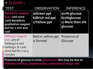
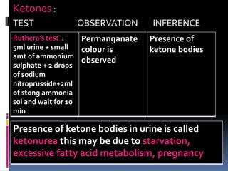
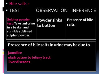
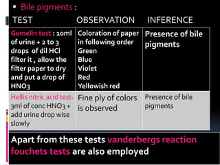
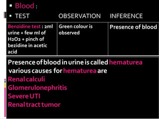
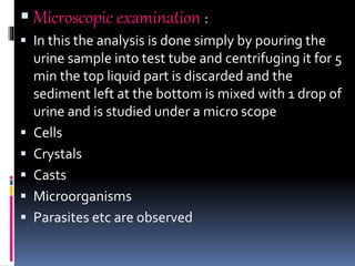
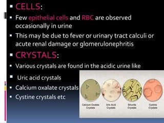
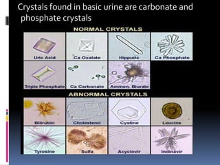
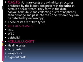
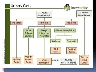
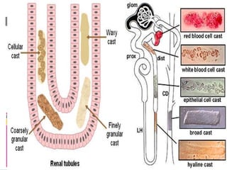
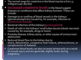
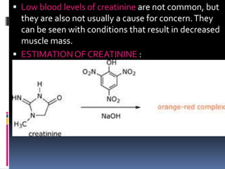
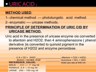
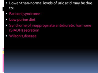
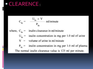
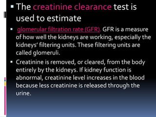
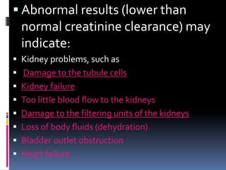
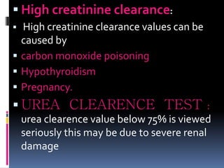
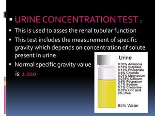
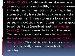
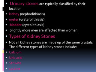
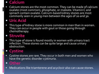
KIDNEY
FUNCTION
TESTS - navya
Y14PHD0120
Introduction
 The main functional unit of kidney is nephron
each kidney consists of millions of nephrons
 Urine is formed by the following mechanism
1.glomerular filtration
2.tubular reabsorbtion
3.tubular secretion
PURPOSE
 To diagnose the disease or disorders of
kidneys and urinary tract and other systematic
diseases that effect the kidney functioning
 Urine examination is usually employed to
diagnose most of the kidney disorders
 Urine examination is classified into 3 types
1.macroscopic examination
2.microscopic examination
3.chemical examination
MACROSCOPICEXAMINATION
 Color
 Odor
 PH
 Specific gravity
 Volume
 Color: normal color of urine is pale yellow due to
urocrome
 Abnormal colors are observed during diseased
state
 Various colors and corresponding diseases are as
follows
RED – hematurea, beetroot juice intake
 BLUE- cholera
 YELLOW-BROWN – bilirubin JAUNDCE
 YELLOW - GREEN – biliverdin
 ORANGE – excessive sweat,fever
 BLACK – melanin
 Odor: smell of urine is usually light pungent
 Various other smells are also observed
 AROMATIC :volatile fatty acids
 AMMONICAL: due to bacterial action
 FRUITY ODOR : ketonurea
Volume: normal value is 1000-
2000/24hrs(depending upon water intake)
Increase in urine levels – POLYUREA (more than
2000 ml)
This may be due to certain drugs, IV
solutions,diabetis mellitus etc
 Decrease in urine levels: (less than 400ml/24hrs)
This is called OLIGUREA
ANUREA : less than 100 ml of urine in 24 hrs
These conditions may be due to dehydration,
congestive heart failure , due to renal tract
obstruction etc
PH: Normal PH of urine is 6 it may be between 5-9
depending upon diet
High ph : (classic renal alkalosis) due to sodium
carbonate containing drugs, strict vegetarian
Lower ph:(classic renal acidosis) due to
ammoniumchloridecontainingdrugs,starvation,keto
sis,fever,diabetis
Specific gravity: normal value of specific gravity is
1.001-1.040
Increase in specific gravity is due to dehydration,
fever, vomiting, diarrhea ,congestive heart failure,
diabetes mellitus
Decrease in specific gravity is due to diabetes
insipidus
CHEMICAL EXAMINATION
Test for various abnormalities like :
Proteins
Sugars
Ketones
Bile salts and bile pigments
Blood
PROTIENS :
TEST OBSERVATION INFERENCE
Sulphosalicylic acid
test: 3ml of urine +
sulphosalicyclic
acid drop by drop
White precipitate
is formed
Presence of
proteins
Helis nitric acid ring
test :3ml conc nitric
acid+urine sample
drop wise from sides
of test tube
White ring is
formed at the
junction of two
layers
Presence of
proteins
Heat coagulation:
boil 5ml of urine
sample for 5 min
Turbidity is
observed
Presence of
proteins
Presence of protein in urine
is called proteinurea
Various causes are
 Severe exercise
 High protein diet
 Pregnancy
 Kidney disease
 Damage to lower urinary tract
 Fasting
 SUGARS :
TEST OBSERVATION INFERENCE
Benedicts reagent
test : 2ml urine
+2ml benedicts
qualitative reagent
boil for 2 min and
cool
a)Green ppt
b)Brick red ppt
c)Yellow ppt
a)1% glucose
b)2%glucose
c) More than 2%
glucose
Fehling's reagent
test: 2ml of
Fehling's A and
Fehling’s B +2ml
urine boil for 2 to 5
minutes
Red or yellow ppt
is formed
Presence of
Glucose
Presence of glucose in urine glucosurea this may be due to
diabetes mellitus ,hypertension, corticosterol drugs etc
Ketones :
TEST OBSERVATION INFERENCE
Ruthera’s test :
5ml urine + small
amt of ammonium
sulphate + 2 drops
of sodium
nitroprusside+2ml
of stong ammonia
sol and wait for 10
min
Permanganate
colour is
observed
Presence of
ketone bodies
Presence of ketone bodies in urine is called
ketonurea this may be due to starvation,
excessive fatty acid metabolism, pregnancy
 Bile salts :
 TEST OBSERVATION INFERENCE
Sulphur powder
test:Take 5ml urine
in a beaker and
sprinkle sublimed
sulphur powder
Powder sinks
to bottom
Presence of bile
salts
Prescence of bilesaltsinurinemaybedueto
jaundice
obstructiontobiliarytract
liverdiseases
 Bile pigments :
TEST OBSERVATION INFERENCE
Gemelin test : 10ml
of urine + 2 to 3
drops of dil HCl
filter it , allow the
filter paper to dry
and put a drop of
HNO3
Coloration of paper
in following order
Green
Blue
Violet
Red
Yellowish red
Presence of bile
pigments
Hellis nitric acid test:
3ml of conc HNO3 +
add urine drop wise
slowly
Fine ply of colors
is observed
Presence of bile
pigments
Apart from these tests vanderbergs reaction
fouchets tests are also employed
 Prescence of bile salts in urine may be
due to
 Jaundice (hemolytic or obstructive )
 obstruction to biliary tract
 liver diseases
 Blood :
 TEST OBSERVATION INFERENCE
Benzidine test : 2ml
urine + few ml of
H2O2 + pinch of
bezidine in acetic
acid
Green colour is
observed
Presence of blood
Presenceofbloodinurineiscalledhematurea
variouscauses forhematureaare
Renalcalculi
Glomerulonephritis
SevereUTI
Renaltracttumor
 Microscopic examination :
 In this the analysis is done simply by pouring the
urine sample into test tube and centrifuging it for 5
min the top liquid part is discarded and the
sediment left at the bottom is mixed with 1 drop of
urine and is studied under a micro scope
 Cells
 Crystals
 Casts
 Microorganisms
 Parasites etc are observed
 CELLS:
 Few epithelial cells and RBC are observed
occasionally in urine
 This may be due to fever or urinary tract calculi or
acute renal damage or glomerulonephritis
 CRYSTALS:
 Various crystals are found in the acidic urine like
 Uric acid crystals
 Calcium oxalate crystals
 Cystine crystals etc
Crystals found in basic urine are carbonate and
phosphate crystals
 CASTS: Urinary casts are cylindrical structures
produced by the kidney and present in the urine in
certain disease states.They form in the distal
convoluted tubule and collecting ducts of nephrons,
then dislodge and pass into the urine, where they can
be detected by microscopy.
 These casts are of two types
 CELLULAR CASTS
 RBC
 WBC
 epithelial
 ACELLULAR CASTS
 Hyaline casts
 fatty casts
 waxy casts
 pigment casts
NPN CONSTITUENTS OF URIne: NPN
stands for non protein nitrogenous compounds
 The nitrogen content of substances other
than protein in blood, tissues, and waste materials.
 i.e nitrogen associated with
 Urea
 uric acid
 creatine
 polypeptides.
 Approximately one half of the nonprotein nitrogen
in the blood is associated with urea
 Clinically significant NPN constituents along with
approximate plasma conc are listed below
 Urea - 45%
 Amino acids - 20%
 Uric acid - 20%
 Creatinine - 5%
 Creatine - 2%
 Ammonia - 0.2%
 UREA: BUN stands for blood urea nitrogen. Urea
nitrogen is formed when protein breaks down.
 A test can be done to measure the amount of urea
nitrogen in the blood.The BUN test is often done to
check kidney function.
 The normal result is generally 6 - 20 mg/dL.
 Higher-than-normal levels(Azotemia)
may be due to:
 Congestive heart faliure
 Excessive protein levels in the gastrointestinal tract
 Gastrointestinal bleeding
 Hypovolemia (dehydration)
 Heart attack
 Kidney disease, including glomerulonephritis,
pyelonephritis, and acute tubular necrosis
 Kidney failure (uremia)
 Shock
 Urinary tract obstruction
 Lower-than-normal levels may be due
to:
 Liver failure
 Low protein diet
 Malnutrition
 Over-hydration
ESTIMATION OF UREA :
 CREATININE : A creatinine test reveals important
information about your kidneys.
 Creatinine is a chemical waste product that's
produced by your muscle metabolism and to a
smaller extent by eating meat. Healthy kidneys
filter creatinine and other waste products from your
blood.The filtered waste products leave your body
in your urine.
 If your kidneys aren't functioning properly, an
increased level of creatinine may accumulate in your
blood. A serum creatinine test measures the level of
creatinine in your blood and gives you an estimate
of how well your kidneys filter (glomerular filtration
rate). A creatinine urine test can measure creatinine
in your urine.
 The normal range for creatinine in the blood may be 0.6 to 1.3
milligrams per deciliter
 Increased creatinine levels in the blood suggest
diseases or conditions that affect kidney function.These can
include:
 Damage to or swelling of blood vessels in the kidneys
(glomerulonephritis) caused by, for example, infection or
autoimmune diseases
 Bacterial infection of the kidneys (pyelonephritis)
 Death of cells in the kidneys' small tubes (acute tubular necrosis)
caused by, for example, drugs or toxins
 Prostate disease, kidney stone, or other causes of urinary tract
obstruction
 Reduced blood flow to the kidney due to
shock, dehydration, congestive heart failure, atherosclerosis, or
complications of diabetes
 Creatinine blood levels can also increase temporarily as a result
of muscle injury and are generally slightly lower during
pregnancy.
 Low blood levels of creatinine are not common, but
they are also not usually a cause for concern.They
can be seen with conditions that result in decreased
muscle mass.
 ESTIMATION OF CREATININE :
 URICACID :
 Greater-than-normal levels of uric acid
(hyperuricemia) may be due to:
 Acidosis
 Alcoholism
 Chemotherapy-related side effects
 Diabetes
 Excessive exercise
 Gout
 Hypoparathyroidism
 Lead poisoning
 Leukemia
 Medullary cystic kidney disease
 Nephrolithiasis
 Polycythemia vera
 Purine-rich diet
 Renal failure
 Toxemia of pregnancy
 Lower-than-normal levels of uric acid may be due
to:
 Fanconi syndrome
 Low purine diet
 Syndrome of inappropriate antidiuretic hormone
(SIADH) secretion
 Wilson's disease
 CLEARENCE:
 The creatinine clearance test is
used to estimate
 glomerular filtration rate (GFR). GFR is a measure
of how well the kidneys are working, especially the
kidneys’ filtering units.These filtering units are
called glomeruli.
 Creatinine is removed, or cleared, from the body
entirely by the kidneys. If kidney function is
abnormal, creatinine level increases in the blood
because less creatinine is released through the
urine.
 Abnormal results (lower than
normal creatinine clearance) may
indicate:
 Kidney problems, such as
 Damage to the tubule cells
 Kidney failure
 Too little blood flow to the kidneys
 Damage to the filtering units of the kidneys
 Loss of body fluids (dehydration)
 Bladder outlet obstruction
 Heart failure
 High creatinine clearance:
 High creatinine clearance values can be
caused by
 carbon monoxide poisoning
 Hypothyroidism
 Pregnancy.
 UREA CLEARENCE TEST :
urea clearence value below 75% is viewed
seriously this may be due to severe renal
damage
 URINE CONCENTRATIONTEST :
 This is used to asses the renal tubular function
 This test includes the measurement of specific
gravity which depends on concentration of solute
present in urine
 Normal specific gravity value
is 1.020
 RENAL CALCULI : A kidney stone, also known as
a renal calculus or nephrolith, is a calculus formed
in the kidneys from minerals in the urine. Kidney
stones typically leave the body by passage in the
urine stream, and many stones are formed and
passed without causing symptoms. If stones grow
to sufficient size (usually at least 3 millimeters
(0.12 in)) they can cause blockage of the ureter.
This leads to pain, most commonly beginning in
the flank or lower back and often radiating to
the genitals.This pain is often known as renal
colic and typically comes in waves lasting 20 to 60
minutes.
 Urinary stones are typically classified by their
location
 kidney (nephrolithiasis)
 ureter (ureterolithiasis)
 bladder (cystolithiasis)
 Slightly more men are affected than women.
 Types of Kidney Stones
 Not all kidney stones are made up of the same crystals.
The different types of kidney stones include:
 Calcium
 Uric acid
 Struvite
 cyctine
 Calcium
 Calcium stones are the most common.They can be made of calcium
oxalate (most common), phosphate, or maleate.Vitamin C and
spinach contain oxalate. Calcium-based kidney stones are most
commonly seen in young men between the ages of 20 and 30.
 Uric Acid
 This type of kidney stone is more common in men than in women.
They can occur in people with gout or those going through
chemotherapy.
 Struvite
 This type of stone is found mostly in women with urinary tract
infection.These stones can be quite large and cause urinary
obstruction.
 Cystine
 Cystine stones are rare.They occur in both men and women who
have the genetic disorder cystinuria.
 Other
 Medications like triamterene and acyclovir also can cause stones.
 CAUSES:
 various causes for kidney stones are
 Dehydration
 Protein(High dietary intake of animal protein )
 Sodium
 Refined sugars
 Fructose
 High fructose corn syrup
 Oxalate
 Grapefruit juice
 Apple juice increase the risk of kidney stone
formation.
 SYMPTOMS :
 blood in the urine
 vomiting
 nausea
 discolored or foul-smelling urine
 chills
 Fever
 DIAGNOSIS :
 abdominal X-rays
 ultrasound of the kidney
 MRI of the abdomen and kidneys
 abdominal CT scan
 TREATMENT :
 Drinking six to eight glasses of water a day increases
urine flow. People who are dehydrated or have severe
nausea and vomiting may need intravenous fluids.
Other treatment options include:
 Medication
 Pain relief may require narcotic medications.The
presence of infection requires treatment with
antibiotics. Other medications include:
 allopurinol for uric acid stones
 diuretics
 sodium bicarbonate or sodium citrate
 phosphorus solutions
 Lithotripsy:
 Extracorporeal shock wave lithotripsy uses sound waves
to break up large stones so they can more easily pass
down the ureters into your bladder.This procedure can
be uncomfortable and may require light anesthesia. It
can cause bruising on the abdomen and back and
bleeding around the kidney and nearby organs.
 Tunnel Surgery :
 (Percutaneous Nephrolithotomy)
 Stones are removed through a small incision in your
back and may be needed when:
 the stone causes obstruction and infection or is
damaging the kidneys
 the stone has grown too large to pass
 pain cannot be controlled
 Ureteroscopy
 When a stone is stuck in the ureter or bladder,
your doctor may use an instrument called a
ureteroscope to remove it. A small wire with a
camera attached is inserted into the urethra and
passed into the bladder.A small cage is used to
snag the stone and remove it.The stone is then
sent to the laboratory for analysis.
kidney function tests

kidney function tests

  • 1.
  • 2.
    Introduction  The mainfunctional unit of kidney is nephron each kidney consists of millions of nephrons  Urine is formed by the following mechanism 1.glomerular filtration 2.tubular reabsorbtion 3.tubular secretion
  • 4.
    PURPOSE  To diagnosethe disease or disorders of kidneys and urinary tract and other systematic diseases that effect the kidney functioning  Urine examination is usually employed to diagnose most of the kidney disorders  Urine examination is classified into 3 types 1.macroscopic examination 2.microscopic examination 3.chemical examination
  • 6.
    MACROSCOPICEXAMINATION  Color  Odor PH  Specific gravity  Volume
  • 7.
     Color: normalcolor of urine is pale yellow due to urocrome  Abnormal colors are observed during diseased state  Various colors and corresponding diseases are as follows RED – hematurea, beetroot juice intake  BLUE- cholera  YELLOW-BROWN – bilirubin JAUNDCE  YELLOW - GREEN – biliverdin  ORANGE – excessive sweat,fever  BLACK – melanin
  • 8.
     Odor: smellof urine is usually light pungent  Various other smells are also observed  AROMATIC :volatile fatty acids  AMMONICAL: due to bacterial action  FRUITY ODOR : ketonurea Volume: normal value is 1000- 2000/24hrs(depending upon water intake) Increase in urine levels – POLYUREA (more than 2000 ml) This may be due to certain drugs, IV solutions,diabetis mellitus etc
  • 9.
     Decrease inurine levels: (less than 400ml/24hrs) This is called OLIGUREA ANUREA : less than 100 ml of urine in 24 hrs These conditions may be due to dehydration, congestive heart failure , due to renal tract obstruction etc PH: Normal PH of urine is 6 it may be between 5-9 depending upon diet High ph : (classic renal alkalosis) due to sodium carbonate containing drugs, strict vegetarian Lower ph:(classic renal acidosis) due to ammoniumchloridecontainingdrugs,starvation,keto sis,fever,diabetis
  • 10.
    Specific gravity: normalvalue of specific gravity is 1.001-1.040 Increase in specific gravity is due to dehydration, fever, vomiting, diarrhea ,congestive heart failure, diabetes mellitus Decrease in specific gravity is due to diabetes insipidus CHEMICAL EXAMINATION Test for various abnormalities like : Proteins Sugars Ketones Bile salts and bile pigments Blood
  • 11.
    PROTIENS : TEST OBSERVATIONINFERENCE Sulphosalicylic acid test: 3ml of urine + sulphosalicyclic acid drop by drop White precipitate is formed Presence of proteins Helis nitric acid ring test :3ml conc nitric acid+urine sample drop wise from sides of test tube White ring is formed at the junction of two layers Presence of proteins Heat coagulation: boil 5ml of urine sample for 5 min Turbidity is observed Presence of proteins
  • 12.
    Presence of proteinin urine is called proteinurea Various causes are  Severe exercise  High protein diet  Pregnancy  Kidney disease  Damage to lower urinary tract  Fasting
  • 13.
     SUGARS : TESTOBSERVATION INFERENCE Benedicts reagent test : 2ml urine +2ml benedicts qualitative reagent boil for 2 min and cool a)Green ppt b)Brick red ppt c)Yellow ppt a)1% glucose b)2%glucose c) More than 2% glucose Fehling's reagent test: 2ml of Fehling's A and Fehling’s B +2ml urine boil for 2 to 5 minutes Red or yellow ppt is formed Presence of Glucose Presence of glucose in urine glucosurea this may be due to diabetes mellitus ,hypertension, corticosterol drugs etc
  • 14.
    Ketones : TEST OBSERVATIONINFERENCE Ruthera’s test : 5ml urine + small amt of ammonium sulphate + 2 drops of sodium nitroprusside+2ml of stong ammonia sol and wait for 10 min Permanganate colour is observed Presence of ketone bodies Presence of ketone bodies in urine is called ketonurea this may be due to starvation, excessive fatty acid metabolism, pregnancy
  • 15.
     Bile salts:  TEST OBSERVATION INFERENCE Sulphur powder test:Take 5ml urine in a beaker and sprinkle sublimed sulphur powder Powder sinks to bottom Presence of bile salts Prescence of bilesaltsinurinemaybedueto jaundice obstructiontobiliarytract liverdiseases
  • 16.
     Bile pigments: TEST OBSERVATION INFERENCE Gemelin test : 10ml of urine + 2 to 3 drops of dil HCl filter it , allow the filter paper to dry and put a drop of HNO3 Coloration of paper in following order Green Blue Violet Red Yellowish red Presence of bile pigments Hellis nitric acid test: 3ml of conc HNO3 + add urine drop wise slowly Fine ply of colors is observed Presence of bile pigments Apart from these tests vanderbergs reaction fouchets tests are also employed
  • 17.
     Prescence ofbile salts in urine may be due to  Jaundice (hemolytic or obstructive )  obstruction to biliary tract  liver diseases
  • 18.
     Blood : TEST OBSERVATION INFERENCE Benzidine test : 2ml urine + few ml of H2O2 + pinch of bezidine in acetic acid Green colour is observed Presence of blood Presenceofbloodinurineiscalledhematurea variouscauses forhematureaare Renalcalculi Glomerulonephritis SevereUTI Renaltracttumor
  • 19.
     Microscopic examination:  In this the analysis is done simply by pouring the urine sample into test tube and centrifuging it for 5 min the top liquid part is discarded and the sediment left at the bottom is mixed with 1 drop of urine and is studied under a micro scope  Cells  Crystals  Casts  Microorganisms  Parasites etc are observed
  • 20.
     CELLS:  Fewepithelial cells and RBC are observed occasionally in urine  This may be due to fever or urinary tract calculi or acute renal damage or glomerulonephritis  CRYSTALS:  Various crystals are found in the acidic urine like  Uric acid crystals  Calcium oxalate crystals  Cystine crystals etc
  • 21.
    Crystals found inbasic urine are carbonate and phosphate crystals
  • 22.
     CASTS: Urinarycasts are cylindrical structures produced by the kidney and present in the urine in certain disease states.They form in the distal convoluted tubule and collecting ducts of nephrons, then dislodge and pass into the urine, where they can be detected by microscopy.  These casts are of two types  CELLULAR CASTS  RBC  WBC  epithelial  ACELLULAR CASTS  Hyaline casts  fatty casts  waxy casts  pigment casts
  • 25.
    NPN CONSTITUENTS OFURIne: NPN stands for non protein nitrogenous compounds  The nitrogen content of substances other than protein in blood, tissues, and waste materials.  i.e nitrogen associated with  Urea  uric acid  creatine  polypeptides.  Approximately one half of the nonprotein nitrogen in the blood is associated with urea
  • 26.
     Clinically significantNPN constituents along with approximate plasma conc are listed below  Urea - 45%  Amino acids - 20%  Uric acid - 20%  Creatinine - 5%  Creatine - 2%  Ammonia - 0.2%  UREA: BUN stands for blood urea nitrogen. Urea nitrogen is formed when protein breaks down.  A test can be done to measure the amount of urea nitrogen in the blood.The BUN test is often done to check kidney function.
  • 27.
     The normalresult is generally 6 - 20 mg/dL.  Higher-than-normal levels(Azotemia) may be due to:  Congestive heart faliure  Excessive protein levels in the gastrointestinal tract  Gastrointestinal bleeding  Hypovolemia (dehydration)  Heart attack  Kidney disease, including glomerulonephritis, pyelonephritis, and acute tubular necrosis  Kidney failure (uremia)  Shock  Urinary tract obstruction
  • 28.
     Lower-than-normal levelsmay be due to:  Liver failure  Low protein diet  Malnutrition  Over-hydration ESTIMATION OF UREA :
  • 29.
     CREATININE :A creatinine test reveals important information about your kidneys.  Creatinine is a chemical waste product that's produced by your muscle metabolism and to a smaller extent by eating meat. Healthy kidneys filter creatinine and other waste products from your blood.The filtered waste products leave your body in your urine.  If your kidneys aren't functioning properly, an increased level of creatinine may accumulate in your blood. A serum creatinine test measures the level of creatinine in your blood and gives you an estimate of how well your kidneys filter (glomerular filtration rate). A creatinine urine test can measure creatinine in your urine.
  • 30.
     The normalrange for creatinine in the blood may be 0.6 to 1.3 milligrams per deciliter  Increased creatinine levels in the blood suggest diseases or conditions that affect kidney function.These can include:  Damage to or swelling of blood vessels in the kidneys (glomerulonephritis) caused by, for example, infection or autoimmune diseases  Bacterial infection of the kidneys (pyelonephritis)  Death of cells in the kidneys' small tubes (acute tubular necrosis) caused by, for example, drugs or toxins  Prostate disease, kidney stone, or other causes of urinary tract obstruction  Reduced blood flow to the kidney due to shock, dehydration, congestive heart failure, atherosclerosis, or complications of diabetes  Creatinine blood levels can also increase temporarily as a result of muscle injury and are generally slightly lower during pregnancy.
  • 31.
     Low bloodlevels of creatinine are not common, but they are also not usually a cause for concern.They can be seen with conditions that result in decreased muscle mass.  ESTIMATION OF CREATININE :
  • 32.
  • 33.
     Greater-than-normal levelsof uric acid (hyperuricemia) may be due to:  Acidosis  Alcoholism  Chemotherapy-related side effects  Diabetes  Excessive exercise  Gout  Hypoparathyroidism  Lead poisoning  Leukemia  Medullary cystic kidney disease  Nephrolithiasis  Polycythemia vera  Purine-rich diet  Renal failure  Toxemia of pregnancy
  • 34.
     Lower-than-normal levelsof uric acid may be due to:  Fanconi syndrome  Low purine diet  Syndrome of inappropriate antidiuretic hormone (SIADH) secretion  Wilson's disease
  • 35.
  • 36.
     The creatinineclearance test is used to estimate  glomerular filtration rate (GFR). GFR is a measure of how well the kidneys are working, especially the kidneys’ filtering units.These filtering units are called glomeruli.  Creatinine is removed, or cleared, from the body entirely by the kidneys. If kidney function is abnormal, creatinine level increases in the blood because less creatinine is released through the urine.
  • 37.
     Abnormal results(lower than normal creatinine clearance) may indicate:  Kidney problems, such as  Damage to the tubule cells  Kidney failure  Too little blood flow to the kidneys  Damage to the filtering units of the kidneys  Loss of body fluids (dehydration)  Bladder outlet obstruction  Heart failure
  • 38.
     High creatinineclearance:  High creatinine clearance values can be caused by  carbon monoxide poisoning  Hypothyroidism  Pregnancy.  UREA CLEARENCE TEST : urea clearence value below 75% is viewed seriously this may be due to severe renal damage
  • 39.
     URINE CONCENTRATIONTEST:  This is used to asses the renal tubular function  This test includes the measurement of specific gravity which depends on concentration of solute present in urine  Normal specific gravity value is 1.020
  • 41.
     RENAL CALCULI: A kidney stone, also known as a renal calculus or nephrolith, is a calculus formed in the kidneys from minerals in the urine. Kidney stones typically leave the body by passage in the urine stream, and many stones are formed and passed without causing symptoms. If stones grow to sufficient size (usually at least 3 millimeters (0.12 in)) they can cause blockage of the ureter. This leads to pain, most commonly beginning in the flank or lower back and often radiating to the genitals.This pain is often known as renal colic and typically comes in waves lasting 20 to 60 minutes.
  • 42.
     Urinary stonesare typically classified by their location  kidney (nephrolithiasis)  ureter (ureterolithiasis)  bladder (cystolithiasis)  Slightly more men are affected than women.  Types of Kidney Stones  Not all kidney stones are made up of the same crystals. The different types of kidney stones include:  Calcium  Uric acid  Struvite  cyctine
  • 43.
     Calcium  Calciumstones are the most common.They can be made of calcium oxalate (most common), phosphate, or maleate.Vitamin C and spinach contain oxalate. Calcium-based kidney stones are most commonly seen in young men between the ages of 20 and 30.  Uric Acid  This type of kidney stone is more common in men than in women. They can occur in people with gout or those going through chemotherapy.  Struvite  This type of stone is found mostly in women with urinary tract infection.These stones can be quite large and cause urinary obstruction.  Cystine  Cystine stones are rare.They occur in both men and women who have the genetic disorder cystinuria.  Other  Medications like triamterene and acyclovir also can cause stones.
  • 44.
     CAUSES:  variouscauses for kidney stones are  Dehydration  Protein(High dietary intake of animal protein )  Sodium  Refined sugars  Fructose  High fructose corn syrup  Oxalate  Grapefruit juice  Apple juice increase the risk of kidney stone formation.
  • 45.
     SYMPTOMS : blood in the urine  vomiting  nausea  discolored or foul-smelling urine  chills  Fever  DIAGNOSIS :  abdominal X-rays  ultrasound of the kidney  MRI of the abdomen and kidneys  abdominal CT scan
  • 46.
     TREATMENT : Drinking six to eight glasses of water a day increases urine flow. People who are dehydrated or have severe nausea and vomiting may need intravenous fluids. Other treatment options include:  Medication  Pain relief may require narcotic medications.The presence of infection requires treatment with antibiotics. Other medications include:  allopurinol for uric acid stones  diuretics  sodium bicarbonate or sodium citrate  phosphorus solutions
  • 47.
     Lithotripsy:  Extracorporealshock wave lithotripsy uses sound waves to break up large stones so they can more easily pass down the ureters into your bladder.This procedure can be uncomfortable and may require light anesthesia. It can cause bruising on the abdomen and back and bleeding around the kidney and nearby organs.  Tunnel Surgery :  (Percutaneous Nephrolithotomy)  Stones are removed through a small incision in your back and may be needed when:  the stone causes obstruction and infection or is damaging the kidneys  the stone has grown too large to pass  pain cannot be controlled
  • 48.
     Ureteroscopy  Whena stone is stuck in the ureter or bladder, your doctor may use an instrument called a ureteroscope to remove it. A small wire with a camera attached is inserted into the urethra and passed into the bladder.A small cage is used to snag the stone and remove it.The stone is then sent to the laboratory for analysis.