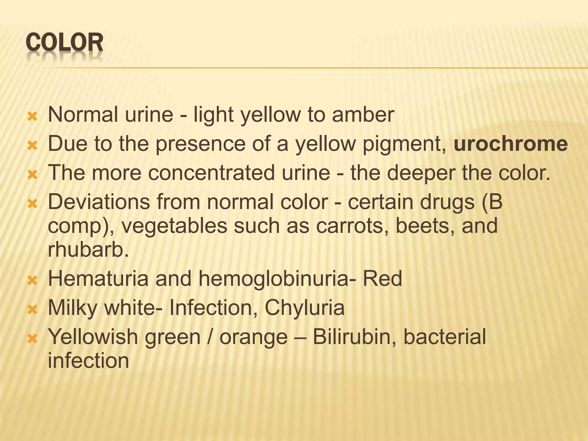
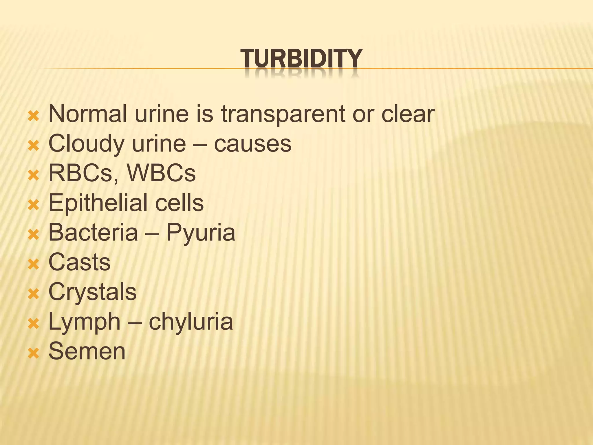
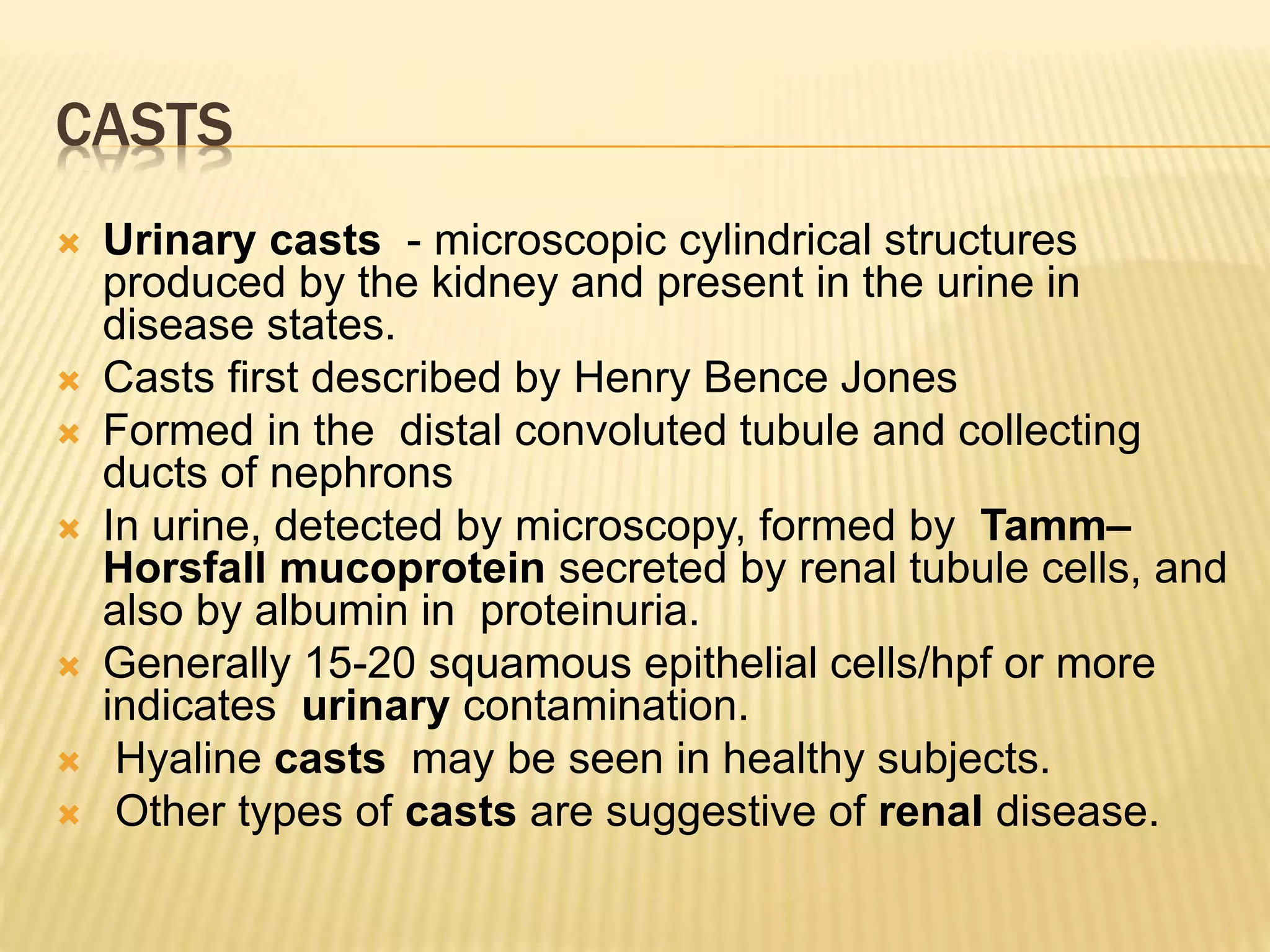
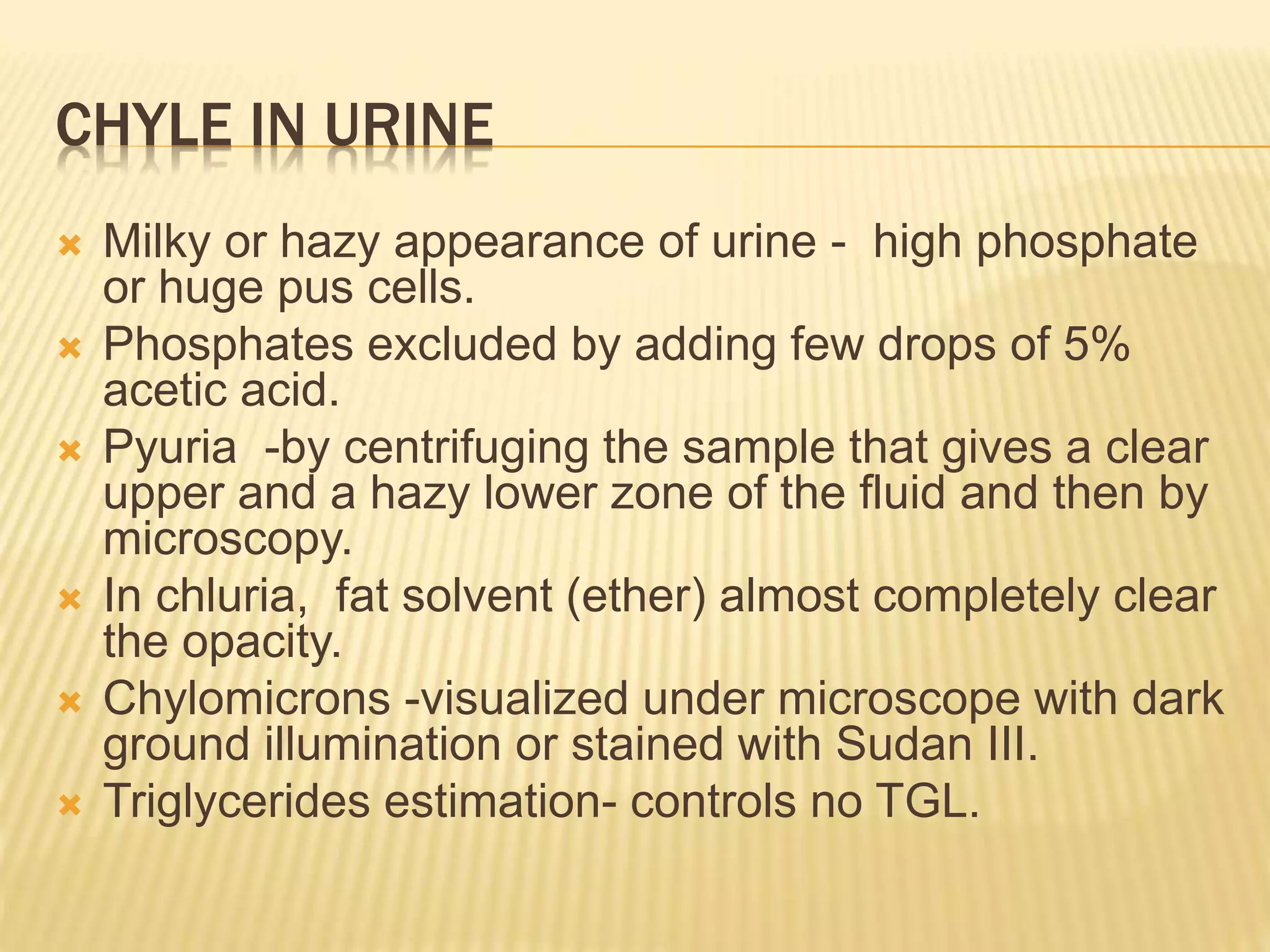
URINE EXAMINATION
Dr.S.Sethupathy,M.D.,Ph.D.,
Professor of Biochemistry,
Rajah Muthiah Medical College,
Annnamalai University
KIDNEY FUNCTIONS
 Excretion waste products such as urea,
Creatinine, uric acid
 Maintenance of acid base balance
 Maintenance of water and electrolyte
balance
 Hormone function – Erythropoietin
 Regulation of blood calcium level –
Formation of calcitriol by 1 α – hydroxylase
URINE EXAMINATION
A complete physical, chemical and
microscopic examination plays a an
important role in diagnosis of renal
and urinary tract diseases.
COLOR
 Normal urine - light yellow to amber
 Due to the presence of a yellow pigment, urochrome
 The more concentrated urine - the deeper the color.
 Deviations from normal color - certain drugs (B
comp), vegetables such as carrots, beets, and
rhubarb.
 Hematuria and hemoglobinuria- Red
 Milky white- Infection, Chyluria
 Yellowish green / orange – Bilirubin, bacterial
infection
ABNORMAL COLORS AND CAUSES
ODOR
Slightly aromatic, characteristic
of freshly voided urine.
Urine becomes more ammonia-
like upon standing due to
bacterial activity.
Odor Condition
Aromatic odor Normal
Fruity smell ( acetone) Diabeteic ketosis (Type 1)
Fishy odor UTI
Ammoniacal odor UTI caused by
pseudomonas & proteus
Mousy odor Phenyl ketonuria
Burnt sugar Maple syrup urine
disease
TURBIDITY
 Normal urine is transparent or clear
 Cloudy urine – causes
 RBCs, WBCs
 Epithelial cells
 Bacteria – Pyuria
 Casts
 Crystals
 Lymph – chyluria
 Semen
CASTS
 Urinary casts - microscopic cylindrical structures
produced by the kidney and present in the urine in
disease states.
 Casts first described by Henry Bence Jones
 Formed in the distal convoluted tubule and collecting
ducts of nephrons
 In urine, detected by microscopy, formed by Tamm–
Horsfall mucoprotein secreted by renal tubule cells, and
also by albumin in proteinuria.
 Generally 15-20 squamous epithelial cells/hpf or more
indicates urinary contamination.
 Hyaline casts may be seen in healthy subjects.
 Other types of casts are suggestive of renal disease.
URINARY CASTS
RBC CASTS
LEUCOCYTE (WBC) CASTS
HYALINE CASTS
GRANULAR CASTS
PIGMENT CASTS
CRYSTALS
CHYLE IN URINE
 Milky or hazy appearance of urine - high phosphate
or huge pus cells.
 Phosphates excluded by adding few drops of 5%
acetic acid.
 Pyuria -by centrifuging the sample that gives a clear
upper and a hazy lower zone of the fluid and then by
microscopy.
 In chluria, fat solvent (ether) almost completely clear
the opacity.
 Chylomicrons -visualized under microscope with dark
ground illumination or stained with Sudan III.
 Triglycerides estimation- controls no TGL.
SEMEN IN URINE
 In retrograde ejaculation semen goes
backward into the bladder.
 It is not a harmful condition.
 The semen mixes with urine and
passes out of the body the next time the
man urinates.
PH
 Ranges from 4.5 - 8.0.
 Average is 6.0, slightly acidic.
 Under 5.0 is acidic, higher than 8.0 is
alkaline
 Vegetarian diets increase alkalinity.
 Bacterial infections also increase alkalinity.
ACIDIC URINE
 High protein diets increase acidity.
 Dehydration
 Diabetic ketoacidosis
 Diarrhea
 starvation
ALKALINE URINE
 gastric suctioning , pyloric obstruction,
vomiting - that takes away stomach acids
 kidney failure
 kidney tubular acidosis
 respiratory alkalosis
 urinary tract infection
SPECIFIC GRAVITY
 Specific gravity measures the ability of the kidneys to
concentrate or dilute urine depending on fluid intake.
 Normal range 1.005 - 1.030 , average range 1.010 -
1.025.
 Low specific gravity is associated with conditions
like diabetes insipidus, excessive water intake,
diuretic use or chronic renal failure.
 High specific gravity levels are associated with
diabetes mellitus, adrenal abnormalities or excessive
water loss due to vomiting, diarrhea or kidney
inflammation. A specific gravity that never varies is
indicative of severe renal failure.
VOLUME
HIGH S.G
 Volume loss (dehydration, vomiting, diarrhea,
fever)
 Heart failure
 Renal artery stenosis
 Shock
 Syndrome of inappropriate antidiuretic
hormone
 Diabetes mellitus-glucosuria
LOW S.G
 Diabetes insipidus
 Excessive water intake
 Diuretic use
 Chronic renal failure (fixed S.G- 1.010).
 Renal failure , Pyelonephritis ,
 Glomerulonephritis
 Psychogenic polydipsia
 Malignant hypertension
ABNORMAL CONSTITUENTS
 Glucose – Glycosuria
 Normally the filtered glucose is reabsorbed by the
renal tubules
 Renal threshold for glucose- Blood glucose level
160-180 mg/dl
 If blood glucose levels exceeds renal threshold level,
glucose spills over into the urine.
 Main cause: diabetes mellitus
 Renal glucosuria- reduced renal threshold level
 Blood glucose level-normal but urine glucose
positive
PROTEIN (ALBUMIN)
 Albumin do not pass through glomerulus.
 Presence indicates abnormal increased
permeability of the glomerulus membrane.
 Non-pathological causes are: pregnancy,
physical exertion, increased protein
consumption.
 Pathological causes are: glomerulonephritis ,
bacterial toxins, chemical poisons.
URINARY PROTEIN -ALBUMINURIA
 Less than 30 mg /24 hrs or 30 mg/gm
Creatinine or 30 µg/mg creatinie is normal.
 30 – 300 mg /24 hrs or 30 -300 mg/gm
creatinine or 30-300 µg/mg Creatinine
is microalbuminuria
 More than 300 mg / day – frank albuminuria
 Healthy albumin to Creatinine ratio:
 less than or equal to 3.5 mg/mmol ( 30mg / gm
Creatinine) - Creatinine mol.wt: 113
URINARY KETONES
 Ketone bodies -acetoacetic acid, beta-
hydroxybutyric acid,
 Causes: diabetes mellitus, starvation, ketogenic
diet
 drinking excess alcohol
 excessive vomiting , pregnancy
 illness or infection
 heart attack , emotional or physical trauma
 medications, such as corticosteroids and
diuretics
URINARY KETONES RANGE
 Under 0.6 millimoles per liter or 6mg/dl -
Normal
 ketone level 0.6 to 1.5 millimoles per liter
or 6-15 mg/dl higher than normal; test again
in 2 to 4 hours
 1.6 to 3.0 millimoles per liter or 16- 30 mg/dl
–Moderate
 Above 3.0 millimoles per liter or > 30mg/dl -
high
BILIRUBIN
 Fouchet’s test: This is a simple and sensitive test.
Five ml of fresh urine in a test tube is mxed with 2.5 ml of
10% of barium chloride .A precipitate - barium sulphate-
bilirubin complex forms
 Filter and spread the precipitate on a filter paper.
 one drop of Fouchet’s reagent is added to the precipitate.
(Fouchet’s reagent consists of 25 grams of trichloroacetic
acid, 10 ml of 10% ferric chloride, and distilled water 100
ml).
 Immediate development of blue-green color around the
drop indicates presence of bilirubin .
 Causes: liver disorders, cirrhosis, hepatitis, obstruction of
bile duct
FOUCHET’S TEST - POSITIVE
Reagent strips or tablets impregnated with diazo
reagent: :Based on reaction of bilirubin with diazo reagent;
Color change is proportional to the concentration of bilirubin.
Tablets (Ictotest) detect 0.05-0.1 mg of bilirubin/dl of urine;
Reagent strip tests are less sensitive (0.5 mg/dl).
UROBILINOGEN
 Normal – 0.2 mg/dl - 1mg/dl
 Less than 0.2 mg/dl - low
 More than 1 mg /dl – high
 High urobilinogen level
 Hemolytic anemias, malaria
 False Positive Results - Elevated nitrate in the
urine
 High carbohydrate intake
 Timing (test is done later in the afternoon)
 Drugs that make the urine red, such as
phenazopyridine (Pyridium)
 Low urobilinogen-- cholestasis - causes
include :
 Bile duct blockage (gallstones, cysts, and
tumors)
 Liver disease/damage
 Pregnancy
 Severe infection
 Pancreatic cancer
 Broad-spectrum antibiotics
 False Negative Results
 Exposure of urine sample to direct sunlight–
urobilinogen breaks down
HEMATURIA- CAUSES
 Kidney infections (pyelonephritis).
 A bladder or kidney stone.
 Enlarged prostate.
 Kidney disease. .
 Cancer.
 Inherited disorders. Sickle cell anemia
 Alport syndrome, which affects the filtering membranes in the
glomeruli of the kidneys.
 Kidney injury. A blow or other injury to your kidneys from an
accident or contact sports can cause
 Medications. drug cyclophosphamide and penicillin , aspirin,
heparin can cause urinary bleeding.
 Strenuous exercise. It's rare for strenuous exercise to lead to
gross hematuria,
HEMOGLOBINURIA -CAUSES
 Acute glomerulonephritis
 Burns , Renal cancer , Malaria
 Microangiopathies,
 Transfusion reactions
 IgM autoimmune hemolytic anemia
 Glucose-6-phosphate dehydrogenase deficiency
 Pyelonephritis
 Sickle cell anemia
 Tuberculosis of the urinary tract
 March hemoglobinuria secondary to repetitive impacts on the
body, usually the feet
 Athletic nephritis secondary to strenuous exercise
 Acute lead poisoning
DIP-STICK TEST –HEMATURIA
 A nondiagnostic screening test.
 A positive result is due to oxidation of a test-strip
reagent; it does not confirm that blood cells are
present.
 Factors that can cause a positive result on a dipstick
test include hemoglobinuria, myoglobinuria,
concentrated urine, menstrual blood in the urine
sample, and rigorous exercise
 False-positive results can occur (certain dyes or
drugs, beets, oxalates).
 The strip is read in a min. (sensitivity of 100% and a
specificity of 99% in detecting 1-5 RBCs per high-
power field (hpf).
DIP-STICK TEST FOR HEMATURIA
 Confirm presence of RBCs microscopically.
 If the specific gravity of the urine is very low
(< 1.007), microscopy can fail to detect owing
to cell lysis.
 Repeat the test after restricting the patient’s
fluid intake
 Other investigations should be avoided, and
the dipstick and microscopic urinalysis
should be repeated twice within 2 weeks.
LEUKOCYTES AND NITRITES
 Normal - 0-5 WBCs per high power field
(wbc/hpf)
 High number of leukocytes
may indicate the presence of urinary tract
infection
 If the test for leukocyte esterase is positive
but finds no nitrite, an infection may still be
present.
 E. coli bacteria are most commonly
associated with nitrites in the urine.
Test Report format Results
Physical- color and odor
Turbidity
Specific gravity
pH
Chemical - protein
Glucose
ketones
urobilinogen
bilirubin
blood
leukocytes
nitrites
Microscopic - RBCs
Leukocytes
Crystals
Casts
Bacteria /yeast
Thank you

Urine examination

  • 1.
    URINE EXAMINATION Dr.S.Sethupathy,M.D.,Ph.D., Professor ofBiochemistry, Rajah Muthiah Medical College, Annnamalai University
  • 2.
    KIDNEY FUNCTIONS  Excretionwaste products such as urea, Creatinine, uric acid  Maintenance of acid base balance  Maintenance of water and electrolyte balance  Hormone function – Erythropoietin  Regulation of blood calcium level – Formation of calcitriol by 1 α – hydroxylase
  • 3.
    URINE EXAMINATION A completephysical, chemical and microscopic examination plays a an important role in diagnosis of renal and urinary tract diseases.
  • 4.
    COLOR  Normal urine- light yellow to amber  Due to the presence of a yellow pigment, urochrome  The more concentrated urine - the deeper the color.  Deviations from normal color - certain drugs (B comp), vegetables such as carrots, beets, and rhubarb.  Hematuria and hemoglobinuria- Red  Milky white- Infection, Chyluria  Yellowish green / orange – Bilirubin, bacterial infection
  • 5.
  • 7.
    ODOR Slightly aromatic, characteristic offreshly voided urine. Urine becomes more ammonia- like upon standing due to bacterial activity.
  • 8.
    Odor Condition Aromatic odorNormal Fruity smell ( acetone) Diabeteic ketosis (Type 1) Fishy odor UTI Ammoniacal odor UTI caused by pseudomonas & proteus Mousy odor Phenyl ketonuria Burnt sugar Maple syrup urine disease
  • 9.
    TURBIDITY  Normal urineis transparent or clear  Cloudy urine – causes  RBCs, WBCs  Epithelial cells  Bacteria – Pyuria  Casts  Crystals  Lymph – chyluria  Semen
  • 10.
    CASTS  Urinary casts- microscopic cylindrical structures produced by the kidney and present in the urine in disease states.  Casts first described by Henry Bence Jones  Formed in the distal convoluted tubule and collecting ducts of nephrons  In urine, detected by microscopy, formed by Tamm– Horsfall mucoprotein secreted by renal tubule cells, and also by albumin in proteinuria.  Generally 15-20 squamous epithelial cells/hpf or more indicates urinary contamination.  Hyaline casts may be seen in healthy subjects.  Other types of casts are suggestive of renal disease.
  • 11.
  • 12.
  • 13.
  • 14.
  • 15.
  • 16.
  • 17.
  • 18.
    CHYLE IN URINE Milky or hazy appearance of urine - high phosphate or huge pus cells.  Phosphates excluded by adding few drops of 5% acetic acid.  Pyuria -by centrifuging the sample that gives a clear upper and a hazy lower zone of the fluid and then by microscopy.  In chluria, fat solvent (ether) almost completely clear the opacity.  Chylomicrons -visualized under microscope with dark ground illumination or stained with Sudan III.  Triglycerides estimation- controls no TGL.
  • 19.
    SEMEN IN URINE In retrograde ejaculation semen goes backward into the bladder.  It is not a harmful condition.  The semen mixes with urine and passes out of the body the next time the man urinates.
  • 20.
    PH  Ranges from4.5 - 8.0.  Average is 6.0, slightly acidic.  Under 5.0 is acidic, higher than 8.0 is alkaline  Vegetarian diets increase alkalinity.  Bacterial infections also increase alkalinity.
  • 21.
    ACIDIC URINE  Highprotein diets increase acidity.  Dehydration  Diabetic ketoacidosis  Diarrhea  starvation
  • 22.
    ALKALINE URINE  gastricsuctioning , pyloric obstruction, vomiting - that takes away stomach acids  kidney failure  kidney tubular acidosis  respiratory alkalosis  urinary tract infection
  • 23.
    SPECIFIC GRAVITY  Specificgravity measures the ability of the kidneys to concentrate or dilute urine depending on fluid intake.  Normal range 1.005 - 1.030 , average range 1.010 - 1.025.  Low specific gravity is associated with conditions like diabetes insipidus, excessive water intake, diuretic use or chronic renal failure.  High specific gravity levels are associated with diabetes mellitus, adrenal abnormalities or excessive water loss due to vomiting, diarrhea or kidney inflammation. A specific gravity that never varies is indicative of severe renal failure.
  • 24.
  • 25.
    HIGH S.G  Volumeloss (dehydration, vomiting, diarrhea, fever)  Heart failure  Renal artery stenosis  Shock  Syndrome of inappropriate antidiuretic hormone  Diabetes mellitus-glucosuria
  • 26.
    LOW S.G  Diabetesinsipidus  Excessive water intake  Diuretic use  Chronic renal failure (fixed S.G- 1.010).  Renal failure , Pyelonephritis ,  Glomerulonephritis  Psychogenic polydipsia  Malignant hypertension
  • 27.
    ABNORMAL CONSTITUENTS  Glucose– Glycosuria  Normally the filtered glucose is reabsorbed by the renal tubules  Renal threshold for glucose- Blood glucose level 160-180 mg/dl  If blood glucose levels exceeds renal threshold level, glucose spills over into the urine.  Main cause: diabetes mellitus  Renal glucosuria- reduced renal threshold level  Blood glucose level-normal but urine glucose positive
  • 28.
    PROTEIN (ALBUMIN)  Albumindo not pass through glomerulus.  Presence indicates abnormal increased permeability of the glomerulus membrane.  Non-pathological causes are: pregnancy, physical exertion, increased protein consumption.  Pathological causes are: glomerulonephritis , bacterial toxins, chemical poisons.
  • 29.
    URINARY PROTEIN -ALBUMINURIA Less than 30 mg /24 hrs or 30 mg/gm Creatinine or 30 µg/mg creatinie is normal.  30 – 300 mg /24 hrs or 30 -300 mg/gm creatinine or 30-300 µg/mg Creatinine is microalbuminuria  More than 300 mg / day – frank albuminuria  Healthy albumin to Creatinine ratio:  less than or equal to 3.5 mg/mmol ( 30mg / gm Creatinine) - Creatinine mol.wt: 113
  • 30.
    URINARY KETONES  Ketonebodies -acetoacetic acid, beta- hydroxybutyric acid,  Causes: diabetes mellitus, starvation, ketogenic diet  drinking excess alcohol  excessive vomiting , pregnancy  illness or infection  heart attack , emotional or physical trauma  medications, such as corticosteroids and diuretics
  • 31.
    URINARY KETONES RANGE Under 0.6 millimoles per liter or 6mg/dl - Normal  ketone level 0.6 to 1.5 millimoles per liter or 6-15 mg/dl higher than normal; test again in 2 to 4 hours  1.6 to 3.0 millimoles per liter or 16- 30 mg/dl –Moderate  Above 3.0 millimoles per liter or > 30mg/dl - high
  • 32.
    BILIRUBIN  Fouchet’s test:This is a simple and sensitive test. Five ml of fresh urine in a test tube is mxed with 2.5 ml of 10% of barium chloride .A precipitate - barium sulphate- bilirubin complex forms  Filter and spread the precipitate on a filter paper.  one drop of Fouchet’s reagent is added to the precipitate. (Fouchet’s reagent consists of 25 grams of trichloroacetic acid, 10 ml of 10% ferric chloride, and distilled water 100 ml).  Immediate development of blue-green color around the drop indicates presence of bilirubin .  Causes: liver disorders, cirrhosis, hepatitis, obstruction of bile duct
  • 33.
    FOUCHET’S TEST -POSITIVE Reagent strips or tablets impregnated with diazo reagent: :Based on reaction of bilirubin with diazo reagent; Color change is proportional to the concentration of bilirubin. Tablets (Ictotest) detect 0.05-0.1 mg of bilirubin/dl of urine; Reagent strip tests are less sensitive (0.5 mg/dl).
  • 34.
    UROBILINOGEN  Normal –0.2 mg/dl - 1mg/dl  Less than 0.2 mg/dl - low  More than 1 mg /dl – high  High urobilinogen level  Hemolytic anemias, malaria  False Positive Results - Elevated nitrate in the urine  High carbohydrate intake  Timing (test is done later in the afternoon)  Drugs that make the urine red, such as phenazopyridine (Pyridium)
  • 35.
     Low urobilinogen--cholestasis - causes include :  Bile duct blockage (gallstones, cysts, and tumors)  Liver disease/damage  Pregnancy  Severe infection  Pancreatic cancer  Broad-spectrum antibiotics  False Negative Results  Exposure of urine sample to direct sunlight– urobilinogen breaks down
  • 36.
    HEMATURIA- CAUSES  Kidneyinfections (pyelonephritis).  A bladder or kidney stone.  Enlarged prostate.  Kidney disease. .  Cancer.  Inherited disorders. Sickle cell anemia  Alport syndrome, which affects the filtering membranes in the glomeruli of the kidneys.  Kidney injury. A blow or other injury to your kidneys from an accident or contact sports can cause  Medications. drug cyclophosphamide and penicillin , aspirin, heparin can cause urinary bleeding.  Strenuous exercise. It's rare for strenuous exercise to lead to gross hematuria,
  • 37.
    HEMOGLOBINURIA -CAUSES  Acuteglomerulonephritis  Burns , Renal cancer , Malaria  Microangiopathies,  Transfusion reactions  IgM autoimmune hemolytic anemia  Glucose-6-phosphate dehydrogenase deficiency  Pyelonephritis  Sickle cell anemia  Tuberculosis of the urinary tract  March hemoglobinuria secondary to repetitive impacts on the body, usually the feet  Athletic nephritis secondary to strenuous exercise  Acute lead poisoning
  • 38.
    DIP-STICK TEST –HEMATURIA A nondiagnostic screening test.  A positive result is due to oxidation of a test-strip reagent; it does not confirm that blood cells are present.  Factors that can cause a positive result on a dipstick test include hemoglobinuria, myoglobinuria, concentrated urine, menstrual blood in the urine sample, and rigorous exercise  False-positive results can occur (certain dyes or drugs, beets, oxalates).  The strip is read in a min. (sensitivity of 100% and a specificity of 99% in detecting 1-5 RBCs per high- power field (hpf).
  • 39.
    DIP-STICK TEST FORHEMATURIA  Confirm presence of RBCs microscopically.  If the specific gravity of the urine is very low (< 1.007), microscopy can fail to detect owing to cell lysis.  Repeat the test after restricting the patient’s fluid intake  Other investigations should be avoided, and the dipstick and microscopic urinalysis should be repeated twice within 2 weeks.
  • 40.
    LEUKOCYTES AND NITRITES Normal - 0-5 WBCs per high power field (wbc/hpf)  High number of leukocytes may indicate the presence of urinary tract infection  If the test for leukocyte esterase is positive but finds no nitrite, an infection may still be present.  E. coli bacteria are most commonly associated with nitrites in the urine.
  • 41.
    Test Report formatResults Physical- color and odor Turbidity Specific gravity pH Chemical - protein Glucose ketones urobilinogen bilirubin blood leukocytes nitrites Microscopic - RBCs Leukocytes Crystals Casts Bacteria /yeast
  • 42.