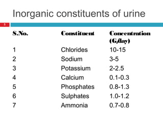
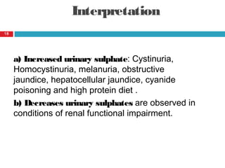
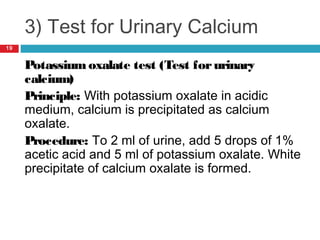
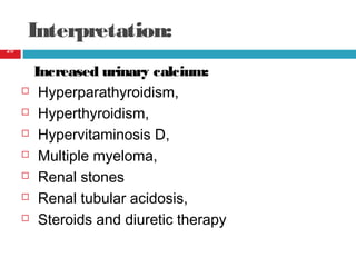
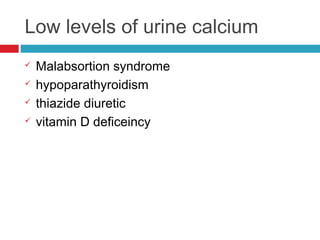
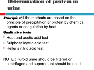
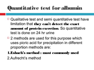
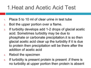
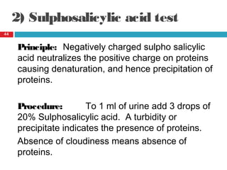
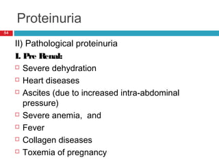
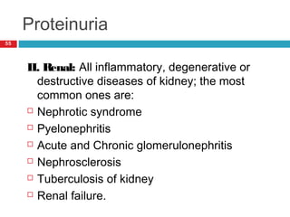
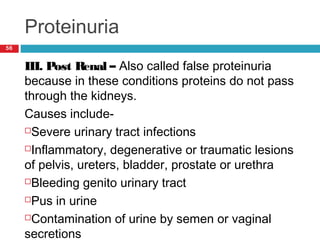
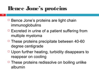
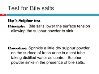
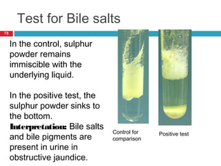
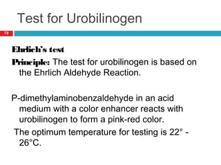
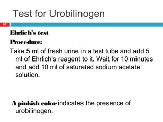
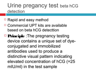
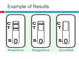
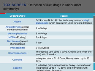
URINE BIO CHEMICALURINE BIO CHEMICAL
ANALYSIS & ITS CLINICALANALYSIS & ITS CLINICAL
SIGNIFICANCESIGNIFICANCE
Presented by
Dr Neeraj Nirala
GUIDE:
Dr Neera Samar
UNIT HEAD:
Dr R.L Meena
SEMINAR PRESENTATION
RNT MEDICAL COLLEGE &
MB GOVT HOSPITAL
Introduction
URINE : is excretory product
from body
The basic steps in urine
formation are :
1. Glomerular Filtration
2. Tubular Reabsorption
3. Tubular Secretion
4. Excretion.
Composition of normal urine
3
 Normal urine contains 90-
95% water and about 60
G/day of solid constituents
which may be organic or
inorganic in nature
Organic constituents of urine
4
S.No. Constituent Concentration
1. Urea 10-45 mg/dl
2. Uric acid 2.5- 7.0 mg/dl
3. Creatinine 0.6-1.2 mg/dl
Inorganic constituents of urine
5
S.No. Constituent Concentration
(G/day)
1 Chlorides 10-15
2 Sodium 3-5
3 Potassium 2-2.5
4 Calcium 0.1-0.3
5 Phosphates 0.8-1.3
6 Sulphates 1.0-1.2
7 Ammonia 0.7-0.8
Purpose
 General evaluation of health
 Diagnosis of disease or disorders of the
kidneys or urinary tract
 Diagnosis of other systemic disease that affect
kidney function
 Monitoring of patients e.g like diabetes
 Screening for drug abuse eg. amphetamines
Collection of urine specimens
 The first voided morning urine (the most
common)
 Random urine (for emergency)
 Clean-catch, midstream urine (for urine
culture)
 Urinary catheter
 Suprapubic aspiration
Attention
 Need to be examined within 1-2 hour
MIDSTREAMURINE COLLECTION
 In both sexes atleast 200ml of urine should
be passed before collecting a midstream
urine specimen without interrupting the
urine flow
 Gloves should be weared for urinary
collection
 A sterile urine container should be used
 Should be clean catch
HOW TO COLLECT
 MALES
1. Foreskin is retracted and held back
throughout the collection
2. Urethral meatus is cleansed by moist gauze
FEMALE
1. Where possible use vaginal tampon
2. Hold labia well separated during collection
3. Gently cleanse the periurethral area with
several moistened gauze pieces from
anterior to posterior
URINARY CATHETERSPECIMEN: Nearly
200ml urine should be passed before
collection of urine
Specimen
Collection
Supra-pubic Needle Aspiration
A fine needle is
passed through
sterilized
suprapubic skin
directly into a
full bladder.
Uncontaminated
urine can then
be aspirated
Sample preservation
12
 There is no single all purpose preservative
 For determination of urea, ammonia, nitrogen and
calcium- Hydrochloric acid is used (2 N or
concentrated HCL)
 For determination of sodium, potassium, chloride,
bicarbonate, calcium, phosphorus, urea,
ammonia, amino acids, creatinine, proteins,
reducing substances and ketone bodies- Thymol
is used
 Toluene is a very satisfactory preservative for
urine
Bio-Chemical AnalysisBio-Chemical Analysis
Chemical examination
 Urine PH
 Protein
 Reducing sugars
 Ketones
 Bilirubin
 Urobilinogen
 Nitrites
 Occult blood
 Beta hcG
 Certains drugs
Normal constituents Abnormal constituents
Inorganic constituents
Chloride
Sulphate
Calcium
ammonia
Organic constituents
Urea
Creatinine
Uric acid
Analysis of normal constituents
A) Inorganic constituents
i) Test forUrinary chlorides(silver nitrate test)
Principle- Silver chloride is precipitated in the
presence of nitric acid and silver nitrate.
Procedure-Take 2 ml of urine and add 0.5 ml of
concentrated nitric acid and 1 ml of silver
nitrate. A white precipitate of silver chloride
appears.
15
Interpretation:
a) Increased Urinary chlorides:
Polydipsia, use of diuretics
and Addison's disease.
b) Decreased urinary chlorides:
Excessive sweating, fasting,
diarrhea, excessive vomiting,
edema, diabetes Insipidus,
infections and adrenocortical
hyper function (Cushing's
syndrome).
16
2) Test for urinary sulphates
Bariumchloride test (test for
sulphates)
Principle: Urinary sulphate is
precipitated as barium
sulphate on reaction with
barium chloride solution.
Procedure: Take 3 ml of urine
and add 1 ml of conc. HCl
and 2 ml of 10% barium
chloride. White precipitate
indicates the presence of
sulphates.
17
Interpretation
a) Increased urinary sulphate: Cystinuria,
Homocystinuria, melanuria, obstructive
jaundice, hepatocellular jaundice, cyanide
poisoning and high protein diet .
b) Decreases urinary sulphates are observed in
conditions of renal functional impairment.
18
3) Test for Urinary Calcium
Potassium oxalate test (Test forurinary
calcium)
Principle: With potassium oxalate in acidic
medium, calcium is precipitated as calcium
oxalate.
Procedure: To 2 ml of urine, add 5 drops of 1%
acetic acid and 5 ml of potassium oxalate. White
precipitate of calcium oxalate is formed.
19
Interpretation:
Increased urinary calcium:
 Hyperparathyroidism,
 Hyperthyroidism,
 Hypervitaminosis D,
 Multiple myeloma,
 Renal stones
 Renal tubular acidosis,
 Steroids and diuretic therapy
20
Low levels of urine calcium
 Malabsortion syndrome
 hypoparathyroidism
 thiazide diuretic
 vitamin D deficeincy
4) Test for Ammonia
Principle : ammonia is evaporated
when made alkaline
TEST: To 5ml urine add 2% sodium
carbonate till solution is alkaline to
litmus
Boil the solution and place a piece of
litmus paper at the mouth of test
tube.
Colourchanges to blue
Damp red litmus paper turns blue
on exposure to fumes of ammonia.
22
Interpretation:
a) Increased urinary ammonia:
 Diabetic keto acidosis
 ingestion of acid forming foods
 urinary tract infections.
b) Decreased urinary ammonia:
 Alkalosis
 Nephritis
B) Tests for Organic
Constituents
1) Test forUrea
(Sodium hypobromite test)
Principle: When urea is treated with
sodium hypobromite, it
decomposes to give nitrogen.
Procedure: To 2 ml of urine in a test
tube, add 4-5 drops of sodium
hypobromite.
Observe the effervescence of
nitrogen gas.
24
B) Tests for Organic
Constituents
2) Urease test forurea
Principle: Soyabean powder contains the
enzyme urease. This enzyme under pH 7-8
and temperature 550
C decomposes urea in to
ammonia and carbon dioxide which together
form ammonium carbonate (alkaline
component) which changes the slightly acidic
reaction(yellow color) to alkaline reaction(pink
color).
25
B) Tests for Organic
Constituents
3.Biuret test forurea
Principle : Urea when heated decomposes with
the liberation of ammonia and the formation of
biuret. Biuret is dissolved in water and
develops a violet color forming a complex with
alkaline copper sulphate solution.
26
Interpretation:
a) Increased urinary urea: Fever, diabetes
mellitus, excess of adrenocortical activity
b) Decreased urinary urea: Liver diseases,
metabolic or respiratory acidosis, nephritis
27
ii) Tests for Creatinine
a) Jaffe’s Reaction (Test for
creatinine)
Principle: Creatinine reacts with picric
acid in the alkaline medium to form a
reddish colored complex of creatinine
picrate
Procedure: Take 5 ml of urine and add
an aqueous solution of picric acid.
Make the mixture alkaline with NaOH
solution. A red color is produced.
28
ii) Tests for Creatinine
b) Nitroprusside test
To 5 ml of urine add a few drops of sodium
nitroprusside and make the solution alkaline
with sodium hydroxide (NaOH).
A ruby red color is formed that turns yellow.
This test is also called Wey’s test.
29
ii) Tests for Creatinine
c) Nitroprusside -Acetic acid test (Salkowaski
test)
Procedure : Take 5 ml of urine, add a few drops
of sodium nitroprusside and then make the
solution alkaline with NaOH. A ruby red color is
formed that turns yellow.
To the yellow precipitate, add an excess of
acetic acid and heat the solution.
A green color is obtained that turns blue upon
standing.
30
ii) Test for Creatinine
Interpretation:
a) Creatinuria- Creatinuria occurs in uncontrolled
diabetes mellitus, thyrotoxicosis, myasthenia
gravis, starvation, infancy, pregnancy, muscular
disorders and in growing period.
b) Decreased urinary creatinine: Renal failure
31
Creatinine clearance ( CLcr)
 Measurement of GFR
 Done on 24 urine sample
 GFR can be estimated by using Cockcroft-
Gault formula for creatinine clearance
CLcr(ml/min)= 140 – age/ serum creatinine x
body weight in kg/72 (females multply wt by
0.85)
USES :
 for adjusting of drug doses
 To predict the remaining renal function
iii) Tests for Uric acid
a) Phospho tungstic acid test (Foruric
acid)
Principle: Uric acid is a reducing agent in
alkaline medium. It reduces phospho
tungstic acid to tungsten blue.
Procedure: Take 2 ml of urine, add a few
drops of phospho tungstic acid reagent
followed by a few drops 20% sodium
carbonate. Observe the appearance of
blue color
33
iii)Tests for Uric acid
b) Benedict’s test
Principle- Uric acid is soluble in alkali. The blue
color is developed due to the reduction of
phospho tungstic acid by uric acid.
Procedure : To 2 ml of urine , add a few drops of
Benedict’s uric acid reagent and add a pinch
of anhydrous sodium carbonate and mix. A
deep blue color indicates the presence of uric
acid.
34
SIGINIFICANCE
HIGHLEVELS
 metastasis
 Disease that results in breakdown of muscle fibers
(rhabdomyolysis)
 Disorders that affect the bone marrow (myeloproliferative
disorder)
 Fanconi syndrome
 Gout andHigh-purine diet
LOWLEVELS
Kidney disease both AKI and CKD
Abnormal constituents
Urinary pH/reaction
 Reaction reflects ability of kidney to maintain
normal hydrogen ion concentration in plasma
& ECF
 Normal urine is acidic, pH ranges
between 4.5-8.0 with a mean of 6.0 in 24
hours
Measurement of urinary pH
37
Urinary pH is measured
by-
 pH papers
 Litmus papers
Variations of urinary pH
A) Acidic urine-
Physiologically, It is found after
 A protein rich diet
 Heavy exercise
Pathologically
 Ketosis-diabetes, starvation, fever
 Systemic acidosis
 UTI- E.coli
 Acidification therapy
Alkaline urine
 Strict vegetarian
 Systemic alkalosis
 UTI- Proteus
 Alkalization therapy
Determination of protein in
urine
Principle:All the methods are based on the
principle of precipitation of protein by chemical
agents or coagulation by heat.
Qualitative tests
 Heat and acetic acid test
 Sulphosalicyclic acid test
 Heller’s nitric acid test
NOTE : Turbid urine should be filtered or
centrifuged and supernatant should be used
Quantitative test foralbumin
 Qualitative test and semi quantitative test have
limitation that they can’t detect the exact
amount of protein excretion. So quantitative
test is done on 24 hr urine
 2 methods are used for this purpose which
uses picric acid for precipitation in different
proportion methods are: 
1.Esbach’s method : most commonly used
2.Aufrecht’s method
1.Heat and Acetic Acid Test 
1. Place 5 to 10 ml of clear urine in test tube
2. Boil the upper portion over a flame.
3. If turbidity develops add 1-2 drops of glacial acetic
acid. Sometimes turbidity may be due to
phosphate or carbonate precipitation.it is so then
glacial acetic acid clear up the turbidity if it is due
to protein then precipitation will be there after the
addition of acetic acid
4. Reboil the specimen
5. If turbidity is present protein is present .if there is
no turbidity at upper portion then protein is absent
Grading o f turbidity
 Negative : No cloudiness
 Trace: Barely visible cloudiness.
 1+ : definite cloud without granular flocculation
 2+ : heavy and granular cloud without granular
flocculation
 3+ : densed cloud with marked flocculation.
 4+ : thick curdy precipitation and coagulation-
2) Sulphosalicylic acid test
44
Principle: Negatively charged sulpho salicylic
acid neutralizes the positive charge on proteins
causing denaturation, and hence precipitation of
proteins.
Procedure: To 1 ml of urine add 3 drops of
20% Sulphosalicylic acid. A turbidity or
precipitate indicates the presence of proteins.
Absence of cloudiness means absence of
proteins.
Test for proteins
45
S.No. Observation Inference
(Approximate
protein
concentration)
mg/100 ml
1) Barely visible turbidity 5
2) Distinct turbidity 10-30
3) Moderate turbidity 40-100
4) Heavy Turbidity 200-500
5) Heavy
flocculent/precipitation
500
3) Heller’s Nitric acid ring test
46
Principle: Concentrated HNO3 causes
denaturation and hence precipitation of proteins.
Procedure: Take 3-5 ml of concentrated nitric
acid. Incline the tube and to it add carefully, 2-3
ml of urine, so that it forms the upper layer
without disturbing the lower HNO3 layer.
In a positive reaction, a white zone of
precipitate protein will appear at the junction of
two liquids.
Quantitative test: ESBACK’S
test
Reagent : dissolve 5g of picric acid with 10g of
citric acid in 500ml water
Procedure: fill an Esbacks albuminometer with
urine to mark U. Add reagent to mark R. Close
with a rubber stopper, invert several times znd
set aside in cool place for 24 hrs. Read off the
results according to the marking on the tube
which shows albumin in g/L .
Principle: albumin + picric acid= albumin piariate
(get deposited)
Precautions
1. If SG of urine > 1.030 urine
should be diluted with
water, otherwise albumin
picrate formed will not
settle down
2. If turbid urine, it should be
filtered
3. If urine alkaline it should be
acidifiedwith 3% acetic acid
otherwise acidity of
esback’s reagent will be
ESBACK’S
ALBUMINOMETER
MARKING’S IN ESBACK’S
ALBUMINOMETER
LOWER
PART
UPPER
PART
Tests for proteins
50
Interpretation- Insignificant amounts of proteins
are excreted in urine in normal health not
exceeding 20-80 mg/dl. This small amount is not
detectable by routine methods.
Under certain conditions, as much as 20 G or
more proteins may be excreted per day in urine.
The most common type of proteinuria is
albuminuria; hence proteinuria and albuminuria
are used synonymously.
Proteinuria
51
When proteins appear in urine in detectable
amounts, it is called proteinuria. It can be
caused by-
a) Increased glomerular permeability
b) Reduced tubular reabsorption
c) Increased secretion of proteins
d) Increased concentration of low molecular
weight proteins in the plasma
Proteinuria
 Urine protein composition ( total 150mg/day in
adults )
1. Tamm-horsfall protein 70 mg
2. Blood group related antigen 35 mg
3. Albumin 15 mg
4. Mucopolysaccharide 15 mg
5. Immunoglobulins 5 mg
6. Rest hormones and enzymes 10 mg
Proteinuria
53
Proteinuria may be- Physiological or
Pathological
I) Physiological Proteinuria
Causes include-
 Violent exercise
 Pregnancy
 Postural
 Alimentary
 Exposure to cold
Proteinuria
54
II) Pathological proteinuria
I. Pre Renal:
 Severe dehydration
 Heart diseases
 Ascites (due to increased intra-abdominal
pressure)
 Severe anemia, and
 Fever
 Collagen diseases
 Toxemia of pregnancy
Proteinuria
55
II. Renal: All inflammatory, degenerative or
destructive diseases of kidney; the most
common ones are:
 Nephrotic syndrome
 Pyelonephritis
 Acute and Chronic glomerulonephritis
 Nephrosclerosis
 Tuberculosis of kidney
 Renal failure.
Proteinuria
56
III. Post Renal – Also called false proteinuria
because in these conditions proteins do not pass
through the kidneys.
Causes include-
Severe urinary tract infections
Inflammatory, degenerative or traumatic lesions
of pelvis, ureters, bladder, prostate or urethra
Bleeding genito urinary tract
Pus in urine
Contamination of urine by semen or vaginal
secretions
During heat and acetic acid
 If cloudiness is seen it may be due to
phosphate or carbonates confirmby adding
3% glacial acetic acid. if it is due to protein
cloudiness persist and if it is due to the
phosphate the cloudiness disappears and if it
is due to carbonate cloudiness disappears
with effervescence.
 If cloudiness is disappeared when nitric acid
is added then it is due to mucin and
nucleoprotein. 
 If cloudiness appears with the tube is being
Bence Jone’s proteins
58
 Bence Jone’s proteins are light chain
immunoglobulins
 Excreted in urine of a patient suffering from
multiple myeloma
 These proteins precipitate between 40-60
degree centigrade
 Upon further heating, turbidity disappears to
reappear on cooling
 These proteins redissolve on boiling unlike
albumin
Tests for reducing sugars
59
1) Benedict’s Test
Principle :Benedict's reagent contains sodium
carbonate, coppersulphate and sodium
citrate.
In the alkaline medium provided by Sodium
carbonate, the copper remains as cupric
hydroxide. Sodium citrate acts as a stabilizing
agent to prevent precipitation of cupric
hydroxide.
In alkaline medium, sugars form enediols,
cupric ions are reduced, and corresponding
Tests for reducing sugars
60
Benedict’s test
Procedure:
Take 5 ml of Benedict's
reagent, add 8 drops of
urine. Boil for 2 minutes or
keep it in the boiling water
bath for 5 minutes. A light
green, yellow or brick red
color is produced
depending on
concentration of urinary
glucose.
Negative
test
Positive
test
Tests for reducing sugars
61
Observations
Benedict‘s test is a semi quantitative test. The
color of the precipitate gives a rough estimate of
the reducing sugars present in the given sample.
Green color- Up to 0.5 g%
Green precipitate - 0.5-1.0 g%(+)
Yellow precipitate -1.0-1.5 g% (++)
Orange precipitate- 1.5-2.0 g% (+++)
Brickred precipitate- >2.0 g% (++++)
Tests for reducing sugars
62
2) Fehling Test
 Another reduction test
 Contains KOH and Sodium potassium Tartrate
in place of Sodium carbonate and sodium
citrate in Benedict’s reagent
 Not used any more, since it is less sensitive,
less specific and the strong alkali causes
caramelisation of the sugars present in the
given sample.
Tests for reducing sugars
63
Interpretation:
 Positive Benedict's test signifies Glycosuria.
 Glycosuria is a non-specific term. Any reducing
sugar found in urine is denoted by glycosuria
 Lactosuria - in lactose intolerance
 Galactosuria - in galactosemia
 Fructosuria - in hereditary fructose
intolerance
 Pentosuria - in essential Pentosuria
Glycosuria
64
Causes of Glucosuria are: (Glucosuria and
Glycosuria are used synonymously)
a. Renal glycosuria- pregnancy, hereditary,
diseases of renal tubules, heavy metal poisoning .
b. Diabetes mellitus
c. Alimentary glucosuria
d. Hyperthyroidism, hyperpituitarism and
hyperadrenalism
e. Stress, severe infections, increased intracranial
pressure
Glycosuria
65
Examples of non-carbohydrate substances
which give a positive Benedict's reaction are:
a) Creatinine
b) Ascorbic acid
c) Glucuronates
d) Drugs: Salicylates, PAS and Isoniazid
Tests for Ketone bodies
66
Rothera’s Test
Principle: Nitroprusside in
alkaline medium reacts with a
ketone group to form a purple
ring. It is given by acetone
and acetoacetate, but not by
Beta hydroxy butyric acid.
Tests for Ketone bodies
67
Procedure:
 Saturate 5 ml of urine with solid ammonium
sulphate and add 0.5 ml of freshly prepared
sodium nitroprusside (5%).
 Mix well and add liquor ammonia from the
side of tube.
 A purple ring at the junction of the liquid
indicates the presence of ketone bodies.
2) Gerhardt’s ferric chloride
test68
Principle: A purplish color is given by aceto
acetate. On boiling acetoacetate is converted to
acetone and does not give this test positive.
This test is only given by acetoacetate and not
by beta hydroxy butyric acid directly.
Tests for Ketone bodies
69
Procedure- Add 10% ferric chloride solution drop
by drop to 5 ml of urine in a test tube. If
phosphates are present, precipitates of ferric
phosphates may form, that should be filtered off
and the ferric chloride is added.
False positive Gerhardt’s test may be obtained
with Salicylic acid and Salicylates.
Tests for Ketone bodies
70
3) Test forβ- OHbutyric acid
• No direct test for β- OH butyric acid
• Indirect test is performed
Procedure : Add a few drops of Acetic acid to
urine diluted 1:1 with distilled water. Boil for a
few minutes to remove acetone and aceto
acetic acid.
Add about 1.0 ml of H2O2, warm gently, cool, and
perform Rothera’s test .
Tests for Ketone bodies
71
Acetone, acetoacetate and beta hydroxy butyrate
are the ketone bodies. Ketonemia and hence
ketonuria occurs mostly in conditions of glucose
deprivation.
Causes of Ketonuria:
1) Uncontrolled diabetes mellitus
2) Starvation
3) High fat feeding
4) Heavy exercise
5) Toxemia of pregnancy
Tests for bile pigments
72
1) Fouchet’s test
Principle: BaCl2 reacts with sulphate in urine to
form barium sulphate. If bilirubin is present in
urine, it adheres to precipitate and is
detected by oxidation to form biliverdin
(Green) with FeCl3 in the presence of
trichloro acetic acid. Nitric acid oxidizes
bilirubin to biliverdin giving different colors
from green to violet.
Tests for bile pigments
73
1) Fouchet’s test
Procedure:
Take 5 ml of 10% BaCl2 to 10 ml
of urine and filter. Dry the filter
paper and add a few drops of
Fouchet's reagent (Prepared
by adding 10 mg of 10% FeCl3
to 100 ml of 25% TCA). A
green color is obtained due to
oxidation of bilirubin to
biliverdin.
Tests for bile pigments
74
2) Gmelin’s test
Principle: Nitric acid oxidizes Bilirubin to
Biliverdin giving different colors from green to
violet.
Procedure: To about 5 ml of concentrated HNO3
in a test tube, add an equal volume of urine
carefully so that the two liquids do not mix.
At the junction of two liquids various colored
rings (Green, blue, red, violet etc.) will be
formed.
Tests for bile pigments
75
3) Iodine test
Procedure : Dilute some tincture of iodine with
one to two volumes of water and layer it
carefully on to some urine in a test tube, a
green ring at the junction of two fluids
indicates the presence of Bilirubin.
It is not a sensitive test, can not detect small
amount of bilirubin present in the given
sample.
Tests for bile pigments
76
Interpretation
Bilirubin in urine means increased amount of
conjugated bilirubin because unconjugated
bilirubin is water insoluble and is also bound to
albumin, hence cannot cross the glomerular
membrane.
Causes of bilirubinuria are:
1) Moderate to severe hepatocellular damage
2) Obstruction of bile duct- Intra or extra hepatic
In prehepatic jaundice, bilirubin is absent in urine.
Test for Bile salts
77
Hay’s Sulphurtest
Principle: Bile salts lower the surface tension
allowing the sulphur powder to sink
Procedure: Sprinkle a little dry sulphur powder
on the surface of fresh urine in a test tube
taking distilled water as control. Sulphur
powder sinks in the presence of bile salts.
Test for Bile salts
78
Control for
comparison
Positive test
In the control, sulphur
powder remains
immiscible with the
underlying liquid.
In the positive test, the
sulphur powder sinks to
the bottom.
Interpretation: Bile salts
and bile pigments are
present in urine in
obstructive jaundice.
Test for Urobilinogen
79
Ehrlich’s test
Principle: The test for urobilinogen is based on
the Ehrlich Aldehyde Reaction.
P-dimethylaminobenzaldehyde in an acid
medium with a color enhancer reacts with
urobilinogen to form a pink-red color.
The optimum temperature for testing is 22° -
26°C.
Test for Urobilinogen
80
Ehrlich’s test
Procedure:
Take 5 ml of fresh urine in a test tube and add 5
ml of Ehrlich's reagent to it. Wait for 10 minutes
and add 10 ml of saturated sodium acetate
solution.
A pinkish colorindicates the presence of
urobilinogen.
Test for Urobilinogen
81
Interpretation:
Urobilinogen is found in urine in hepatic and
prehepatic jaundice.
It is present in excessive amount in prehepatic
jaundice and is completely absent in post
hepatic jaundice.
An increased urobilinogen concentration in
urine is a sensitive index of liver dysfunction or
hemolytic disorders.
Test for blood
82
Benzidine Test
Principle: Hydrogen peroxide liberated from Hb
oxidizes Benzidine to form a colored derivative.
Procedure:
To 3 ml of saturated Benzidine solution in
glacial acetic acid, add 2 ml of urine and add 1
ml of 3% H2O2. A blue or green color develops
within 10 minutes indicating the presence of
blood. Color developing after 10 minutes is not
a positive test but it is due to oxidation of
Benzidine by atmospheric oxygen.
Test for blood
83
Interpretation:
Presence of blood in urine is called hematuria.
a. Gross hematuria:
Urine appears reddish in gross hematuria and
this is observed in renal stones, malignancies,
trauma, tuberculosis and acute
glomerulonephritis.
Test for blood
84
b. Microscopic hematuria:
Blood is not visible to naked eyes. It is observed in:
 Malignant hypertension,
Sickle cell anemia,
Coagulation disorders,
Polycystic kidney disease,
Incompatible blood transfusion,
Auto immune hemolytic anemia.
Biochemistry dept MBGH Central
lab URINE
ANALYZER
URINE DIPSTICK READ AT 60
SEC
URINE REPORT BY CENTRAL
LAB
PARAMETER
S
1.SG
2.PH
3.LEUCOCYTES
4.NITROGEN
5.PROTEIN
6.GLUCOSE
7.KETONES
8.UROBILINOGEN
9.BILIRUBIN
10.ERYTHROCYTES
11.COLOUR
Urine pregancy test beta hCG
detection
 Rapid and easy method
 Commercial UPT kits are available
based on beta hCG detecttion
 Principle :The pregnancy testing
device contains a unique set of dye-
conjugated and immobilized
antibodies used to produce a
distinctive visual pattern indicating
elevated concentration of hCG (=25
mIU/ml) in the test sample
Example of Results
SUBSTANCE URINE
Alcohol
6–24 hours Note: Alcohol tests may measure ethyl
glucuronide, which can stay in urine for up to 80 hours
Amphetamines(except
methamphetamine)
1 to 3 days
Methamphetamine 3 to 5 days
MDMA (Ecstasy) 3 – 4 days
Barbiturates(except
phenobarbital)
1 day
Phenobarbital 2 to 3 weeks
Benzodiazepines
Therapeutic use: up to 7 days. Chronic use (over one
year): 4 to 6 weeks
Cannabis
Infrequent users: 7-10 Days; Heavy users: up to 30
days;
Cocaine
2 to 5 days (with exceptions for heavy users who can
test positive up to 7–10 days, and individuals with
TOX SCREEN :Detection of illicit drugs in urine( most
commonly)
ThankThank youyou

biochemical Urinalysis Dr Neeraj

  • 1.
    URINE BIO CHEMICALURINEBIO CHEMICAL ANALYSIS & ITS CLINICALANALYSIS & ITS CLINICAL SIGNIFICANCESIGNIFICANCE Presented by Dr Neeraj Nirala GUIDE: Dr Neera Samar UNIT HEAD: Dr R.L Meena SEMINAR PRESENTATION RNT MEDICAL COLLEGE & MB GOVT HOSPITAL
  • 2.
    Introduction URINE : isexcretory product from body The basic steps in urine formation are : 1. Glomerular Filtration 2. Tubular Reabsorption 3. Tubular Secretion 4. Excretion.
  • 3.
    Composition of normalurine 3  Normal urine contains 90- 95% water and about 60 G/day of solid constituents which may be organic or inorganic in nature
  • 4.
    Organic constituents ofurine 4 S.No. Constituent Concentration 1. Urea 10-45 mg/dl 2. Uric acid 2.5- 7.0 mg/dl 3. Creatinine 0.6-1.2 mg/dl
  • 5.
    Inorganic constituents ofurine 5 S.No. Constituent Concentration (G/day) 1 Chlorides 10-15 2 Sodium 3-5 3 Potassium 2-2.5 4 Calcium 0.1-0.3 5 Phosphates 0.8-1.3 6 Sulphates 1.0-1.2 7 Ammonia 0.7-0.8
  • 6.
    Purpose  General evaluationof health  Diagnosis of disease or disorders of the kidneys or urinary tract  Diagnosis of other systemic disease that affect kidney function  Monitoring of patients e.g like diabetes  Screening for drug abuse eg. amphetamines
  • 7.
    Collection of urinespecimens  The first voided morning urine (the most common)  Random urine (for emergency)  Clean-catch, midstream urine (for urine culture)  Urinary catheter  Suprapubic aspiration Attention  Need to be examined within 1-2 hour
  • 8.
    MIDSTREAMURINE COLLECTION  Inboth sexes atleast 200ml of urine should be passed before collecting a midstream urine specimen without interrupting the urine flow  Gloves should be weared for urinary collection  A sterile urine container should be used  Should be clean catch
  • 9.
    HOW TO COLLECT MALES 1. Foreskin is retracted and held back throughout the collection 2. Urethral meatus is cleansed by moist gauze
  • 10.
    FEMALE 1. Where possibleuse vaginal tampon 2. Hold labia well separated during collection 3. Gently cleanse the periurethral area with several moistened gauze pieces from anterior to posterior URINARY CATHETERSPECIMEN: Nearly 200ml urine should be passed before collection of urine
  • 11.
    Specimen Collection Supra-pubic Needle Aspiration Afine needle is passed through sterilized suprapubic skin directly into a full bladder. Uncontaminated urine can then be aspirated
  • 12.
    Sample preservation 12  Thereis no single all purpose preservative  For determination of urea, ammonia, nitrogen and calcium- Hydrochloric acid is used (2 N or concentrated HCL)  For determination of sodium, potassium, chloride, bicarbonate, calcium, phosphorus, urea, ammonia, amino acids, creatinine, proteins, reducing substances and ketone bodies- Thymol is used  Toluene is a very satisfactory preservative for urine
  • 13.
  • 14.
    Chemical examination  UrinePH  Protein  Reducing sugars  Ketones  Bilirubin  Urobilinogen  Nitrites  Occult blood  Beta hcG  Certains drugs Normal constituents Abnormal constituents Inorganic constituents Chloride Sulphate Calcium ammonia Organic constituents Urea Creatinine Uric acid
  • 15.
    Analysis of normalconstituents A) Inorganic constituents i) Test forUrinary chlorides(silver nitrate test) Principle- Silver chloride is precipitated in the presence of nitric acid and silver nitrate. Procedure-Take 2 ml of urine and add 0.5 ml of concentrated nitric acid and 1 ml of silver nitrate. A white precipitate of silver chloride appears. 15
  • 16.
    Interpretation: a) Increased Urinarychlorides: Polydipsia, use of diuretics and Addison's disease. b) Decreased urinary chlorides: Excessive sweating, fasting, diarrhea, excessive vomiting, edema, diabetes Insipidus, infections and adrenocortical hyper function (Cushing's syndrome). 16
  • 17.
    2) Test forurinary sulphates Bariumchloride test (test for sulphates) Principle: Urinary sulphate is precipitated as barium sulphate on reaction with barium chloride solution. Procedure: Take 3 ml of urine and add 1 ml of conc. HCl and 2 ml of 10% barium chloride. White precipitate indicates the presence of sulphates. 17
  • 18.
    Interpretation a) Increased urinarysulphate: Cystinuria, Homocystinuria, melanuria, obstructive jaundice, hepatocellular jaundice, cyanide poisoning and high protein diet . b) Decreases urinary sulphates are observed in conditions of renal functional impairment. 18
  • 19.
    3) Test forUrinary Calcium Potassium oxalate test (Test forurinary calcium) Principle: With potassium oxalate in acidic medium, calcium is precipitated as calcium oxalate. Procedure: To 2 ml of urine, add 5 drops of 1% acetic acid and 5 ml of potassium oxalate. White precipitate of calcium oxalate is formed. 19
  • 20.
    Interpretation: Increased urinary calcium: Hyperparathyroidism,  Hyperthyroidism,  Hypervitaminosis D,  Multiple myeloma,  Renal stones  Renal tubular acidosis,  Steroids and diuretic therapy 20
  • 21.
    Low levels ofurine calcium  Malabsortion syndrome  hypoparathyroidism  thiazide diuretic  vitamin D deficeincy
  • 22.
    4) Test forAmmonia Principle : ammonia is evaporated when made alkaline TEST: To 5ml urine add 2% sodium carbonate till solution is alkaline to litmus Boil the solution and place a piece of litmus paper at the mouth of test tube. Colourchanges to blue Damp red litmus paper turns blue on exposure to fumes of ammonia. 22
  • 23.
    Interpretation: a) Increased urinaryammonia:  Diabetic keto acidosis  ingestion of acid forming foods  urinary tract infections. b) Decreased urinary ammonia:  Alkalosis  Nephritis
  • 24.
    B) Tests forOrganic Constituents 1) Test forUrea (Sodium hypobromite test) Principle: When urea is treated with sodium hypobromite, it decomposes to give nitrogen. Procedure: To 2 ml of urine in a test tube, add 4-5 drops of sodium hypobromite. Observe the effervescence of nitrogen gas. 24
  • 25.
    B) Tests forOrganic Constituents 2) Urease test forurea Principle: Soyabean powder contains the enzyme urease. This enzyme under pH 7-8 and temperature 550 C decomposes urea in to ammonia and carbon dioxide which together form ammonium carbonate (alkaline component) which changes the slightly acidic reaction(yellow color) to alkaline reaction(pink color). 25
  • 26.
    B) Tests forOrganic Constituents 3.Biuret test forurea Principle : Urea when heated decomposes with the liberation of ammonia and the formation of biuret. Biuret is dissolved in water and develops a violet color forming a complex with alkaline copper sulphate solution. 26
  • 27.
    Interpretation: a) Increased urinaryurea: Fever, diabetes mellitus, excess of adrenocortical activity b) Decreased urinary urea: Liver diseases, metabolic or respiratory acidosis, nephritis 27
  • 28.
    ii) Tests forCreatinine a) Jaffe’s Reaction (Test for creatinine) Principle: Creatinine reacts with picric acid in the alkaline medium to form a reddish colored complex of creatinine picrate Procedure: Take 5 ml of urine and add an aqueous solution of picric acid. Make the mixture alkaline with NaOH solution. A red color is produced. 28
  • 29.
    ii) Tests forCreatinine b) Nitroprusside test To 5 ml of urine add a few drops of sodium nitroprusside and make the solution alkaline with sodium hydroxide (NaOH). A ruby red color is formed that turns yellow. This test is also called Wey’s test. 29
  • 30.
    ii) Tests forCreatinine c) Nitroprusside -Acetic acid test (Salkowaski test) Procedure : Take 5 ml of urine, add a few drops of sodium nitroprusside and then make the solution alkaline with NaOH. A ruby red color is formed that turns yellow. To the yellow precipitate, add an excess of acetic acid and heat the solution. A green color is obtained that turns blue upon standing. 30
  • 31.
    ii) Test forCreatinine Interpretation: a) Creatinuria- Creatinuria occurs in uncontrolled diabetes mellitus, thyrotoxicosis, myasthenia gravis, starvation, infancy, pregnancy, muscular disorders and in growing period. b) Decreased urinary creatinine: Renal failure 31
  • 32.
    Creatinine clearance (CLcr)  Measurement of GFR  Done on 24 urine sample  GFR can be estimated by using Cockcroft- Gault formula for creatinine clearance CLcr(ml/min)= 140 – age/ serum creatinine x body weight in kg/72 (females multply wt by 0.85) USES :  for adjusting of drug doses  To predict the remaining renal function
  • 33.
    iii) Tests forUric acid a) Phospho tungstic acid test (Foruric acid) Principle: Uric acid is a reducing agent in alkaline medium. It reduces phospho tungstic acid to tungsten blue. Procedure: Take 2 ml of urine, add a few drops of phospho tungstic acid reagent followed by a few drops 20% sodium carbonate. Observe the appearance of blue color 33
  • 34.
    iii)Tests for Uricacid b) Benedict’s test Principle- Uric acid is soluble in alkali. The blue color is developed due to the reduction of phospho tungstic acid by uric acid. Procedure : To 2 ml of urine , add a few drops of Benedict’s uric acid reagent and add a pinch of anhydrous sodium carbonate and mix. A deep blue color indicates the presence of uric acid. 34
  • 35.
    SIGINIFICANCE HIGHLEVELS  metastasis  Diseasethat results in breakdown of muscle fibers (rhabdomyolysis)  Disorders that affect the bone marrow (myeloproliferative disorder)  Fanconi syndrome  Gout andHigh-purine diet LOWLEVELS Kidney disease both AKI and CKD
  • 36.
    Abnormal constituents Urinary pH/reaction Reaction reflects ability of kidney to maintain normal hydrogen ion concentration in plasma & ECF  Normal urine is acidic, pH ranges between 4.5-8.0 with a mean of 6.0 in 24 hours
  • 37.
    Measurement of urinarypH 37 Urinary pH is measured by-  pH papers  Litmus papers
  • 38.
    Variations of urinarypH A) Acidic urine- Physiologically, It is found after  A protein rich diet  Heavy exercise Pathologically  Ketosis-diabetes, starvation, fever  Systemic acidosis  UTI- E.coli  Acidification therapy
  • 39.
    Alkaline urine  Strictvegetarian  Systemic alkalosis  UTI- Proteus  Alkalization therapy
  • 40.
    Determination of proteinin urine Principle:All the methods are based on the principle of precipitation of protein by chemical agents or coagulation by heat. Qualitative tests  Heat and acetic acid test  Sulphosalicyclic acid test  Heller’s nitric acid test NOTE : Turbid urine should be filtered or centrifuged and supernatant should be used
  • 41.
    Quantitative test foralbumin Qualitative test and semi quantitative test have limitation that they can’t detect the exact amount of protein excretion. So quantitative test is done on 24 hr urine  2 methods are used for this purpose which uses picric acid for precipitation in different proportion methods are:  1.Esbach’s method : most commonly used 2.Aufrecht’s method
  • 42.
    1.Heat and AceticAcid Test  1. Place 5 to 10 ml of clear urine in test tube 2. Boil the upper portion over a flame. 3. If turbidity develops add 1-2 drops of glacial acetic acid. Sometimes turbidity may be due to phosphate or carbonate precipitation.it is so then glacial acetic acid clear up the turbidity if it is due to protein then precipitation will be there after the addition of acetic acid 4. Reboil the specimen 5. If turbidity is present protein is present .if there is no turbidity at upper portion then protein is absent
  • 43.
    Grading o fturbidity  Negative : No cloudiness  Trace: Barely visible cloudiness.  1+ : definite cloud without granular flocculation  2+ : heavy and granular cloud without granular flocculation  3+ : densed cloud with marked flocculation.  4+ : thick curdy precipitation and coagulation-
  • 44.
    2) Sulphosalicylic acidtest 44 Principle: Negatively charged sulpho salicylic acid neutralizes the positive charge on proteins causing denaturation, and hence precipitation of proteins. Procedure: To 1 ml of urine add 3 drops of 20% Sulphosalicylic acid. A turbidity or precipitate indicates the presence of proteins. Absence of cloudiness means absence of proteins.
  • 45.
    Test for proteins 45 S.No.Observation Inference (Approximate protein concentration) mg/100 ml 1) Barely visible turbidity 5 2) Distinct turbidity 10-30 3) Moderate turbidity 40-100 4) Heavy Turbidity 200-500 5) Heavy flocculent/precipitation 500
  • 46.
    3) Heller’s Nitricacid ring test 46 Principle: Concentrated HNO3 causes denaturation and hence precipitation of proteins. Procedure: Take 3-5 ml of concentrated nitric acid. Incline the tube and to it add carefully, 2-3 ml of urine, so that it forms the upper layer without disturbing the lower HNO3 layer. In a positive reaction, a white zone of precipitate protein will appear at the junction of two liquids.
  • 47.
    Quantitative test: ESBACK’S test Reagent: dissolve 5g of picric acid with 10g of citric acid in 500ml water Procedure: fill an Esbacks albuminometer with urine to mark U. Add reagent to mark R. Close with a rubber stopper, invert several times znd set aside in cool place for 24 hrs. Read off the results according to the marking on the tube which shows albumin in g/L . Principle: albumin + picric acid= albumin piariate (get deposited)
  • 48.
    Precautions 1. If SGof urine > 1.030 urine should be diluted with water, otherwise albumin picrate formed will not settle down 2. If turbid urine, it should be filtered 3. If urine alkaline it should be acidifiedwith 3% acetic acid otherwise acidity of esback’s reagent will be ESBACK’S ALBUMINOMETER
  • 49.
  • 50.
    Tests for proteins 50 Interpretation-Insignificant amounts of proteins are excreted in urine in normal health not exceeding 20-80 mg/dl. This small amount is not detectable by routine methods. Under certain conditions, as much as 20 G or more proteins may be excreted per day in urine. The most common type of proteinuria is albuminuria; hence proteinuria and albuminuria are used synonymously.
  • 51.
    Proteinuria 51 When proteins appearin urine in detectable amounts, it is called proteinuria. It can be caused by- a) Increased glomerular permeability b) Reduced tubular reabsorption c) Increased secretion of proteins d) Increased concentration of low molecular weight proteins in the plasma
  • 52.
    Proteinuria  Urine proteincomposition ( total 150mg/day in adults ) 1. Tamm-horsfall protein 70 mg 2. Blood group related antigen 35 mg 3. Albumin 15 mg 4. Mucopolysaccharide 15 mg 5. Immunoglobulins 5 mg 6. Rest hormones and enzymes 10 mg
  • 53.
    Proteinuria 53 Proteinuria may be-Physiological or Pathological I) Physiological Proteinuria Causes include-  Violent exercise  Pregnancy  Postural  Alimentary  Exposure to cold
  • 54.
    Proteinuria 54 II) Pathological proteinuria I.Pre Renal:  Severe dehydration  Heart diseases  Ascites (due to increased intra-abdominal pressure)  Severe anemia, and  Fever  Collagen diseases  Toxemia of pregnancy
  • 55.
    Proteinuria 55 II. Renal: Allinflammatory, degenerative or destructive diseases of kidney; the most common ones are:  Nephrotic syndrome  Pyelonephritis  Acute and Chronic glomerulonephritis  Nephrosclerosis  Tuberculosis of kidney  Renal failure.
  • 56.
    Proteinuria 56 III. Post Renal– Also called false proteinuria because in these conditions proteins do not pass through the kidneys. Causes include- Severe urinary tract infections Inflammatory, degenerative or traumatic lesions of pelvis, ureters, bladder, prostate or urethra Bleeding genito urinary tract Pus in urine Contamination of urine by semen or vaginal secretions
  • 57.
    During heat andacetic acid  If cloudiness is seen it may be due to phosphate or carbonates confirmby adding 3% glacial acetic acid. if it is due to protein cloudiness persist and if it is due to the phosphate the cloudiness disappears and if it is due to carbonate cloudiness disappears with effervescence.  If cloudiness is disappeared when nitric acid is added then it is due to mucin and nucleoprotein.   If cloudiness appears with the tube is being
  • 58.
    Bence Jone’s proteins 58 Bence Jone’s proteins are light chain immunoglobulins  Excreted in urine of a patient suffering from multiple myeloma  These proteins precipitate between 40-60 degree centigrade  Upon further heating, turbidity disappears to reappear on cooling  These proteins redissolve on boiling unlike albumin
  • 59.
    Tests for reducingsugars 59 1) Benedict’s Test Principle :Benedict's reagent contains sodium carbonate, coppersulphate and sodium citrate. In the alkaline medium provided by Sodium carbonate, the copper remains as cupric hydroxide. Sodium citrate acts as a stabilizing agent to prevent precipitation of cupric hydroxide. In alkaline medium, sugars form enediols, cupric ions are reduced, and corresponding
  • 60.
    Tests for reducingsugars 60 Benedict’s test Procedure: Take 5 ml of Benedict's reagent, add 8 drops of urine. Boil for 2 minutes or keep it in the boiling water bath for 5 minutes. A light green, yellow or brick red color is produced depending on concentration of urinary glucose. Negative test Positive test
  • 61.
    Tests for reducingsugars 61 Observations Benedict‘s test is a semi quantitative test. The color of the precipitate gives a rough estimate of the reducing sugars present in the given sample. Green color- Up to 0.5 g% Green precipitate - 0.5-1.0 g%(+) Yellow precipitate -1.0-1.5 g% (++) Orange precipitate- 1.5-2.0 g% (+++) Brickred precipitate- >2.0 g% (++++)
  • 62.
    Tests for reducingsugars 62 2) Fehling Test  Another reduction test  Contains KOH and Sodium potassium Tartrate in place of Sodium carbonate and sodium citrate in Benedict’s reagent  Not used any more, since it is less sensitive, less specific and the strong alkali causes caramelisation of the sugars present in the given sample.
  • 63.
    Tests for reducingsugars 63 Interpretation:  Positive Benedict's test signifies Glycosuria.  Glycosuria is a non-specific term. Any reducing sugar found in urine is denoted by glycosuria  Lactosuria - in lactose intolerance  Galactosuria - in galactosemia  Fructosuria - in hereditary fructose intolerance  Pentosuria - in essential Pentosuria
  • 64.
    Glycosuria 64 Causes of Glucosuriaare: (Glucosuria and Glycosuria are used synonymously) a. Renal glycosuria- pregnancy, hereditary, diseases of renal tubules, heavy metal poisoning . b. Diabetes mellitus c. Alimentary glucosuria d. Hyperthyroidism, hyperpituitarism and hyperadrenalism e. Stress, severe infections, increased intracranial pressure
  • 65.
    Glycosuria 65 Examples of non-carbohydratesubstances which give a positive Benedict's reaction are: a) Creatinine b) Ascorbic acid c) Glucuronates d) Drugs: Salicylates, PAS and Isoniazid
  • 66.
    Tests for Ketonebodies 66 Rothera’s Test Principle: Nitroprusside in alkaline medium reacts with a ketone group to form a purple ring. It is given by acetone and acetoacetate, but not by Beta hydroxy butyric acid.
  • 67.
    Tests for Ketonebodies 67 Procedure:  Saturate 5 ml of urine with solid ammonium sulphate and add 0.5 ml of freshly prepared sodium nitroprusside (5%).  Mix well and add liquor ammonia from the side of tube.  A purple ring at the junction of the liquid indicates the presence of ketone bodies.
  • 68.
    2) Gerhardt’s ferricchloride test68 Principle: A purplish color is given by aceto acetate. On boiling acetoacetate is converted to acetone and does not give this test positive. This test is only given by acetoacetate and not by beta hydroxy butyric acid directly.
  • 69.
    Tests for Ketonebodies 69 Procedure- Add 10% ferric chloride solution drop by drop to 5 ml of urine in a test tube. If phosphates are present, precipitates of ferric phosphates may form, that should be filtered off and the ferric chloride is added. False positive Gerhardt’s test may be obtained with Salicylic acid and Salicylates.
  • 70.
    Tests for Ketonebodies 70 3) Test forβ- OHbutyric acid • No direct test for β- OH butyric acid • Indirect test is performed Procedure : Add a few drops of Acetic acid to urine diluted 1:1 with distilled water. Boil for a few minutes to remove acetone and aceto acetic acid. Add about 1.0 ml of H2O2, warm gently, cool, and perform Rothera’s test .
  • 71.
    Tests for Ketonebodies 71 Acetone, acetoacetate and beta hydroxy butyrate are the ketone bodies. Ketonemia and hence ketonuria occurs mostly in conditions of glucose deprivation. Causes of Ketonuria: 1) Uncontrolled diabetes mellitus 2) Starvation 3) High fat feeding 4) Heavy exercise 5) Toxemia of pregnancy
  • 72.
    Tests for bilepigments 72 1) Fouchet’s test Principle: BaCl2 reacts with sulphate in urine to form barium sulphate. If bilirubin is present in urine, it adheres to precipitate and is detected by oxidation to form biliverdin (Green) with FeCl3 in the presence of trichloro acetic acid. Nitric acid oxidizes bilirubin to biliverdin giving different colors from green to violet.
  • 73.
    Tests for bilepigments 73 1) Fouchet’s test Procedure: Take 5 ml of 10% BaCl2 to 10 ml of urine and filter. Dry the filter paper and add a few drops of Fouchet's reagent (Prepared by adding 10 mg of 10% FeCl3 to 100 ml of 25% TCA). A green color is obtained due to oxidation of bilirubin to biliverdin.
  • 74.
    Tests for bilepigments 74 2) Gmelin’s test Principle: Nitric acid oxidizes Bilirubin to Biliverdin giving different colors from green to violet. Procedure: To about 5 ml of concentrated HNO3 in a test tube, add an equal volume of urine carefully so that the two liquids do not mix. At the junction of two liquids various colored rings (Green, blue, red, violet etc.) will be formed.
  • 75.
    Tests for bilepigments 75 3) Iodine test Procedure : Dilute some tincture of iodine with one to two volumes of water and layer it carefully on to some urine in a test tube, a green ring at the junction of two fluids indicates the presence of Bilirubin. It is not a sensitive test, can not detect small amount of bilirubin present in the given sample.
  • 76.
    Tests for bilepigments 76 Interpretation Bilirubin in urine means increased amount of conjugated bilirubin because unconjugated bilirubin is water insoluble and is also bound to albumin, hence cannot cross the glomerular membrane. Causes of bilirubinuria are: 1) Moderate to severe hepatocellular damage 2) Obstruction of bile duct- Intra or extra hepatic In prehepatic jaundice, bilirubin is absent in urine.
  • 77.
    Test for Bilesalts 77 Hay’s Sulphurtest Principle: Bile salts lower the surface tension allowing the sulphur powder to sink Procedure: Sprinkle a little dry sulphur powder on the surface of fresh urine in a test tube taking distilled water as control. Sulphur powder sinks in the presence of bile salts.
  • 78.
    Test for Bilesalts 78 Control for comparison Positive test In the control, sulphur powder remains immiscible with the underlying liquid. In the positive test, the sulphur powder sinks to the bottom. Interpretation: Bile salts and bile pigments are present in urine in obstructive jaundice.
  • 79.
    Test for Urobilinogen 79 Ehrlich’stest Principle: The test for urobilinogen is based on the Ehrlich Aldehyde Reaction. P-dimethylaminobenzaldehyde in an acid medium with a color enhancer reacts with urobilinogen to form a pink-red color. The optimum temperature for testing is 22° - 26°C.
  • 80.
    Test for Urobilinogen 80 Ehrlich’stest Procedure: Take 5 ml of fresh urine in a test tube and add 5 ml of Ehrlich's reagent to it. Wait for 10 minutes and add 10 ml of saturated sodium acetate solution. A pinkish colorindicates the presence of urobilinogen.
  • 81.
    Test for Urobilinogen 81 Interpretation: Urobilinogenis found in urine in hepatic and prehepatic jaundice. It is present in excessive amount in prehepatic jaundice and is completely absent in post hepatic jaundice. An increased urobilinogen concentration in urine is a sensitive index of liver dysfunction or hemolytic disorders.
  • 82.
    Test for blood 82 BenzidineTest Principle: Hydrogen peroxide liberated from Hb oxidizes Benzidine to form a colored derivative. Procedure: To 3 ml of saturated Benzidine solution in glacial acetic acid, add 2 ml of urine and add 1 ml of 3% H2O2. A blue or green color develops within 10 minutes indicating the presence of blood. Color developing after 10 minutes is not a positive test but it is due to oxidation of Benzidine by atmospheric oxygen.
  • 83.
    Test for blood 83 Interpretation: Presenceof blood in urine is called hematuria. a. Gross hematuria: Urine appears reddish in gross hematuria and this is observed in renal stones, malignancies, trauma, tuberculosis and acute glomerulonephritis.
  • 84.
    Test for blood 84 b.Microscopic hematuria: Blood is not visible to naked eyes. It is observed in:  Malignant hypertension, Sickle cell anemia, Coagulation disorders, Polycystic kidney disease, Incompatible blood transfusion, Auto immune hemolytic anemia.
  • 85.
    Biochemistry dept MBGHCentral lab URINE ANALYZER
  • 86.
  • 87.
    URINE REPORT BYCENTRAL LAB PARAMETER S 1.SG 2.PH 3.LEUCOCYTES 4.NITROGEN 5.PROTEIN 6.GLUCOSE 7.KETONES 8.UROBILINOGEN 9.BILIRUBIN 10.ERYTHROCYTES 11.COLOUR
  • 88.
    Urine pregancy testbeta hCG detection  Rapid and easy method  Commercial UPT kits are available based on beta hCG detecttion  Principle :The pregnancy testing device contains a unique set of dye- conjugated and immobilized antibodies used to produce a distinctive visual pattern indicating elevated concentration of hCG (=25 mIU/ml) in the test sample
  • 89.
  • 90.
    SUBSTANCE URINE Alcohol 6–24 hours Note:Alcohol tests may measure ethyl glucuronide, which can stay in urine for up to 80 hours Amphetamines(except methamphetamine) 1 to 3 days Methamphetamine 3 to 5 days MDMA (Ecstasy) 3 – 4 days Barbiturates(except phenobarbital) 1 day Phenobarbital 2 to 3 weeks Benzodiazepines Therapeutic use: up to 7 days. Chronic use (over one year): 4 to 6 weeks Cannabis Infrequent users: 7-10 Days; Heavy users: up to 30 days; Cocaine 2 to 5 days (with exceptions for heavy users who can test positive up to 7–10 days, and individuals with TOX SCREEN :Detection of illicit drugs in urine( most commonly)
  • 91.