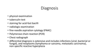
Tuberculosis Of Peripheral
Lymph nodes
-Ladi Anudeep
ISM-IUK
• Also known as Tuberculous Lymphadenitis
• Lymphadenitis is the most common extrapulmonary manifestation of
tuberculosis
• TB lymphadenitis is seen in nearly 35 per cent of extrapulmonary TB
which constituted about 15 to 20 percent of all cases of TB
• Cervical lymph nodes are the most common site of involvement and
reported in 60% to 90% patients and referred to as “scrofula.”
• has also been found at intrathoracic, intraabdominal (mesenteric
lymph nodes or paraaortic), and occasionally, axillary, inguinal, and
intramammary sites.
• Cervical lymphadenitis, which is also referred to as scrofula, may be
manifestation of a systemic tuberculous disease or a unique clinical
entity localized to neck.
• Mycobacterium tuberculosis is the most common causative agent in
India
• Lymphadenopathy due to non-tuberculous mycobacterial (NTM) is
uncommonly reported from India
• Mycobacterium avium-intracellulare complex is the most common
causative agent of non tuberculous peripheral lymph node TB
• Mycobacterial lymphadenitis most frequently affects patients in their
second decade but may afflict patients of any age.
• There is a female predominance (approximately 2:1) in most of the
studies
• Infection with the human immunodeficiency virus (HIV) is associated
with an increased frequency of both pulmonary and extrapulmonary
tuberculosis particularly lymphadenitis.
• A higher incidence of TB lymphadenitis has been noted in countries
with a high prevalence of both TB and HIV
• In India, children under 14 years of age have incidence rates of 4.4
cases per 1000
Pathogenesis
• The infection of lymph nodes by mycobacterium occurs either
through hematogenous dissemination following primary tuberculosis
or as a local extension from tuberculous infection of the tonsils or
adenoids
• Extrapulmonary tuberculosis in HIV infection is seen when CD4 counts
are below 300/ml
• It may occur during primary tuberculous infection or as a result of
reactivation of dormant foci or direct extension from a contiguous
focus
• Primary infection occurs on initial exposure to tubercle bacilli.
• Inhaled droplet nuclei are small enough to pass muco-ciliary defenses
of bronchi and lodge in terminal alveoli of lungs.
• The bacilli multiply in the lung which is called Ghon focus.
• The lymphatics drain the bacilli to the hilar lymph nodes.
• The Ghon focus and related hilar lymphadenopathy form the primary
complex.
• The infection may spread from primary focus to regional lymph
nodes.
• From the regional nodes, organism may continue to spread via the
lymphatic system to other nodes or may pass through the nodes to
reach blood stream, from where it can spread to virtually all organ of
the body.
• Hilar, mediastinal and paratracheal lymph nodes are the first site of
spread of infection from the lung parenchyma
• In initial stage of superficial lymph node involvement progressive
multiplication of the M. tuberculosis occurs, the onset of delayed
hypersensitivity is accompanied by marked hyperemia, swelling,
necrosis and caseation of the center of the nodes.
• This can be followed by inflammation, progressive swelling and
matting with other nodes within a group
• Adhesion to the adjacent skin may result in induration and purplish
discoloration.
• The center of the enlarging gland becomes soft and caseous material
may rupture into surrounding tissue or through skin with sinus
formation
• The lymphadenitis due to non-tuberculous mycobacteria is
transmitted from environment by ingestion, inhalation, inoculation
• The portal of entry for NTM may be the oral mucosa or gingiva.
• This is particularly important in children, because deciduous teeth
may harbor the NTM that may reach the neck sites around the
mandible through the lymphatics.
Clinical Presentation
• Lymphadenitis is the most common clinical presentation of
extrapulmonary tuberculosis
• frequently involves the cervical lymph nodes, followed in frequency
by mediastinal, axillary, mesenteric, hepatic portal, perihepatic and
inguinal lymph nodes
• Fistula formation was seen in nearly 10% of the mycobacterial cervical
lymphadenitis
• low grade fever, weight loss and fatigue and less frequently with night
sweats.
• Cough is not a prominent feature of tuberculous lymphadenitis.
• Jones and Campbell classified peripheral tuberculous lymph nodes
into following five stages.
stage 1: enlarged, firm, mobile, discrete nodes showing non-specific
reactive hyperplasia
stage 2: large rubbery nodes fixed to surrounding tissue owing to peri
adenitis
stage 3: central softening due to abscess formation
stage 4: collar-stud abscess formation
stage 5: sinus tract formation.
Young girl with inflamed, swollen, soft
and fluctuant cervical
lymph node leading to abscess
formation
Patient with scrofula–cervical
tuberculous lymphadenitis
Patient with axillary tuberculous
lymphadenitis with draining sinuses
Diagnosis
• physical examination
• tuberculin test
• staining for acid-fast bacilli
• radiologic examination
• fine-needle aspiration cytology (FNAC)
• Polymerase chain reaction (PCR)
• Chest radiograph
• differential diagnosis is extensive and includes infections (viral, bacterial or
fungal), and neoplasms (lymphoma or sarcoma, metastatic carcinoma),
non-specific reactive hyperplasia
Treatment
• Antituberculosis treatment is the mainstay in the management of TB
lymphadenitis
• The National Tuberculosis Programmes worldwide follow the World
Health Organization’s guidelines

Tuberculosis of peripheral lymph nodes

  • 1.
    Tuberculosis Of Peripheral Lymphnodes -Ladi Anudeep ISM-IUK
  • 2.
    • Also knownas Tuberculous Lymphadenitis • Lymphadenitis is the most common extrapulmonary manifestation of tuberculosis • TB lymphadenitis is seen in nearly 35 per cent of extrapulmonary TB which constituted about 15 to 20 percent of all cases of TB • Cervical lymph nodes are the most common site of involvement and reported in 60% to 90% patients and referred to as “scrofula.” • has also been found at intrathoracic, intraabdominal (mesenteric lymph nodes or paraaortic), and occasionally, axillary, inguinal, and intramammary sites.
  • 3.
    • Cervical lymphadenitis,which is also referred to as scrofula, may be manifestation of a systemic tuberculous disease or a unique clinical entity localized to neck. • Mycobacterium tuberculosis is the most common causative agent in India • Lymphadenopathy due to non-tuberculous mycobacterial (NTM) is uncommonly reported from India • Mycobacterium avium-intracellulare complex is the most common causative agent of non tuberculous peripheral lymph node TB
  • 4.
    • Mycobacterial lymphadenitismost frequently affects patients in their second decade but may afflict patients of any age. • There is a female predominance (approximately 2:1) in most of the studies • Infection with the human immunodeficiency virus (HIV) is associated with an increased frequency of both pulmonary and extrapulmonary tuberculosis particularly lymphadenitis. • A higher incidence of TB lymphadenitis has been noted in countries with a high prevalence of both TB and HIV • In India, children under 14 years of age have incidence rates of 4.4 cases per 1000
  • 5.
    Pathogenesis • The infectionof lymph nodes by mycobacterium occurs either through hematogenous dissemination following primary tuberculosis or as a local extension from tuberculous infection of the tonsils or adenoids • Extrapulmonary tuberculosis in HIV infection is seen when CD4 counts are below 300/ml • It may occur during primary tuberculous infection or as a result of reactivation of dormant foci or direct extension from a contiguous focus
  • 6.
    • Primary infectionoccurs on initial exposure to tubercle bacilli. • Inhaled droplet nuclei are small enough to pass muco-ciliary defenses of bronchi and lodge in terminal alveoli of lungs. • The bacilli multiply in the lung which is called Ghon focus. • The lymphatics drain the bacilli to the hilar lymph nodes. • The Ghon focus and related hilar lymphadenopathy form the primary complex. • The infection may spread from primary focus to regional lymph nodes.
  • 7.
    • From theregional nodes, organism may continue to spread via the lymphatic system to other nodes or may pass through the nodes to reach blood stream, from where it can spread to virtually all organ of the body. • Hilar, mediastinal and paratracheal lymph nodes are the first site of spread of infection from the lung parenchyma • In initial stage of superficial lymph node involvement progressive multiplication of the M. tuberculosis occurs, the onset of delayed hypersensitivity is accompanied by marked hyperemia, swelling, necrosis and caseation of the center of the nodes. • This can be followed by inflammation, progressive swelling and matting with other nodes within a group
  • 8.
    • Adhesion tothe adjacent skin may result in induration and purplish discoloration. • The center of the enlarging gland becomes soft and caseous material may rupture into surrounding tissue or through skin with sinus formation • The lymphadenitis due to non-tuberculous mycobacteria is transmitted from environment by ingestion, inhalation, inoculation • The portal of entry for NTM may be the oral mucosa or gingiva. • This is particularly important in children, because deciduous teeth may harbor the NTM that may reach the neck sites around the mandible through the lymphatics.
  • 9.
    Clinical Presentation • Lymphadenitisis the most common clinical presentation of extrapulmonary tuberculosis • frequently involves the cervical lymph nodes, followed in frequency by mediastinal, axillary, mesenteric, hepatic portal, perihepatic and inguinal lymph nodes • Fistula formation was seen in nearly 10% of the mycobacterial cervical lymphadenitis • low grade fever, weight loss and fatigue and less frequently with night sweats. • Cough is not a prominent feature of tuberculous lymphadenitis.
  • 10.
    • Jones andCampbell classified peripheral tuberculous lymph nodes into following five stages. stage 1: enlarged, firm, mobile, discrete nodes showing non-specific reactive hyperplasia stage 2: large rubbery nodes fixed to surrounding tissue owing to peri adenitis stage 3: central softening due to abscess formation stage 4: collar-stud abscess formation stage 5: sinus tract formation.
  • 11.
    Young girl withinflamed, swollen, soft and fluctuant cervical lymph node leading to abscess formation Patient with scrofula–cervical tuberculous lymphadenitis Patient with axillary tuberculous lymphadenitis with draining sinuses
  • 12.
    Diagnosis • physical examination •tuberculin test • staining for acid-fast bacilli • radiologic examination • fine-needle aspiration cytology (FNAC) • Polymerase chain reaction (PCR) • Chest radiograph • differential diagnosis is extensive and includes infections (viral, bacterial or fungal), and neoplasms (lymphoma or sarcoma, metastatic carcinoma), non-specific reactive hyperplasia
  • 13.
    Treatment • Antituberculosis treatmentis the mainstay in the management of TB lymphadenitis • The National Tuberculosis Programmes worldwide follow the World Health Organization’s guidelines