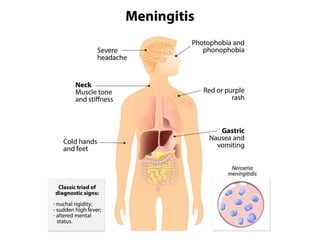
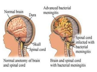
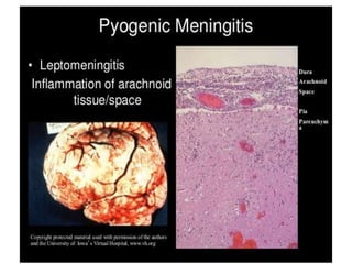
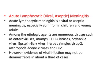
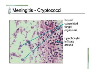
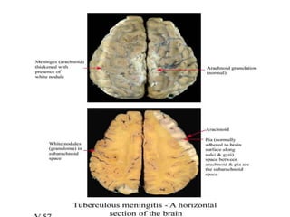
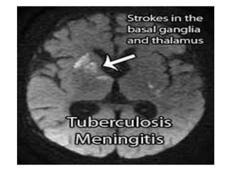
MENINGITIS
-Ladi Anudeep
ISM-IUK
โ€ข Meningitis is inflammatory involvement of the
meninges.
โ€ข Meningitis may involve the dura called
pachymeningitis, or leptomeninges (pia-arachnoid)
termed leptomeningitis.
โ€ข The latter is far more common, and unless
otherwise specified, meningitis would mean
leptomeningitis.
โ€ข Pachymeningitis is invariably an extension of the
inflammation from chronic suppurative otitis media
or from fracture of the skull
โ€ข Acute Pyogenic Meningitis
โ€ข Acute pyogenic or acute purulent meningitis is
acute infection of the pia-arachnoid and of the CSF
enclosed in the subarachnoid space.
โ€ข Since the subarachnoid space is continuous around
the brain, spinal cord and the optic nerves, infection
spreads immediately to whole of the cerebrospinal
meninges as well as to the ventricles.
ETIOPATHOGENESIS. The causative organisms vary with
age of the patient:
1. Escherichia coli infection is common in neonates with
neural tube defects.
2. Haemophilus influenzae is commonly responsible for
infection in infants and children.
3. Neisseria meningitidis causes meningitis in adolescent
and
young adults and is causative for epidemic meningitis.
4. Streptococcus pneumoniae is causative for infection at
extremes of age and following trauma
MORPHOLOGIC FEATURES.
โ€ข Grossly, pus accumulate in the subarachnoid space so
that normally clear CSF becomes turbid or frankly
purulent.
โ€ข The turbid fluid is particularly seen in the sulci and at
the base of the brain where the space is wide. In
fulminant cases, some degree of ventriculitis is also
present having a fibrinous coating on their walls and
containing turbid CSF.
โ€ข In addition, purulent material may interfere with CSF
flow and result in obstructive hydrocephalus
Microscopically
โ€ข There is presence of numerous polymorphonuclear
neutrophils in the subarachnoid space as well as in
the meninges, particularly around the blood vessels.
โ€ข Gram-staining reveals varying number of causative
bacteria
CLINICAL FEATURES AND DIAGNOSIS.
โ€ข Acute bacterial meningitis is a medical emergency. The
immediate clinical manifestations are fever, severe
headache, vomiting, drowsiness, stupor, coma, and
occasionally, convulsions.
โ€ข The most important clinical sign is stiffness of the neck
on forward bending.
The diagnosis is confirmed by examining CSF as soon as
possible. The diagnostic alterations in the CSF in acute
pyogenic meningitis are as under:
1. Naked eye appearance of cloudy or frankly purulent
CSF.
2. Elevated CSF pressure (above 180 mm water).
3. Polymorphonuclear neutrophilic leucocytosis in CSF
(between 10-10,000/ฮผl).
4. Raised CSF protein level (higher than 50 mg/dl).
5. Decreased CSF sugar concentration (lower than 40
mg/
dl).
6. Bacteriologic examination by Gramโ€™s stain or by CSF
culture reveals causative organism.
โ€ข Acute Lymphocytic (Viral, Aseptic) Meningitis
โ€ข Acute lymphocytic meningitis is a viral or aseptic
meningitis, especially common in children and young
adults.
โ€ข Among the etiologic agents are numerous viruses such
as enteroviruses, mumps, ECHO viruses, coxsackie
virus, Epstein-Barr virus, herpes simplex virus-2,
arthropode-borne viruses and HIV.
โ€ข However, evidence of viral infection may not be
demonstrable in about a third of cases.
MORPHOLOGIC FEATURES.
โ€ข Grossly, some cases show swelling of the brain while
others show no distinctive change.
Microscopically
โ€ข there is mild lymphocytic infiltrate in the leptomeninges.
CLINICAL FEATURES AND DIAGNOSIS.
โ€ข The clinical manifestations of viral meningitis are much
the same as in bacterial meningitis with features of
acute onset meningeal symptoms and fever.
โ€ข However, viral meningitis has a benign and self-limiting
clinical course of short duration and is invariably
followed by complete recovery without the
lifethreatening complications of bacterial meningitis
The CSF findings in viral meningitis are as under:
1. Naked eye appearance of clear or slightly turbid CSF.
2. CSF pressure increased (above 250 mm water).
3. Lymphocytosis in CSF (10-100 cells/ฮผ)
4. CSF protein usually normal or mildly raised.
5. CSF sugar concentration usually normal.
6. CSF bacteriologically sterile._x0000_
Chronic (Tuberculous and Cryptococcal)
Meningitis)
โ€ข There are two principal types of chronic meningitisโ€”one
bacterial (tuberculous meningitis) and the other fungal
(cryptococcal meningitis). Both types cause chronic
granulomatous reaction and may produce parenchymal
lesions.
โ€ข Tuberculous meningitis occurs in children and adults
through haematogenous spread of infection from
tuberculosis elsewhere in the body, or it may simply be
a manifestation of miliary tuberculosis. Less commonly,
the spread may occur directly from tuberculosis of a
vertebral body.
โ€ข Cryptococcal meningitis develops particularly in
debilitated or immunocompromised persons, usually as
a result of haematogenous dissemination from a
pulmonary lesion. Cryptococcal meningitis is especially
an important cause of meningitis in patients with AIDS.
MORPHOLOGIC FEATURES.
โ€ข Grossly, in tuberculous meningitis, the subarachnoid
space contains thick exudate, particularly abundant in
the sulci and the base of the brain.
โ€ข Tubercles, 1-2 mm in diameter, may be visible, especially
adjacent to the blood vessels.
โ€ข The exudate in cryptococcal meningitis is scanty,
translucent and gelatinous.
โ€ข Microscopically, tuberculous meningitis shows exudate
of acute and chronic inflammatory cells, and granulomas
with or without caseation necrosis and giant cells.
โ€ข Acidfast bacilli may be demonstrated. Late cases show
dense fibrous adhesions in the subarachnoid space and
consequent hydrocephalus.
โ€ข Cryptococcal meningitis is characterised by infiltration by
lymphocytes, plasmacells, an occasional granuloma and
abundant characteristic capsulated cryptococci.
CLINICAL FEATURES AND DIAGNOSIS.
โ€ข Tuberculous meningitis manifests clinically as
headache, confusion, malaise and vomiting.
โ€ข The clinical course in cryptococcal meningitis may,
however, be fulminant and fatal in a few weeks, or be
indolent for months to years
The CSF findings in chronic meningitis are as under:
1. Naked eye appearance of a clear or slightly turbid CSF
which may form fibrin web on standing.
2. Raised CSF pressure (above 300 mm water).
3. Mononuclear leucocytosis consisting mostly of
lymphocytes and some macrophages (100-1000 cells/ฮผl).
4. Raised protein content.
5. Lowered glucose concentration.
6. Tubercle bacilli may be found on microscopy of
centrifuged deposits by ZN staining in tuberculous
meningitis
Meningitis
Meningitis
Meningitis

Meningitis

  • 1.
  • 2.
    โ€ข Meningitis isinflammatory involvement of the meninges. โ€ข Meningitis may involve the dura called pachymeningitis, or leptomeninges (pia-arachnoid) termed leptomeningitis. โ€ข The latter is far more common, and unless otherwise specified, meningitis would mean leptomeningitis. โ€ข Pachymeningitis is invariably an extension of the inflammation from chronic suppurative otitis media or from fracture of the skull
  • 3.
    โ€ข Acute PyogenicMeningitis โ€ข Acute pyogenic or acute purulent meningitis is acute infection of the pia-arachnoid and of the CSF enclosed in the subarachnoid space. โ€ข Since the subarachnoid space is continuous around the brain, spinal cord and the optic nerves, infection spreads immediately to whole of the cerebrospinal meninges as well as to the ventricles.
  • 4.
    ETIOPATHOGENESIS. The causativeorganisms vary with age of the patient: 1. Escherichia coli infection is common in neonates with neural tube defects. 2. Haemophilus influenzae is commonly responsible for infection in infants and children. 3. Neisseria meningitidis causes meningitis in adolescent and young adults and is causative for epidemic meningitis. 4. Streptococcus pneumoniae is causative for infection at extremes of age and following trauma
  • 5.
    MORPHOLOGIC FEATURES. โ€ข Grossly,pus accumulate in the subarachnoid space so that normally clear CSF becomes turbid or frankly purulent. โ€ข The turbid fluid is particularly seen in the sulci and at the base of the brain where the space is wide. In fulminant cases, some degree of ventriculitis is also present having a fibrinous coating on their walls and containing turbid CSF. โ€ข In addition, purulent material may interfere with CSF flow and result in obstructive hydrocephalus
  • 6.
    Microscopically โ€ข There ispresence of numerous polymorphonuclear neutrophils in the subarachnoid space as well as in the meninges, particularly around the blood vessels. โ€ข Gram-staining reveals varying number of causative bacteria
  • 7.
    CLINICAL FEATURES ANDDIAGNOSIS. โ€ข Acute bacterial meningitis is a medical emergency. The immediate clinical manifestations are fever, severe headache, vomiting, drowsiness, stupor, coma, and occasionally, convulsions. โ€ข The most important clinical sign is stiffness of the neck on forward bending.
  • 8.
    The diagnosis isconfirmed by examining CSF as soon as possible. The diagnostic alterations in the CSF in acute pyogenic meningitis are as under: 1. Naked eye appearance of cloudy or frankly purulent CSF. 2. Elevated CSF pressure (above 180 mm water). 3. Polymorphonuclear neutrophilic leucocytosis in CSF (between 10-10,000/ฮผl). 4. Raised CSF protein level (higher than 50 mg/dl). 5. Decreased CSF sugar concentration (lower than 40 mg/ dl). 6. Bacteriologic examination by Gramโ€™s stain or by CSF culture reveals causative organism.
  • 12.
    โ€ข Acute Lymphocytic(Viral, Aseptic) Meningitis โ€ข Acute lymphocytic meningitis is a viral or aseptic meningitis, especially common in children and young adults. โ€ข Among the etiologic agents are numerous viruses such as enteroviruses, mumps, ECHO viruses, coxsackie virus, Epstein-Barr virus, herpes simplex virus-2, arthropode-borne viruses and HIV. โ€ข However, evidence of viral infection may not be demonstrable in about a third of cases.
  • 13.
    MORPHOLOGIC FEATURES. โ€ข Grossly,some cases show swelling of the brain while others show no distinctive change. Microscopically โ€ข there is mild lymphocytic infiltrate in the leptomeninges.
  • 14.
    CLINICAL FEATURES ANDDIAGNOSIS. โ€ข The clinical manifestations of viral meningitis are much the same as in bacterial meningitis with features of acute onset meningeal symptoms and fever. โ€ข However, viral meningitis has a benign and self-limiting clinical course of short duration and is invariably followed by complete recovery without the lifethreatening complications of bacterial meningitis
  • 15.
    The CSF findingsin viral meningitis are as under: 1. Naked eye appearance of clear or slightly turbid CSF. 2. CSF pressure increased (above 250 mm water). 3. Lymphocytosis in CSF (10-100 cells/ฮผ) 4. CSF protein usually normal or mildly raised. 5. CSF sugar concentration usually normal. 6. CSF bacteriologically sterile._x0000_
  • 18.
    Chronic (Tuberculous andCryptococcal) Meningitis) โ€ข There are two principal types of chronic meningitisโ€”one bacterial (tuberculous meningitis) and the other fungal (cryptococcal meningitis). Both types cause chronic granulomatous reaction and may produce parenchymal lesions.
  • 19.
    โ€ข Tuberculous meningitisoccurs in children and adults through haematogenous spread of infection from tuberculosis elsewhere in the body, or it may simply be a manifestation of miliary tuberculosis. Less commonly, the spread may occur directly from tuberculosis of a vertebral body. โ€ข Cryptococcal meningitis develops particularly in debilitated or immunocompromised persons, usually as a result of haematogenous dissemination from a pulmonary lesion. Cryptococcal meningitis is especially an important cause of meningitis in patients with AIDS.
  • 20.
    MORPHOLOGIC FEATURES. โ€ข Grossly,in tuberculous meningitis, the subarachnoid space contains thick exudate, particularly abundant in the sulci and the base of the brain. โ€ข Tubercles, 1-2 mm in diameter, may be visible, especially adjacent to the blood vessels. โ€ข The exudate in cryptococcal meningitis is scanty, translucent and gelatinous.
  • 21.
    โ€ข Microscopically, tuberculousmeningitis shows exudate of acute and chronic inflammatory cells, and granulomas with or without caseation necrosis and giant cells. โ€ข Acidfast bacilli may be demonstrated. Late cases show dense fibrous adhesions in the subarachnoid space and consequent hydrocephalus. โ€ข Cryptococcal meningitis is characterised by infiltration by lymphocytes, plasmacells, an occasional granuloma and abundant characteristic capsulated cryptococci.
  • 22.
    CLINICAL FEATURES ANDDIAGNOSIS. โ€ข Tuberculous meningitis manifests clinically as headache, confusion, malaise and vomiting. โ€ข The clinical course in cryptococcal meningitis may, however, be fulminant and fatal in a few weeks, or be indolent for months to years
  • 23.
    The CSF findingsin chronic meningitis are as under: 1. Naked eye appearance of a clear or slightly turbid CSF which may form fibrin web on standing. 2. Raised CSF pressure (above 300 mm water). 3. Mononuclear leucocytosis consisting mostly of lymphocytes and some macrophages (100-1000 cells/ฮผl). 4. Raised protein content. 5. Lowered glucose concentration. 6. Tubercle bacilli may be found on microscopy of centrifuged deposits by ZN staining in tuberculous meningitis