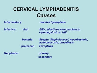
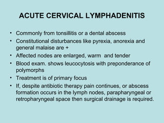
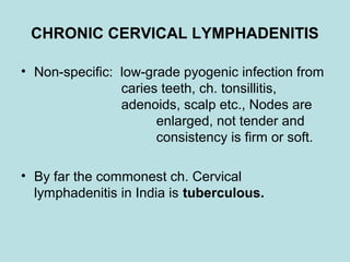
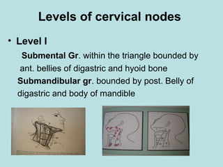
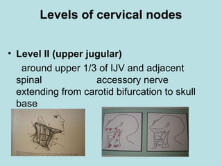
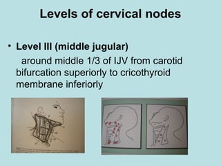
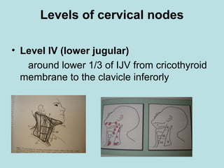
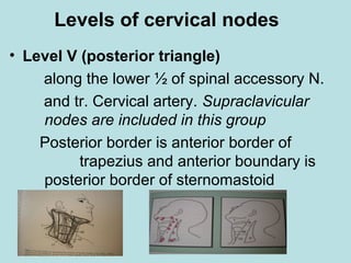
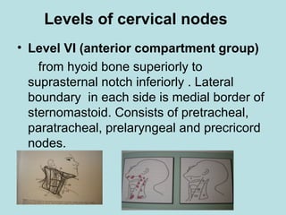
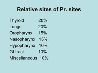
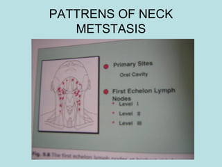
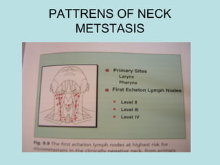
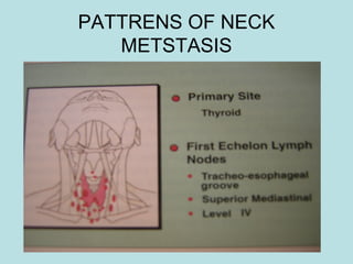
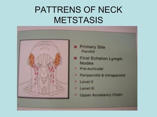
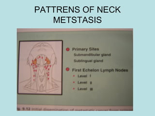
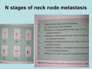
CERVICAL LYMPH NODES
CERVICAL LYMPHADENITIS
Causes
Inflammatory: reactive hyperplasia
Infective: viral EBV, infectious mononucleosis,
cytomegalovirus, HIV
bacteria Strepto, Staphylococci, mycobacteria,
actinomycosis, brucellosis
protozoan Toxoplsma
Neoplastic: primary
secondary
ACUTE CERVICAL LYMPHADENITIS
• Commonly from tonsillitis or a dental abscess
• Constitutional disturbances like pyrexia, anorexia and
general malaise are +
• Affected nodes are enlarged, warm and tender
• Blood exam. shows leucocytosis with preponderance of
polymorphs
• Treatment is of primary focus
• If, despite antibiotic therapy pain continues, or abscess
formation occurs in the lymph nodes, parapharyngeal or
retropharyngeal space then surgical drainage is required.
CHRONIC CERVICAL LYMPHADENITIS
• Non-specific: low-grade pyogenic infection from
caries teeth, ch. tonsillitis,
adenoids, scalp etc., Nodes are
enlarged, not tender and
consistency is firm or soft.
• By far the commonest ch. Cervical
lymphadenitis in India is tuberculous.
Tuberculous cervical lymphadenitis
• Commonest form of extra pulmonary tuberculosis esp. in
children and young adults, but may occur at any age.
• Caused by Mycobacterium tuberculosis; both by bovine
and human strains. Now also by atypical mycobacteria
esp. in immuno deficient pts., such as HIV. In most
instances bacilli enter through the tonsil of the
corresponding side. Deep cervical nodes are commonly
affected, but there may be widespread cervical
lymphadenitis.
• Supraclavicular represents upward extension of hilar and
mediastinal lymphadenopathy. Axillary and inguinal
nodes are involved by haematogenous or perhaps by
retrograde lymphatic spread.
Tuberculous cervical lymphadenitis
• In 80% tuberculous process is limited to
clinically affected group but primary focus in
the lungs must always be suspected and
investigated. As renal and pulmonary TB
occasionally coexist, urine should be examined
carefully .
Rarely, pt. develops natural resistance and the
nodes may be detected as calcification on x-ray
or after appropriate treatment
TB cervical adenitis-Pathology
• Granulomatous inflammation with tubercle
• Undergo caseation, necrosis and destruction
• Spread to adjacent nodes- periadenitis; getting
adherent to each other- “matting”
• Caseous nodes with periadenitis deep to deep
fascia perforates with the escape of caseous
material into subcutaneous space resulting in
“collar-stud abscess”.
• abscess gets adherent to skin and burst in the
surface resulting in abscess or sinus
TB cervical adenitis-Pathology
• Stage-1 The glands are enlarged, mobile, firm, and slightly tender.
Histologically, they show non-specific reactive hyperplasia.
• Stage-2 The nodes are large, firm and fixed to surrounding tissues and
to each other. Histologically, they show periadenitis, typical
tuberculous granulomatous tissue with lymphocytes, epithelioid cells,
and caesation.
• Stage-3 The caesation is extensive giving rise to variable consistency
with soft areas of cold abscesses and firm lymph nodes.
• Stage-4 The abscesses burst out of the lymph node mass and extends
into subcutaneous tissue giving rise to a ‘collar-stud’ abscess
• Stage-5 The abscess bursts and gives rise to a persistent, discharging
sinus. The discharge from sinus may infect the surrounding skin and
cause extensive tuberculous ulcer. Ulcers have a typical pale, flabby,
granulation tissue, under-mined edges, and a seropurulent discharge.
TB cervical adenitis-
Types of clinical manifestations
• Acute type : Seen in infants and children
< 5yrs. The glandular enlargement is painful, tender, and
evolves within few days. The overlying skin is red and
oedematous. The child has moderate fever. The clinical
picture resembles acute septic lymphadenitis.
• Caseating Type: Most common type seen in young
adults: Glands are multiple, moderately enlarged and
matted together. The caseation leads to softening, cold
abscess and sinuses. The patient is anemic and
moderately nourished. Constitutional features like fever
anorexia and weight loss may be present.
TB cervical adenitis-
Types of clinical manifestations
• Hyperplastic type in patients with good general
resistance, lymph nodes show a marked degree of
reactive, reticular and lymphoid hyperplasia. TB
garanulomatous tissue is more productive with least
caseation and periadinitis. The glands are notably
enlarged and appear fleshy, elastic and freely mobile
resembling those of Hodgekin’s disease.
• Atrophic type: Seen in elderly individual in whom
lymphoid tissue undergoes natural process of involution,
glands are comparatively small and soon burst with the
onset of caesation.
TB cervical adenitis-
Diagnosis
Clinical diagnosis is not difficult in classical case, where chronic
iymphadenopathy in young individual is associated with matting
and soft areas of caesation.
• A negative tuberculin test excludes.
A positive test has no diagnostic value.
ESR raised. Serum albumin falls and gamma globulins increase.
• An X-ray chest is mandatory for coexisting pulmonary lesion.
• Positive FNAC and biopsy are essential for confirming diagnosis.
Typical tuberculous granuloma with epithelioid cells, giant cells
and lymphocytes surrounding the area of caseation are
characteristics.
• Besides M.tuberlcosis ,atypical bacteria such as M.scrofulaceum
and M. intercellulare have been recovered from cases.
TB cervical adenitis-
Treatment
• Multi drug regime
• Initial phase for 4 drugs for 2 – 3 months.
Cap Rifampicin - 450-600 mg. daily.
Tab Isoniazid - 300 mg daily.
Tab Ethambutol – 1000 mg daily.
Tab Pyrazinamide – 1500 mg daily.
• Subsequently 2 drugs for 4-6 months.
Cap Rifampicin and Tab Isoniazid.
With full course of anti tuberculosis drug therapy, the glands subside
within 3 – 4 months and response is even quicker in children.
TB cervical adenitis-
Treatment
• Cold absess: repeated non dependent
aspiration; streptomycin may be instilled locally.
• Surgical excision is indicated
(a) When the glands continue to persist after
adequate chemotherapy and localised to one
single group.
(b) for persistent sinuses- secondary infection,
necrotic and calcified material replacing the
lymph nodes, and fibrosis are responsible for
persistence of sinus even after the active
disease has subsided.
SEC. CERVICAL LYMPH NODES
Prognostic factors
• Presence or absence of cl. palpable nodes,
• size, number, location
• Involvement below crico-thyroid - Lower (Level IV) &
posterior (Level V) is ominous
• extra nodal spread to soft tissue
• perivascular and perineural infiltration
• tumour emboli in regional lymphatics
Levels of cervical nodes
• Level I
Submental Gr. within the triangle bounded by
ant. bellies of digastric and hyoid bone
Submandibular gr. bounded by post. Belly of
digastric and body of mandible
Levels of cervical nodes
• Level II (upper jugular)
around upper 1/3 of IJV and adjacent
spinal accessory nerve
extending from carotid bifurcation to skull
base
Levels of cervical nodes
• Level III (middle jugular)
around middle 1/3 of IJV from carotid
bifurcation superiorly to cricothyroid
membrane inferiorly
Levels of cervical nodes
• Level IV (lower jugular)
around lower 1/3 of IJV from cricothyroid
membrane to the clavicle inferorly
Levels of cervical nodes
• Level V (posterior triangle)
along the lower ½ of spinal accessory N.
and tr. Cervical artery. Supraclavicular
nodes are included in this group
Posterior border is anterior border of
trapezius and anterior boundary is
posterior border of sternomastoid
Levels of cervical nodes
• Level VI (anterior compartment group)
from hyoid bone superiorly to
suprasternal notch inferiorly . Lateral
boundary in each side is medial border of
sternomastoid. Consists of pretracheal,
paratracheal, prelaryngeal and precricord
nodes.
OCCULT PRIMARY
• Male : Female - 4 : 1
• Age Peak incidence 65yrs for men 55 for women
• 1/3 to1/2 Sq. cell ca., 1/4 anaplastic ca.
1/4 adeno ca. if supraclavicular is involved followed
by miscellaneous tumours such as melanoma and
thyroid gland tumours
• Primary sites in order of frequency
Head and neck sites
nasopharynx, tonsil, base of tongue, thyroid,
supraglotic larynx, floor of mouth, palate and
pyriform fossa
Non head and neck sites
bronchus, oesophagus, breast and stomach
Relative sites of Pr. sites
Thyroid 20%
Lungs 20%
Oropharynx 15%
Nasopharynx 15%
Hypopharynx 10%
GI tract 10%
Miscellaneous 10%
PATTRENS OF NECK
METSTASIS
PATTRENS OF NECK
METSTASIS
PATTRENS OF NECK
METSTASIS
PATTRENS OF NECK
METSTASIS
PATTRENS OF NECK
METSTASIS
N stages of neck node metastasis

Cervical lymphadenitis

  • 1.
  • 2.
    CERVICAL LYMPHADENITIS Causes Inflammatory: reactivehyperplasia Infective: viral EBV, infectious mononucleosis, cytomegalovirus, HIV bacteria Strepto, Staphylococci, mycobacteria, actinomycosis, brucellosis protozoan Toxoplsma Neoplastic: primary secondary
  • 3.
    ACUTE CERVICAL LYMPHADENITIS •Commonly from tonsillitis or a dental abscess • Constitutional disturbances like pyrexia, anorexia and general malaise are + • Affected nodes are enlarged, warm and tender • Blood exam. shows leucocytosis with preponderance of polymorphs • Treatment is of primary focus • If, despite antibiotic therapy pain continues, or abscess formation occurs in the lymph nodes, parapharyngeal or retropharyngeal space then surgical drainage is required.
  • 4.
    CHRONIC CERVICAL LYMPHADENITIS •Non-specific: low-grade pyogenic infection from caries teeth, ch. tonsillitis, adenoids, scalp etc., Nodes are enlarged, not tender and consistency is firm or soft. • By far the commonest ch. Cervical lymphadenitis in India is tuberculous.
  • 5.
    Tuberculous cervical lymphadenitis •Commonest form of extra pulmonary tuberculosis esp. in children and young adults, but may occur at any age. • Caused by Mycobacterium tuberculosis; both by bovine and human strains. Now also by atypical mycobacteria esp. in immuno deficient pts., such as HIV. In most instances bacilli enter through the tonsil of the corresponding side. Deep cervical nodes are commonly affected, but there may be widespread cervical lymphadenitis. • Supraclavicular represents upward extension of hilar and mediastinal lymphadenopathy. Axillary and inguinal nodes are involved by haematogenous or perhaps by retrograde lymphatic spread.
  • 6.
    Tuberculous cervical lymphadenitis •In 80% tuberculous process is limited to clinically affected group but primary focus in the lungs must always be suspected and investigated. As renal and pulmonary TB occasionally coexist, urine should be examined carefully . Rarely, pt. develops natural resistance and the nodes may be detected as calcification on x-ray or after appropriate treatment
  • 7.
    TB cervical adenitis-Pathology •Granulomatous inflammation with tubercle • Undergo caseation, necrosis and destruction • Spread to adjacent nodes- periadenitis; getting adherent to each other- “matting” • Caseous nodes with periadenitis deep to deep fascia perforates with the escape of caseous material into subcutaneous space resulting in “collar-stud abscess”. • abscess gets adherent to skin and burst in the surface resulting in abscess or sinus
  • 8.
    TB cervical adenitis-Pathology •Stage-1 The glands are enlarged, mobile, firm, and slightly tender. Histologically, they show non-specific reactive hyperplasia. • Stage-2 The nodes are large, firm and fixed to surrounding tissues and to each other. Histologically, they show periadenitis, typical tuberculous granulomatous tissue with lymphocytes, epithelioid cells, and caesation. • Stage-3 The caesation is extensive giving rise to variable consistency with soft areas of cold abscesses and firm lymph nodes. • Stage-4 The abscesses burst out of the lymph node mass and extends into subcutaneous tissue giving rise to a ‘collar-stud’ abscess • Stage-5 The abscess bursts and gives rise to a persistent, discharging sinus. The discharge from sinus may infect the surrounding skin and cause extensive tuberculous ulcer. Ulcers have a typical pale, flabby, granulation tissue, under-mined edges, and a seropurulent discharge.
  • 9.
    TB cervical adenitis- Typesof clinical manifestations • Acute type : Seen in infants and children < 5yrs. The glandular enlargement is painful, tender, and evolves within few days. The overlying skin is red and oedematous. The child has moderate fever. The clinical picture resembles acute septic lymphadenitis. • Caseating Type: Most common type seen in young adults: Glands are multiple, moderately enlarged and matted together. The caseation leads to softening, cold abscess and sinuses. The patient is anemic and moderately nourished. Constitutional features like fever anorexia and weight loss may be present.
  • 10.
    TB cervical adenitis- Typesof clinical manifestations • Hyperplastic type in patients with good general resistance, lymph nodes show a marked degree of reactive, reticular and lymphoid hyperplasia. TB garanulomatous tissue is more productive with least caseation and periadinitis. The glands are notably enlarged and appear fleshy, elastic and freely mobile resembling those of Hodgekin’s disease. • Atrophic type: Seen in elderly individual in whom lymphoid tissue undergoes natural process of involution, glands are comparatively small and soon burst with the onset of caesation.
  • 11.
    TB cervical adenitis- Diagnosis Clinicaldiagnosis is not difficult in classical case, where chronic iymphadenopathy in young individual is associated with matting and soft areas of caesation. • A negative tuberculin test excludes. A positive test has no diagnostic value. ESR raised. Serum albumin falls and gamma globulins increase. • An X-ray chest is mandatory for coexisting pulmonary lesion. • Positive FNAC and biopsy are essential for confirming diagnosis. Typical tuberculous granuloma with epithelioid cells, giant cells and lymphocytes surrounding the area of caseation are characteristics. • Besides M.tuberlcosis ,atypical bacteria such as M.scrofulaceum and M. intercellulare have been recovered from cases.
  • 12.
    TB cervical adenitis- Treatment •Multi drug regime • Initial phase for 4 drugs for 2 – 3 months. Cap Rifampicin - 450-600 mg. daily. Tab Isoniazid - 300 mg daily. Tab Ethambutol – 1000 mg daily. Tab Pyrazinamide – 1500 mg daily. • Subsequently 2 drugs for 4-6 months. Cap Rifampicin and Tab Isoniazid. With full course of anti tuberculosis drug therapy, the glands subside within 3 – 4 months and response is even quicker in children.
  • 13.
    TB cervical adenitis- Treatment •Cold absess: repeated non dependent aspiration; streptomycin may be instilled locally. • Surgical excision is indicated (a) When the glands continue to persist after adequate chemotherapy and localised to one single group. (b) for persistent sinuses- secondary infection, necrotic and calcified material replacing the lymph nodes, and fibrosis are responsible for persistence of sinus even after the active disease has subsided.
  • 14.
    SEC. CERVICAL LYMPHNODES Prognostic factors • Presence or absence of cl. palpable nodes, • size, number, location • Involvement below crico-thyroid - Lower (Level IV) & posterior (Level V) is ominous • extra nodal spread to soft tissue • perivascular and perineural infiltration • tumour emboli in regional lymphatics
  • 15.
    Levels of cervicalnodes • Level I Submental Gr. within the triangle bounded by ant. bellies of digastric and hyoid bone Submandibular gr. bounded by post. Belly of digastric and body of mandible
  • 16.
    Levels of cervicalnodes • Level II (upper jugular) around upper 1/3 of IJV and adjacent spinal accessory nerve extending from carotid bifurcation to skull base
  • 17.
    Levels of cervicalnodes • Level III (middle jugular) around middle 1/3 of IJV from carotid bifurcation superiorly to cricothyroid membrane inferiorly
  • 18.
    Levels of cervicalnodes • Level IV (lower jugular) around lower 1/3 of IJV from cricothyroid membrane to the clavicle inferorly
  • 19.
    Levels of cervicalnodes • Level V (posterior triangle) along the lower ½ of spinal accessory N. and tr. Cervical artery. Supraclavicular nodes are included in this group Posterior border is anterior border of trapezius and anterior boundary is posterior border of sternomastoid
  • 20.
    Levels of cervicalnodes • Level VI (anterior compartment group) from hyoid bone superiorly to suprasternal notch inferiorly . Lateral boundary in each side is medial border of sternomastoid. Consists of pretracheal, paratracheal, prelaryngeal and precricord nodes.
  • 21.
    OCCULT PRIMARY • Male: Female - 4 : 1 • Age Peak incidence 65yrs for men 55 for women • 1/3 to1/2 Sq. cell ca., 1/4 anaplastic ca. 1/4 adeno ca. if supraclavicular is involved followed by miscellaneous tumours such as melanoma and thyroid gland tumours • Primary sites in order of frequency Head and neck sites nasopharynx, tonsil, base of tongue, thyroid, supraglotic larynx, floor of mouth, palate and pyriform fossa Non head and neck sites bronchus, oesophagus, breast and stomach
  • 22.
    Relative sites ofPr. sites Thyroid 20% Lungs 20% Oropharynx 15% Nasopharynx 15% Hypopharynx 10% GI tract 10% Miscellaneous 10%
  • 23.
  • 24.
  • 25.
  • 26.
  • 27.
  • 28.
    N stages ofneck node metastasis