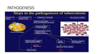
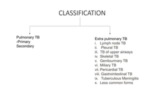
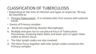
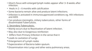
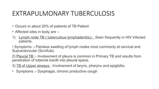
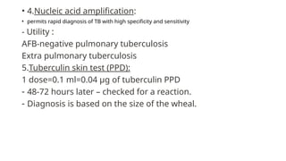
TUBERCULOSIS
TUBERCULOSIS
• Tuberculosis (TB) is a potentially fatal contagious disease that can affect almost
any part of the body but is mainly an infection of the lungs
• Characterized by the formation of tubercles (round nodules) or granulomas in
lungs.
• Causative agent: Mycobacterium tuberculosis complex
• Transmission usually takes place through the airborne spread of droplet nuclei
produced by patients with infectious pulmonary TB.
GLOBAL BURDEN OF TB
CLINICAL FEATURES
• Cough ( 2-3 weeks )
≥
• Hemoptysis
• Chest pain
• Fever
• Night sweats
• Generalized weakness and easy fatiguability
• Weight loss
• Loss of appetite
PATHOGENESIS
CLASSIFICATION
Pulmonary TB
-Primary
Secondary
Extra pulmonary TB
i. Lymph node TB
ii. Pleural TB
iii. TB of upper airways
iv. Skeletal TB
v. Genitourinary TB
vi. Miliary TB
vii. Pericardial TB
viii. Gastrointestinal TB
ix. Tuberculous Meningitis
x. Less common forms
CLASSIFICATION OF TUBERCULOSIS
• Depending on the time of infection and types of response, TB may
be classified as
1. Primary Tuberculosis: It is initiated after first contact with tubercle
bacilli.
• Events of Primary complex :
a. Bacilli are engulfed by alveolar Macrophages
b. Multiply and give rise to sub pleural focus of Tuberculosis
Pneumonia, involving lower lobes and lower part of upper lobes
called as Ghon’s focus
c. The hilar lymph nodes are also involved.
d. The Ghon focus together with hilar lymph nodes constitute the
Primary complex.
• Ghon’s focus with enlarged lymph nodes appear after 3- 8 weeks after
infection.
• Heals in 2 – 6 months with calcification
• Some bacteria remain alive and produce latent infections.
• Infection is activated in immunosuppressed conditions eg. HIV infections
and AIDS
• Can produce meningitis, miliary tuberculosis, other forms of
disseminated Tuberculosis.
2. Secondary Tuberculosis:
• Mainly occurs due to Reactivation of latent infection.
• May also due to Exogenous reinfection
• Differs from Primary Infection in the sense that it
 Leads to cavitation of Lungs
 Enlargement of Lymph nodes
 Expectoration of Bacteria laden sputum.
 Dissemination into Lungs and other extra pulmonary areas.
EXTRAPULMONARY TUBERCULOSIS
• Occurs in about 20% of patients of TB Patient
• Affected sites in body are :-
1) Lymph node TB ( tuberculous lymphadenitis):- Seen frequently in HIV infected
patients.
• Symptoms :- Painless swelling of lymph nodes most commonly at cervical and
Supraclavicular (Scrofula)
2) Pleural TB :- Involvement of pleura is common in Primary TB and results from
penetration of tubercle bacilli into pleural space.
3) TB of Upper airways : Involvement of larynx, pharynx and epiglottis.
• Symptoms :- Dysphagia, chronic productive cough
4) Genitourinary TB :15% of all Extra pulmonary cases.
• Symptoms :- Urinary frequency, Dysuria, Haematuria.
5) Skeletal TB : Involvement of weight bearing parts like spine, hip, knee.
• Symptoms : Pain in hip joints and knees, swelling of knees
6) Gastrointestinal TB :-
Symptoms :- Abdominal pain, diarrhoea, weight loss
7) TB Meningitis & Tuberculoma :- 5% of All Extra pulmonary TB
Results from Hematogenous spread of 1⸰
and 2 ⸰
TB.
8) TB Pericarditis :- Spreads mainly in mediastinal or hilar nodes or from lungs.
9) Miliary or disseminated TB : Results from Hematogenous spread of Tubercle
Bacilli.
• Spread is due to entry of infection into pulmonary vein producing lesions in different
extra pulmonary sites.
10) Less common Extra Pulmonary TB : Uveitis, panophthalmitis, Hypersensitivity
related phlyctenular conjunctivis
DIAGNOSIS
1.Bacteriological test
a. Zeihl-Neelsen stain(ZN stain)
b. Auramine stain(fluorescence microscopy)
Microscopic examination of a specimen of three morning successive
sputum that has been spread on a slide, and stained by the above
mentioned stains
2. Sputum culture test:
a. Lowenstein-Jensen(LJ) solid medium: 4-18 weeks
b. Liquid medium : 8-14 days
c. c. Agar medium : 7 to 14 days
3.Radiography: Chest X-Ray(CXR)
• 4.Nucleic acid amplification:
• permits rapid diagnosis of TB with high specificity and sensitivity
- Utility :
AFB-negative pulmonary tuberculosis
Extra pulmonary tuberculosis
5.Tuberculin skin test (PPD):
1 dose=0.1 ml=0.04 µg of tuberculin PPD
- 48-72 hours later – checked for a reaction.
- Diagnosis is based on the size of the wheal.
TREATMENT

TUBERCULOSIS- clinical features and management

  • 1.
  • 2.
    TUBERCULOSIS • Tuberculosis (TB)is a potentially fatal contagious disease that can affect almost any part of the body but is mainly an infection of the lungs • Characterized by the formation of tubercles (round nodules) or granulomas in lungs. • Causative agent: Mycobacterium tuberculosis complex • Transmission usually takes place through the airborne spread of droplet nuclei produced by patients with infectious pulmonary TB.
  • 3.
  • 4.
    CLINICAL FEATURES • Cough( 2-3 weeks ) ≥ • Hemoptysis • Chest pain • Fever • Night sweats • Generalized weakness and easy fatiguability • Weight loss • Loss of appetite
  • 6.
  • 7.
    CLASSIFICATION Pulmonary TB -Primary Secondary Extra pulmonaryTB i. Lymph node TB ii. Pleural TB iii. TB of upper airways iv. Skeletal TB v. Genitourinary TB vi. Miliary TB vii. Pericardial TB viii. Gastrointestinal TB ix. Tuberculous Meningitis x. Less common forms
  • 8.
    CLASSIFICATION OF TUBERCULOSIS •Depending on the time of infection and types of response, TB may be classified as 1. Primary Tuberculosis: It is initiated after first contact with tubercle bacilli. • Events of Primary complex : a. Bacilli are engulfed by alveolar Macrophages b. Multiply and give rise to sub pleural focus of Tuberculosis Pneumonia, involving lower lobes and lower part of upper lobes called as Ghon’s focus c. The hilar lymph nodes are also involved. d. The Ghon focus together with hilar lymph nodes constitute the Primary complex.
  • 9.
    • Ghon’s focuswith enlarged lymph nodes appear after 3- 8 weeks after infection. • Heals in 2 – 6 months with calcification • Some bacteria remain alive and produce latent infections. • Infection is activated in immunosuppressed conditions eg. HIV infections and AIDS • Can produce meningitis, miliary tuberculosis, other forms of disseminated Tuberculosis. 2. Secondary Tuberculosis: • Mainly occurs due to Reactivation of latent infection. • May also due to Exogenous reinfection • Differs from Primary Infection in the sense that it  Leads to cavitation of Lungs  Enlargement of Lymph nodes  Expectoration of Bacteria laden sputum.  Dissemination into Lungs and other extra pulmonary areas.
  • 10.
    EXTRAPULMONARY TUBERCULOSIS • Occursin about 20% of patients of TB Patient • Affected sites in body are :- 1) Lymph node TB ( tuberculous lymphadenitis):- Seen frequently in HIV infected patients. • Symptoms :- Painless swelling of lymph nodes most commonly at cervical and Supraclavicular (Scrofula) 2) Pleural TB :- Involvement of pleura is common in Primary TB and results from penetration of tubercle bacilli into pleural space. 3) TB of Upper airways : Involvement of larynx, pharynx and epiglottis. • Symptoms :- Dysphagia, chronic productive cough
  • 11.
    4) Genitourinary TB:15% of all Extra pulmonary cases. • Symptoms :- Urinary frequency, Dysuria, Haematuria. 5) Skeletal TB : Involvement of weight bearing parts like spine, hip, knee. • Symptoms : Pain in hip joints and knees, swelling of knees 6) Gastrointestinal TB :- Symptoms :- Abdominal pain, diarrhoea, weight loss 7) TB Meningitis & Tuberculoma :- 5% of All Extra pulmonary TB Results from Hematogenous spread of 1⸰ and 2 ⸰ TB. 8) TB Pericarditis :- Spreads mainly in mediastinal or hilar nodes or from lungs.
  • 12.
    9) Miliary ordisseminated TB : Results from Hematogenous spread of Tubercle Bacilli. • Spread is due to entry of infection into pulmonary vein producing lesions in different extra pulmonary sites. 10) Less common Extra Pulmonary TB : Uveitis, panophthalmitis, Hypersensitivity related phlyctenular conjunctivis
  • 13.
    DIAGNOSIS 1.Bacteriological test a. Zeihl-Neelsenstain(ZN stain) b. Auramine stain(fluorescence microscopy) Microscopic examination of a specimen of three morning successive sputum that has been spread on a slide, and stained by the above mentioned stains 2. Sputum culture test: a. Lowenstein-Jensen(LJ) solid medium: 4-18 weeks b. Liquid medium : 8-14 days c. c. Agar medium : 7 to 14 days 3.Radiography: Chest X-Ray(CXR)
  • 16.
    • 4.Nucleic acidamplification: • permits rapid diagnosis of TB with high specificity and sensitivity - Utility : AFB-negative pulmonary tuberculosis Extra pulmonary tuberculosis 5.Tuberculin skin test (PPD): 1 dose=0.1 ml=0.04 µg of tuberculin PPD - 48-72 hours later – checked for a reaction. - Diagnosis is based on the size of the wheal.
  • 17.