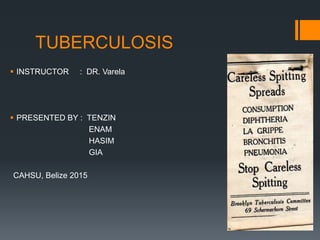
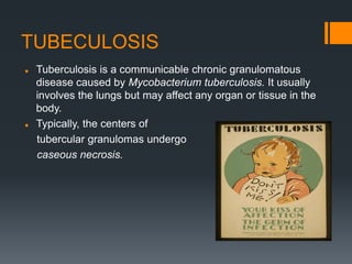
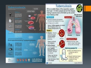
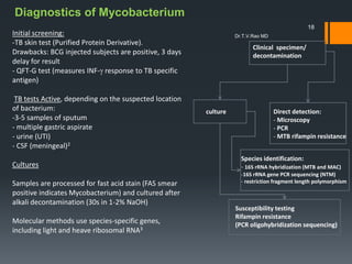
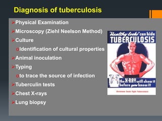
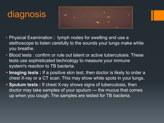
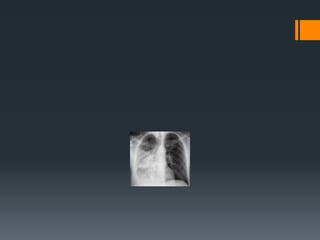
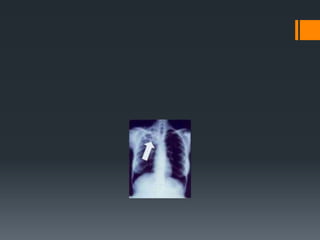
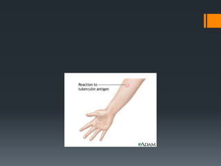
TUBERCULOSIS
 INSTRUCTOR : DR. Varela
 PRESENTED BY : TENZIN
ENAM
HASIM
GIA
CAHSU, Belize 2015
TUBECULOSIS
 Tuberculosis is a communicable chronic granulomatous
disease caused by Mycobacterium tuberculosis. It usually
involves the lungs but may affect any organ or tissue in the
body.
 Typically, the centers of
tubercular granulomas undergo
caseous necrosis.
 Tuberculosis flourishes wherever there is poverty,
crowding, and chronic debilitating illness, elderly
persons, with their weakened defenses, are vulnerable.
 Certain disease states also increase the risk: diabetes
mellitus, Hodgkin disease, chronic lung disease, chronic
renal failure, malnutrition, alcoholism, and
immunosuppression.
 In areas of the world where HIV infection is prevalent, it
has become the single most important risk factor for the
development of tuberculosis.
 It is important that infection be differentiated from
disease. Infection implies seeding of a focus with
organisms, which may or may not cause clinically
significant tissue damage (i.e., disease).
 In most persons, an asymptomatic focus of pulmonary
infection appears that is self-limited.
 Generally, the only evidence of infection, if any remains,
is a tiny, telltale fibrocalcific nodule at the site of the
infection.
 Viable organisms may remain dormant in such loci for
decades, and possibly for the life of the host. Such
persons are infected but do not have active disease and
so cannot transmit organisms to others.
 However when their defenses are lowered, the infection
may reactivate to produce communicable and potentially
life-threatening disease.
Primary tuberculosis
 It is the form of disease that develops in a previously
unexposed, and therefore unsensitized, person. Elderly persons
and profoundly immunosuppressed persons may lose their
sensitivity to the tubercle bacillus and so may develop primary
tuberculosis more than once. With primary tuberculosis, the
source of the organism is exogenous. About 5% of those newly
infected develop significant disease.
The chief implications of primary tuberculosis are that (1) it
induces hypersensitivity and increased resistance; (2) the foci of
scarring may harbor viable bacilli for years, perhaps for life, and
thus be the nidus for reactivation at a later time when host
defenses are compromised; and (3) uncommonly, the disease
may develop without interruption into so-called progressive
primary tuberculosis.
 Progressive primary tuberculosis occurs in individuals
who are immunocompromised such as AIDS( CD4+
counts <200 cells/mm3 ) or in malnourished children or
in the elderly. Certain racial groups, such as Inuit.
 Immunosuppression results in an inability to mount a
CD4+ T cell-mediated immunologic reaction. The lack of
a tissue hypersensitivity reaction results in the absence
of the characteristic caseating granulomas (nonreactive
tuberculosis).
 Progressive primary tuberculosis more often resembles
an acute bacterial pneumonia, with lower and middle
lobe consolidation.
 Lymphohematogenous dissemination is a dreaded
complication and may result in the development of
tuberculous meningitis and miliary tuberculosis.
Symptoms-
 nonproductive cough,
 chest pain,
 fever and
 loss of appetite.
Secondary (or postprimary)
tuberculosis:
 is the pattern of disease that arises in a previously
sensitized host.
 commonly it arises from reactivation of dormant primary
lesions many decades after initial infection, particularly
when host resistance is weakened.
 It may also result from exogenous reinfection because of
waning of the protection afforded by the primary disease
or because of a large inoculum of virulent bacilli.
 Reactivation of endogenous tuberculosis is more common in
low-prevalence areas.
 Whatever the source of the organism, only a few individuals
(less than 5%) with primary disease subsequently develop
secondary tuberculosis.
 Secondary pulmonary tuberculosis is classically localized to
the apex of one or both upper lobes.
 Because of the preexistence of hypersensitivity, the bacilli
excite a prompt and marked tissue response that tends to
wall off the focus. As a result of this localization, the regional
lymph nodes are less prominently involved early in the
developing disease than they are in primary tuberculosis.
 On the other hand, cavitation occurs readily in the
secondary form, resulting in dissemination along the
airways. Indeed, cavitation is almost inevitable in
neglected secondary tuberculosis, and erosion into an
airway becomes an important source of infectivity,
because the person now raises sputum containing
bacilli.
 Symptoms –
 Productive cough
 Hemoptysis
 Fever
 Loss of appetite
 Night sweat
 Localized secondary tuberculosis may be asymptomatic.
 Systemic symptoms,often appear early in the course
include malaise, anorexia, weight loss, and fever.
Commonly, the fever is low grade and remittent
(appearing late each afternoon and then subsiding), and
night sweats occur.
 With progressive pulmonary involvement, increasing
amounts of sputum, at first mucoid and later purulent,
appear. When cavitation is present, the sputum contains
tubercle bacilli. Some degree of hemoptysis is present in
about half of all cases of pulmonary tuberculosis.
Pleuritic pain may result from extension of the infection
to the pleural surfaces.
 Extrapulmonary manifestations of tuberculosis depend
on the organ system involved (for example, tuberculous
salpingitis may present as infertility, tuberculous
meningitis with headache and neurologic deficits, Pott
disease with paraplegia).
 Dx. The diagnosis of pulmonary disease is based in part
on the history and on physical and radiographic findings
of consolidation or cavitation in the apices of the lungs.
Ultimately, however, tubercle bacilli must be identified.
Diagnostic discoveries
24th March 1882 (Robert Koch)
TB Day
 Discovery of staining technique
that identified Tuberculosis bacillus
 Definite diagnosis made possible
and thus treatment could begin
1890 (Robert Koch)
 Tuberculin discovered
 Diagnostic use when injected into
skin
1895 (Roentgen)
 Discovery of X-rays
 Early diagnosis of pulmonary
disease
Diagnostics of Mycobacterium
Initial screening:
-TB skin test (Purified Protein Derivative).
Drawbacks: BCG injected subjects are positive, 3 days
delay for result
- QFT-G test (measures INF- response to TB specific
antigen)
TB tests Active, depending on the suspected location
of bacterium:
-3-5 samples of sputum
- multiple gastric aspirate
- urine (UTI)
- CSF (meningeal)2
Cultures
Samples are processed for fast acid stain (FAS smear
positive indicates Mycobacterium) and cultured after
alkali decontamination (30s in 1-2% NaOH)
Molecular methods use species-specific genes,
including light and heave ribosomal RNA3
Clinical specimen/
decontamination
culture Direct detection:
- Microscopy
- PCR
- MTB rifampin resistance
Species identification:
- 16S rRNA hybridization (MTB and MAC)
-16S rRNA gene PCR sequencing (NTM)
- restriction fragment length polymorphism
Susceptibility testing
Rifampin resistance
(PCR oligohybridization sequencing)
18
Dr.T.V.Rao MD
Diagnosis of tuberculosis
Physical Examination
Microscopy (Ziehl Neelson Method)
Culture
oIdentification of cultural properties
Animal inoculation
Typing
oto trace the source of infection
Tuberculin tests
Chest X-rays
Lung biopsy
diagnosis
• Physical Examination : lymph nodes for swelling and use a
stethoscope to listen carefully to the sounds your lungs make while
you breathe.
• Blood tests : confirm or rule out latent or active tuberculosis. These
tests use sophisticated technology to measure your immune
system's reaction to TB bacteria.
 Imaging tests : If a positive skin test, then doctor is likely to order a
chest X-ray or a CT scan. This may show white spots in your lungs.
 Sputum tests : If chest X-ray shows signs of tuberculosis, then
doctor may take samples of your sputum — the mucus that comes
up when you cough. The samples are tested for TB bacteria.
Treatment
 1st line treatment – ethambutol
(or streptomycin), isoniazid, pyrazinamide and
rifamycins.(rifampin, rifabutin, rifapentine)
 2nd line – aminoglycoside, macrolides, cycloserine,
flouroquinolones.
 Vaccine – bcg(not in US)
Reserve drugs which may be used when first line
drugs have failed are:
Ethionamide
Prothionamide
Amikacin
Kanamycin
Capreomycin
Viomycin
Cycloserine
Quinolones (ofloxacin, ciprofloxacin, sparfloxacin)
 New cases – 4 drugs
 Isoniazid
 Rifampin
 Pyrazinamide
 Ethambutol/streptomycin
02000
month
 4 month of isoniazid and rifampin.
 The prognosis of tuberculosis is generally favorable if infections are
localized to the lungs, but it worsens significantly when the disease
occurs in the setting of aged, debilitated, or immunosuppressed
persons, who are at high risk for developing miliary tuberculosis,
and in those with MDR-TB.
 Amyloidosis may appear in persistent cases.
Medication side effects
 Serious side effects of TB drugs aren't common but can be
dangerous when they do occur. All tuberculosis medications
can be highly toxic to your liver. When taking these
medications, call your doctor immediately if you experience
any of the following:
 Nausea or vomiting
 Loss of appetite
 A yellow color to your skin (jaundice)
 Dark urine
 A fever that lasts three or more days and has no obvious
cause
Prevention Protect your family and friends
 Stay home. Don't go to work or school or sleep in a room with
other people during the first few weeks of treatment for active
tuberculosis.
 Ventilate the room. Tuberculosis germs spread more easily in
small closed spaces where air doesn't move. If it's not too cold
outdoors, open the windows and use a fan to blow indoor air
outside.
 Cover your mouth. Use a tissue to cover your mouth anytime
you laugh, sneeze or cough. Put the dirty tissue in a bag, seal it
and throw it away.
 Wear a mask. Wearing a surgical mask when you're around other
people during the first three weeks of treatment may help lessen
the risk of transmission.
THANK YOU

Tuberculosis

  • 1.
    TUBERCULOSIS  INSTRUCTOR :DR. Varela  PRESENTED BY : TENZIN ENAM HASIM GIA CAHSU, Belize 2015
  • 2.
    TUBECULOSIS  Tuberculosis isa communicable chronic granulomatous disease caused by Mycobacterium tuberculosis. It usually involves the lungs but may affect any organ or tissue in the body.  Typically, the centers of tubercular granulomas undergo caseous necrosis.
  • 3.
     Tuberculosis flourisheswherever there is poverty, crowding, and chronic debilitating illness, elderly persons, with their weakened defenses, are vulnerable.  Certain disease states also increase the risk: diabetes mellitus, Hodgkin disease, chronic lung disease, chronic renal failure, malnutrition, alcoholism, and immunosuppression.  In areas of the world where HIV infection is prevalent, it has become the single most important risk factor for the development of tuberculosis.
  • 4.
     It isimportant that infection be differentiated from disease. Infection implies seeding of a focus with organisms, which may or may not cause clinically significant tissue damage (i.e., disease).  In most persons, an asymptomatic focus of pulmonary infection appears that is self-limited.  Generally, the only evidence of infection, if any remains, is a tiny, telltale fibrocalcific nodule at the site of the infection.
  • 5.
     Viable organismsmay remain dormant in such loci for decades, and possibly for the life of the host. Such persons are infected but do not have active disease and so cannot transmit organisms to others.  However when their defenses are lowered, the infection may reactivate to produce communicable and potentially life-threatening disease.
  • 6.
    Primary tuberculosis  Itis the form of disease that develops in a previously unexposed, and therefore unsensitized, person. Elderly persons and profoundly immunosuppressed persons may lose their sensitivity to the tubercle bacillus and so may develop primary tuberculosis more than once. With primary tuberculosis, the source of the organism is exogenous. About 5% of those newly infected develop significant disease. The chief implications of primary tuberculosis are that (1) it induces hypersensitivity and increased resistance; (2) the foci of scarring may harbor viable bacilli for years, perhaps for life, and thus be the nidus for reactivation at a later time when host defenses are compromised; and (3) uncommonly, the disease may develop without interruption into so-called progressive primary tuberculosis.
  • 7.
     Progressive primarytuberculosis occurs in individuals who are immunocompromised such as AIDS( CD4+ counts <200 cells/mm3 ) or in malnourished children or in the elderly. Certain racial groups, such as Inuit.  Immunosuppression results in an inability to mount a CD4+ T cell-mediated immunologic reaction. The lack of a tissue hypersensitivity reaction results in the absence of the characteristic caseating granulomas (nonreactive tuberculosis).
  • 8.
     Progressive primarytuberculosis more often resembles an acute bacterial pneumonia, with lower and middle lobe consolidation.  Lymphohematogenous dissemination is a dreaded complication and may result in the development of tuberculous meningitis and miliary tuberculosis.
  • 9.
    Symptoms-  nonproductive cough, chest pain,  fever and  loss of appetite.
  • 11.
    Secondary (or postprimary) tuberculosis: is the pattern of disease that arises in a previously sensitized host.  commonly it arises from reactivation of dormant primary lesions many decades after initial infection, particularly when host resistance is weakened.  It may also result from exogenous reinfection because of waning of the protection afforded by the primary disease or because of a large inoculum of virulent bacilli.
  • 12.
     Reactivation ofendogenous tuberculosis is more common in low-prevalence areas.  Whatever the source of the organism, only a few individuals (less than 5%) with primary disease subsequently develop secondary tuberculosis.  Secondary pulmonary tuberculosis is classically localized to the apex of one or both upper lobes.  Because of the preexistence of hypersensitivity, the bacilli excite a prompt and marked tissue response that tends to wall off the focus. As a result of this localization, the regional lymph nodes are less prominently involved early in the developing disease than they are in primary tuberculosis.
  • 13.
     On theother hand, cavitation occurs readily in the secondary form, resulting in dissemination along the airways. Indeed, cavitation is almost inevitable in neglected secondary tuberculosis, and erosion into an airway becomes an important source of infectivity, because the person now raises sputum containing bacilli.
  • 14.
     Symptoms – Productive cough  Hemoptysis  Fever  Loss of appetite  Night sweat
  • 15.
     Localized secondarytuberculosis may be asymptomatic.  Systemic symptoms,often appear early in the course include malaise, anorexia, weight loss, and fever. Commonly, the fever is low grade and remittent (appearing late each afternoon and then subsiding), and night sweats occur.  With progressive pulmonary involvement, increasing amounts of sputum, at first mucoid and later purulent, appear. When cavitation is present, the sputum contains tubercle bacilli. Some degree of hemoptysis is present in about half of all cases of pulmonary tuberculosis. Pleuritic pain may result from extension of the infection to the pleural surfaces.
  • 16.
     Extrapulmonary manifestationsof tuberculosis depend on the organ system involved (for example, tuberculous salpingitis may present as infertility, tuberculous meningitis with headache and neurologic deficits, Pott disease with paraplegia).  Dx. The diagnosis of pulmonary disease is based in part on the history and on physical and radiographic findings of consolidation or cavitation in the apices of the lungs. Ultimately, however, tubercle bacilli must be identified.
  • 17.
    Diagnostic discoveries 24th March1882 (Robert Koch) TB Day  Discovery of staining technique that identified Tuberculosis bacillus  Definite diagnosis made possible and thus treatment could begin 1890 (Robert Koch)  Tuberculin discovered  Diagnostic use when injected into skin 1895 (Roentgen)  Discovery of X-rays  Early diagnosis of pulmonary disease
  • 18.
    Diagnostics of Mycobacterium Initialscreening: -TB skin test (Purified Protein Derivative). Drawbacks: BCG injected subjects are positive, 3 days delay for result - QFT-G test (measures INF- response to TB specific antigen) TB tests Active, depending on the suspected location of bacterium: -3-5 samples of sputum - multiple gastric aspirate - urine (UTI) - CSF (meningeal)2 Cultures Samples are processed for fast acid stain (FAS smear positive indicates Mycobacterium) and cultured after alkali decontamination (30s in 1-2% NaOH) Molecular methods use species-specific genes, including light and heave ribosomal RNA3 Clinical specimen/ decontamination culture Direct detection: - Microscopy - PCR - MTB rifampin resistance Species identification: - 16S rRNA hybridization (MTB and MAC) -16S rRNA gene PCR sequencing (NTM) - restriction fragment length polymorphism Susceptibility testing Rifampin resistance (PCR oligohybridization sequencing) 18 Dr.T.V.Rao MD
  • 19.
    Diagnosis of tuberculosis PhysicalExamination Microscopy (Ziehl Neelson Method) Culture oIdentification of cultural properties Animal inoculation Typing oto trace the source of infection Tuberculin tests Chest X-rays Lung biopsy
  • 20.
    diagnosis • Physical Examination: lymph nodes for swelling and use a stethoscope to listen carefully to the sounds your lungs make while you breathe. • Blood tests : confirm or rule out latent or active tuberculosis. These tests use sophisticated technology to measure your immune system's reaction to TB bacteria.  Imaging tests : If a positive skin test, then doctor is likely to order a chest X-ray or a CT scan. This may show white spots in your lungs.  Sputum tests : If chest X-ray shows signs of tuberculosis, then doctor may take samples of your sputum — the mucus that comes up when you cough. The samples are tested for TB bacteria.
  • 21.
    Treatment  1st linetreatment – ethambutol (or streptomycin), isoniazid, pyrazinamide and rifamycins.(rifampin, rifabutin, rifapentine)  2nd line – aminoglycoside, macrolides, cycloserine, flouroquinolones.  Vaccine – bcg(not in US)
  • 22.
    Reserve drugs whichmay be used when first line drugs have failed are: Ethionamide Prothionamide Amikacin Kanamycin Capreomycin Viomycin Cycloserine Quinolones (ofloxacin, ciprofloxacin, sparfloxacin)
  • 23.
     New cases– 4 drugs
  • 24.
     Isoniazid  Rifampin Pyrazinamide  Ethambutol/streptomycin 02000 month
  • 25.
     4 monthof isoniazid and rifampin.
  • 26.
     The prognosisof tuberculosis is generally favorable if infections are localized to the lungs, but it worsens significantly when the disease occurs in the setting of aged, debilitated, or immunosuppressed persons, who are at high risk for developing miliary tuberculosis, and in those with MDR-TB.  Amyloidosis may appear in persistent cases.
  • 27.
    Medication side effects Serious side effects of TB drugs aren't common but can be dangerous when they do occur. All tuberculosis medications can be highly toxic to your liver. When taking these medications, call your doctor immediately if you experience any of the following:  Nausea or vomiting  Loss of appetite  A yellow color to your skin (jaundice)  Dark urine  A fever that lasts three or more days and has no obvious cause
  • 28.
    Prevention Protect yourfamily and friends  Stay home. Don't go to work or school or sleep in a room with other people during the first few weeks of treatment for active tuberculosis.  Ventilate the room. Tuberculosis germs spread more easily in small closed spaces where air doesn't move. If it's not too cold outdoors, open the windows and use a fan to blow indoor air outside.  Cover your mouth. Use a tissue to cover your mouth anytime you laugh, sneeze or cough. Put the dirty tissue in a bag, seal it and throw it away.  Wear a mask. Wearing a surgical mask when you're around other people during the first three weeks of treatment may help lessen the risk of transmission.
  • 32.